
Blackberry Leaves as New Functional Food? Screening Antioxidant, Anti-Inflammatory and Microbiological Activities in Correlation with Phytochemical Analysis

 , , , , , ,
Abstract
:1. Introduction
2. Materials and Methods
2.1. Chemicals and Reagents
2.2. Preparation and Analysis of Rubi Folium Extract
2.2.1. Plan Material
- −
- Rubus Chester—thornless variety, the mature shrub grows up to a height of 1.5 m high; the variety is susceptible to anthracnose, gray mold and verticillium wilt.
- −
- Rubus Loch Ness—thornless variety, the mature shrub grows up to a height of 5 m, thermal and soil requirements of this variety are low.
- −
- Rubus Loch Tay—thornless variety, the mature shrub grows up to a height of 1.5 m, fruits on two-year-old shoots, average thermal and soil requirements.
- −
- Rubus Ruczaj—thornless variety, the mature shrub grows to a height of approximately 2 m, medium thermal and soil requirements, susceptible to gray mold, medium thermal and soil conditions.
2.2.2. Extract Preparation
2.2.3. Determination of Active Compounds Content in Extracts
2.2.4. Total Phenolic Content (TPC)
2.2.5. Antioxidant Activity
Assay with 2,2-Diphenyl-1-picrylhydrazyl (DPPH)
2,2-Azino-bis(3-ethylbenzothiazoline-6-sulfonic Acid) (ABTS) Radical Cation-Based Assays
Oxygen Radical Absorbance Capacity (ORAC) Assay
Effect on Superoxide Dismutase (SOD) Activity
Cupric Ion Reducing Antioxidant Capacity (CUPRAC) Assay
Ferric Ion Reducing Antioxidant Parameter (FRAP) Assay
Hydroxyl Radical Averting Capacity (HORAC) Assay
Effect on Glutathione Reductase (GR) and Glutathione Peroxidase (GPx) Activity
Inhibition of Lipid Peroxidation
β-Carotene Bleaching Test
2.2.6. Effect on Cholinesterase (ChE) Activity
2.2.7. Anti-Inflammatory Activity
Anti-Hyaluronidase Activity
Effect on Cyclooxygenase-2 (COX-2) Activity
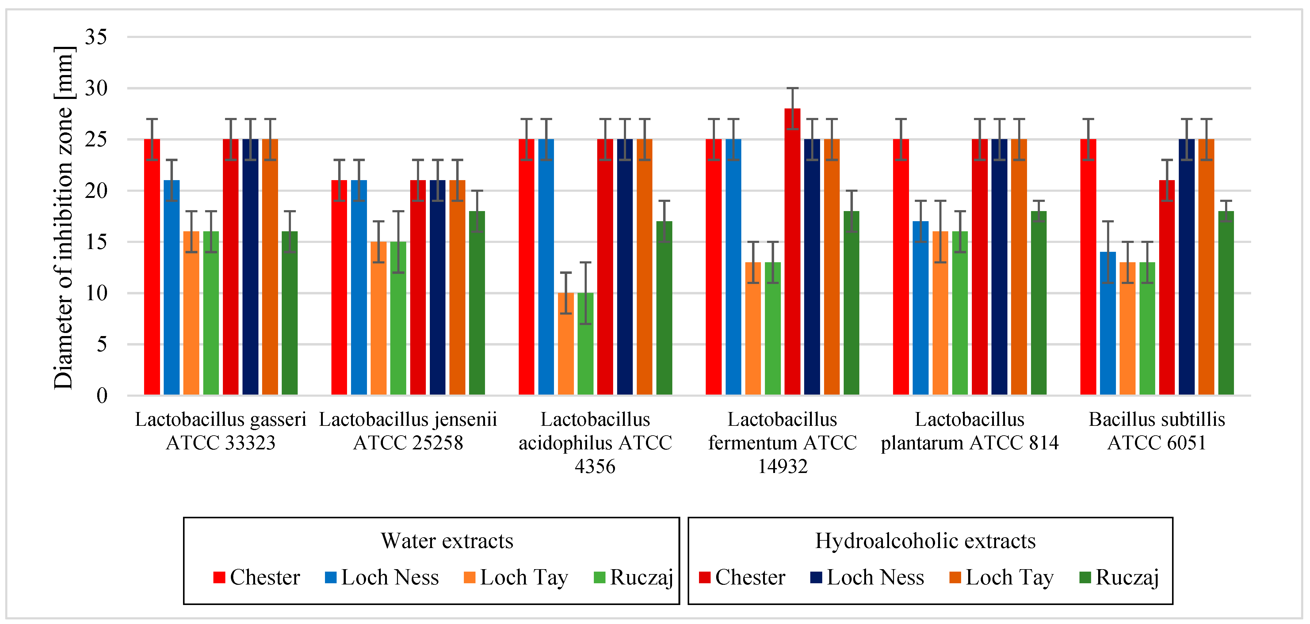
2.2.8. Microbiological Activity
Inoculum Standardization
Assay of Antibacterial Activity Using Agar Well Diffusion Method
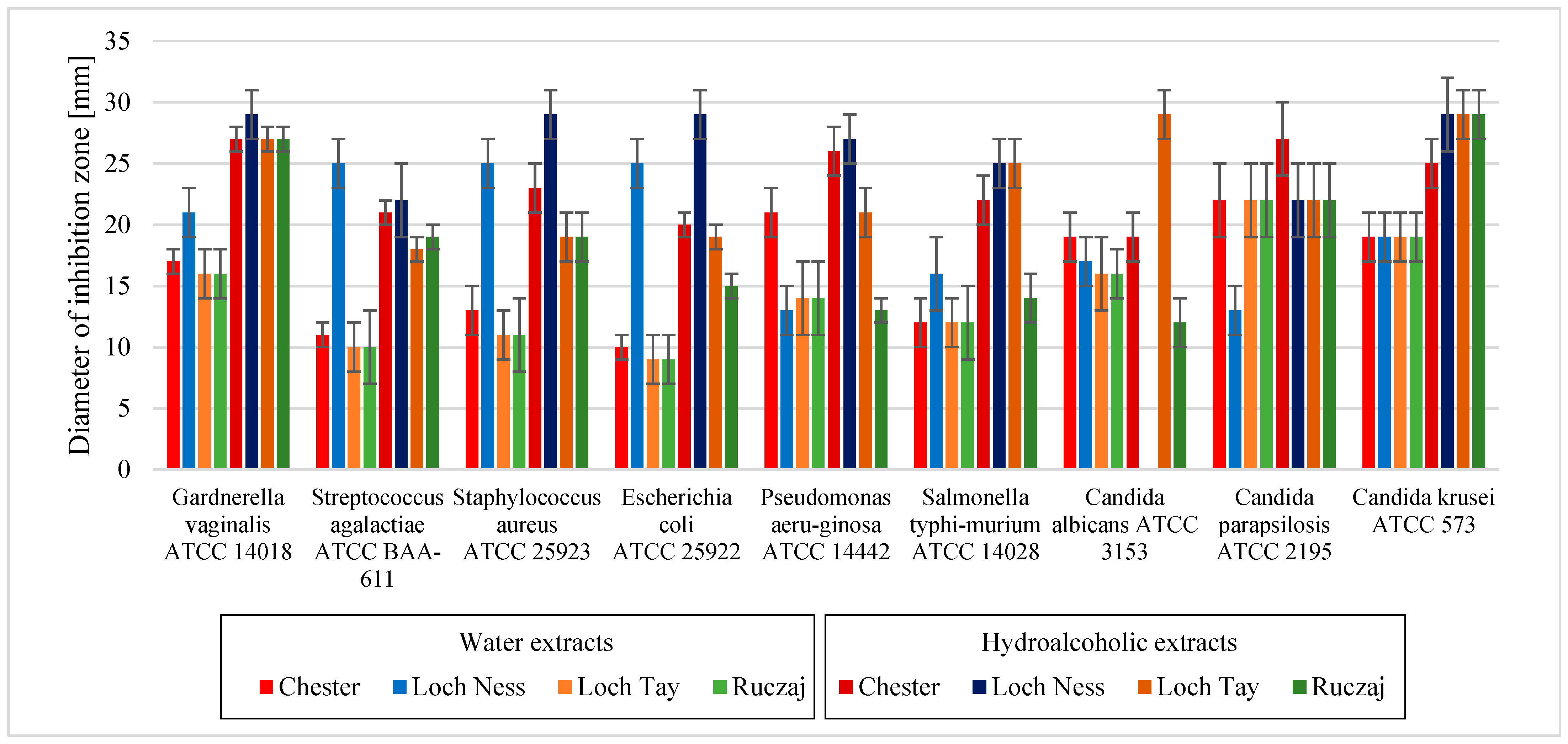
3. Results and Discussion
4. Conclusions
Supplementary Materials
Author Contributions
Funding
Institutional Review Board Statement
Informed Consent Statement
Data Availability Statement
Conflicts of Interest
References
- Hasler, C.M. Functional foods: Benefits, concerns and challenges—A position paper from the american council on science and health. J. Nutr. 2002, 132, 3772–3781. [Google Scholar] [CrossRef] [PubMed] [Green Version]
- Ferlemi, A.V.; Lamari, F.N. Berry Leaves: An Alternative Source of Bioactive Natural Products of Nutritional and Medicinal Value. Antioxidants 2016, 5, 17. [Google Scholar] [CrossRef] [PubMed]
- Kruczek, M.; Kostecka-Gugala, A.; Augustynowicz, J.; Ledwozyw-Smolen, I.; Orzel, A.; Krol-Dyrek, K.; Kaszycki, P. Raspberry and blackberry leaves as a raw material for pharmaceutical industry. Przemysl. Chem. 2015, 94, 1431–1436. [Google Scholar]
- Zia-Ul-Haq, M.; Riaz, M.; De Feo, V.; Jaafar, H.Z.; Moga, M. Rubus fruticosus L.: Constituents, biological activities and health related uses. Molecules 2014, 19, 10998–11029. [Google Scholar] [CrossRef] [PubMed] [Green Version]
- Vilariño, M.V.; Franco, C.; Quarrington, C. Food loss and Waste Reduction as an Integral Part of a Circular Economy. Front. Environ. Sci. 2017, 5, 21. [Google Scholar] [CrossRef] [Green Version]
- Makris, D.P.; Sahin, S. Polyphenolic Antioxidants from Agri-Food Waste Biomass. Antioxidants 2019, 8, 624. [Google Scholar] [CrossRef] [PubMed] [Green Version]
- Osorio, L.L.D.R.; Flórez-López, E.; Grande-Tovar, C.D. The Potential of Selected Agri-Food Loss and Waste to Contribute to a Circular Economy: Applications in the Food, Cosmetic and Pharmaceutical Industries. Molecules 2021, 26, 515. [Google Scholar] [CrossRef]
- Farooq, M.U.; Mumtaz, M.W.; Mukhtar, H.; Rashid, U.; Akhtar, M.T.; Raza, S.A.; Nadeem, M. UHPLC-QTOF-MS/MS based phytochemical characterization and anti-hyperglycemic prospective of hydro-ethanolic leaf extract of Butea monosperma. Sci. Rep. 2020, 10, 3530. [Google Scholar] [CrossRef] [PubMed] [Green Version]
- Faggian, M.; Bernabè, G.; Ferrari, S.; Francescato, S.; Baratto, G.; Castagliuolo, I.; Dall’Acqua, S.; Peron, G. Polyphenol-Rich Larix decidua Bark Extract with Antimicrobial Activity against Respiratory-Tract Pathogens: A Novel Bioactive Ingredient with Potential Pharmaceutical and Nutraceutical Applications. Antibiotics 2021, 10, 789. [Google Scholar] [CrossRef]
- Smeriglio, A.; Denaro, M.; Trombetta, D.; Ragusa, S.; Circosta, C. New Insights on Euphorbia dendroides L. (Euphorbiaceae): Polyphenol Profile and Biological Properties of Hydroalcoholic Extracts from Aerial Parts. Plants 2021, 10, 1621. [Google Scholar] [CrossRef] [PubMed]
- Oszmiański, J.; Wojdyło, A.; Gorzelany, J.; Kapusta, I. Identification and characterization of low molecular weight polyphenols in berry leaf extracts by HPLC-DAD and LC-ESI/MS. J. Agric. Food Chem. 2011, 59, 12830–12835. [Google Scholar] [CrossRef] [PubMed]
- Patel, A.V.; Rojas-Vera, J.; Dacke, C.G. Therapeutic constituents and actions of Rubus species. Curr. Med. Chem. 2004, 11, 1501–1512. [Google Scholar] [CrossRef]
- Abu-Shandi, K.; Al-Rawashdeh, A.; Al-Mazaideh, G.; Abu-Nameh, E.; Al-Amro, A.; Al-Soufi, H.; Al-Ma’abreh, A.; Al-Dawdeyah, A. A Novel Strategy for the Identification of the Medicinal Natural Products in Rubus Fruticosus Plant by Using GC/MS Technique: A Study on Leaves, Stems and Roots of the Plant. Adv. Anal. Chem. 2015, 5, 31–41. [Google Scholar]
- Oszmiański, J.; Wojdyło, A.; Nowicka, P.; Teleszko, M.; Cebulak, T.; Wolanin, M. Determination of phenolic compounds and antioxidant activity in leaves from wild Rubus L. species. Molecules 2015, 20, 4951–4966. [Google Scholar] [CrossRef] [PubMed] [Green Version]
- Commission of the European Comminities. Proceeding of the International Conference of Harmonisation. Validation of analytical procedures Q2(R2). 2018. Available online: https://www.ich.org/page/quality-guidelines (accessed on 3 December 2021).
- Kikowska, M.A.; Chmielewska, M.; Włodarczyk, A.; Studzińska-Sroka, E.; Żuchowski, J.; Stochmal, A.; Kotwicka, M.; Thiem, B. Effect of Pentacyclic Triterpenoids-Rich Callus Extract of Chaenomeles japonica (Thunb.) Lindl. ex Spach on Viability, Morphology, and Proliferation of Normal Human Skin Fibroblasts. Molecules 2018, 23, 3009. [Google Scholar] [CrossRef] [PubMed] [Green Version]
- Denev, P.; Kratchanova, M.; Ciz, M.; Lojek, A.; Vasicek, O.; Nedelcheva, P.; Blazheva, D.; Toshkova, R.; Gardeva, E.; Yossifova, L.; et al. Biological activities of selected polyphenol-rich fruits related to immunity and gastrointestinal health. Food Chem. 2014, 157, 37–44. [Google Scholar] [CrossRef]
- Parschat, K.; Canne, C.; Hüttermann, J.; Kappl, R.; Fetzner, S. Xanthine dehydrogenase from Pseudomonas putida 86: Specificity, oxidation–reduction potentials of its redox-active centers, and first EPR characterization. Biochim. Biophys. Acta. 2001, 1544, 151–165. [Google Scholar] [CrossRef]
- Moreira, P.R.; Maioli, M.A.; Medeiros, H.C.D.; Guelfi, M.; Pereira, F.T.V.; Mingatto, F.E. Protective effect of bixin on carbon tetrachloride-induced hepatotoxicity in rats. Biol. Res. 2014, 47, 49. [Google Scholar] [CrossRef] [PubMed] [Green Version]
- Singh, R.P.; Padmavathi, B.; Rao, A.R. Modulatory influence of Adhatoda vesical (Justicia adhatoda) leaf extract on the enzymes of xenobiotic metabolism, antioxidant status and lipid peroxidation in mice. Mol. Cell. Biochem. 2000, 213, 99–109. [Google Scholar] [CrossRef]
- Kozarski, M.; Klaus, A.; Nikšić, M.; Vrvić, M.M.; Todorović, N.; Jakovljević, D.; Van Griensven, L.J. Antioxidative activities and chemical characterization of polysaccharide extracts from the widely used mushrooms Ganoderma applanatum, Ganoderma lucidum, Lentinus edodes and Trametes versicolor. J. Food Compost. Anal. 2012, 26, 144–153. [Google Scholar] [CrossRef]
- Öztürk, M.; Duru, M.E.; Kivrak, Ş.; Mercan-Doğan, N.; Türkoglu, A.; Özler, M.A. In vitro antioxidant, anticholinesterase and antimicrobial activity studies on three Agaricus species with fatty acid compositions and iron contents: A comparative study on the three most edible mushrooms. Food Chem. Toxicol. 2011, 49, 1353–1360. [Google Scholar] [CrossRef] [PubMed]
- Ellman, G.L.; Lourtney, D.K.; Andres, V.; Gmelin, G.A. New and rapid colorimetric determination of acetylcholinesterase activity. Biochem. Pharmacol. 1961, 7, 88–95. [Google Scholar] [CrossRef]
- Rhee, I.K.; van Rijn, R.M.; Verpoorte, R. Qualitative determination of false-positive effects in the acetylcholinesterase assay using thin layer chromatography. Phytochem. Analysis. 2003, 14, 127–131. [Google Scholar] [CrossRef]
- Szwajgier, D.; Baranowska-Wójcik, E. Terpenes and Phenylpropanoids as Acetyl- and Butyrylcholinesterase Inhibitors: A Comparative Study. Curr. Alzheimer Res. 2019, 16, 963–973. [Google Scholar] [CrossRef]
- Studzińska-Sroka, E.; Dudek-Makuch, M.; Chanaj-Kaczmarek, J.; Czepulis, N.; Korybalska, K.; Rutkowski, R.; Łuczak, J.; Grabowska, K.; Bylka, W.; Witowski, J. Anti-inflammatory Activity and Phytochemical Profile of Galinsoga parviflora Cav. Molecules 2018, 23, 2133. [Google Scholar] [CrossRef] [Green Version]
- Bhattacharya, K.; Chandra, G. Phagodeterrence, larvicidal and oviposition deterrence activity of Tragia involucrata L. (Euphorbiaceae) root extractives against vector of lymphatic filariasis Culex quinquefasciatus (Diptera: Culicidae). Asian Pac. J. Trop. Dis. 2014, 4, 226–232. [Google Scholar] [CrossRef]
- Gudej, J.; Tomczyk, M. Determination of flavonoids, tannins and ellagic acid in leaves from Rubus L. species. Arch. Pharm. Res. 2004, 27, 1114–1119. [Google Scholar] [CrossRef] [PubMed]
- Wang, S.Y.; Lin, H.S. Antioxidant activity in fruits and leaves of blackberry, raspberry, and strawberry varies with cultivar and developmental stage. J. Agric. Food Chem. 2000, 48, 140–146. [Google Scholar] [CrossRef] [PubMed]
- Jazić, M.; Kukrić, Z.; Vulić, J.; Četojević-Simin, D. Polyphenolic composition, antioxidant and antiproliferative effects of wild and cultivated blackberries (Rubus fruticosus L.) pomace. Int. J. Food Sci. Technol. 2019, 54, 194–201. [Google Scholar] [CrossRef] [Green Version]
- Buřičová, L.; Andjelkovic, M.; Čermáková, A.; Réblová, Z.; Jurček, O.; Kolehmainen, E.; Verhé, R.; Kvasnička, F. Antioxidant Capacity and Antioxidants of Strawberry, Blackberry, and Raspberry Leaves. Czech J. Food Sci. 2011, 29, 181–189. [Google Scholar] [CrossRef] [Green Version]
- Priyadarsini, K.I.; Khopde, S.M.; Kumar, S.S.; Mohan, H. Free radical studies of ellagic acid, a natural phenolic antioxidant. J. Agric. Food Chem. 2002, 50, 2200–2206. [Google Scholar] [CrossRef] [PubMed]
- Han, D.H.; Lee, M.J.; Kim, J.H. Antioxidant and apoptosis-inducing activities of ellagic acid. Anticancer Res. 2006, 26, 3601–3606. [Google Scholar] [PubMed]
- Park, J.Y.; Han, X.; Piao, M.J.; Oh, M.C.; Fernando, P.M.; Kang, K.A.; Ryu, Y.S.; Jung, U.; Kim, I.G.; Hyun, J.W. Hyperoside Induces Endogenous Antioxidant System to Alleviate Oxidative Stress. J. Cancer Prev. 2016, 21, 41–47. [Google Scholar] [CrossRef] [PubMed] [Green Version]
- Williamson, M.P.; McCormick, T.G.; Nance, C.L.; Shearer, W.T. Epigallocatechin gallate, the main polyphenol in green tea, binds to the T-cell receptor, CD4: Potential for HIV-1 therapy. J Allergy Clin. Immunol. 2006, 118, 1369–1374. [Google Scholar] [CrossRef]
- Orhan, I.; Kartal, M.; Tosun, F.; Sener, B. Screening of various phenolic acids and flavonoid derivatives for their anticholinesterase potential. Z. Naturforsch. C J. Biosci. 2007, 62, 829–832. [Google Scholar] [CrossRef] [PubMed]
- Szwajgier, D. Anticholinesterase activity of selected phenolic acids and flavonoids—Interaction testing in model solutions. Ann. Agric. Environ. Med. 2015, 22, 690–694. [Google Scholar] [CrossRef] [PubMed]
- Marquina, M.A.; Corao, G.M.; Araujo, L.; Buitrago, D.; Sosa, M. Hyaluronidase inhibitory activity from the polyphenols in the fruit of blackberry (Rubus fruticosus B.). Fitoterapia 2002, 73, 727–729. [Google Scholar] [CrossRef]
- Girish, K.S.; Kemparaju, K.; Nagaraju, S.; Vishwanath, B.S. Hyaluronidase inhibitors: A biological and therapeutic perspective. Curr. Med. Chem. 2009, 16, 2261–2288. [Google Scholar] [CrossRef]
- El-Shitany, N.A.; El-Bastawissy, E.A.; El-Desoky, K. Ellagic acid protects against carrageenan-induced acute inflammation through inhibition of nuclear factor kappa B, inducible cyclooxygenase and proinflammatory cytokines and enhancement of interleukin-10 via an antioxidant mechanism. Int. Immunopharmacol. 2014, 19, 290–299. [Google Scholar] [CrossRef] [PubMed]
- Kawakami, Y.; Hosokawa, T.; Morinaka, T.; Irino, S.; Hirano, S.; Kobayashi, H.; Yoshioka, A.; Suzuki-Yamamoto, T.; Yokoro, M.; Kimoto, M.; et al. Antiatherogenic effect of guava leaf extracts inhibiting leucocyte-type 12-lipoxygenase activity. Food Chem. 2012, 131, 1069–1075. [Google Scholar] [CrossRef]
- Shukla, R.; Pandey, V.; Vadnere, G.P.; Lodhi, S. Chapter 18—Role of Flavonoids in Management of Inflammatory Disorders. In Bioactive Food as Dietary Interventions for Arthritis and Related Inflammatory Diseases, 2nd ed.; Watson, R.R., Preedy, V.R., Eds.; Academic Press: Cambridge, MA, USA, 2019; pp. 293–322. [Google Scholar]
- Kim, S.J.; Um, J.Y.; Lee, J.Y. Anti-inflammatory activity of hyperoside through the suppression of nuclear factor-κB activation in mouse peritoneal macrophages. Am. J. Chin. Med. 2011, 39, 171–181. [Google Scholar] [CrossRef] [PubMed]
- Morrison, M.; van der Heijden, R.; Heeringa, P.; Kaijzel, E.; Verschuren, L.; Blomhoff, R.; Kooistra, T.; Kleemann, R. Epicatechin attenuates atherosclerosis and exerts anti-inflammatory effects on diet-induced human-CRP and NFκB in vivo. Atherosclerosis 2014, 233, 149–156. [Google Scholar] [CrossRef] [PubMed]
- Molan, A.L.; Lila, M.A.; Mawson, J.; De, S. In vitro and in vivo evaluation of the prebiotic activity of water-soluble blueberry extracts. World J. Microbiol. Biotechnol. 2009, 25, 1243–1249. [Google Scholar] [CrossRef]
- Silva, S.; Costa, E.M.; Mendes, M.; Morais, R.M.; Calhau, C.; Pintado, M.M. Antimicrobial, antiadhesive and antibiofilm activity of an ethanolic, anthocyanin-rich blueberry extract purified by solid phase extraction. J. Appl. Microbiol. 2016, 121, 693–703. [Google Scholar] [CrossRef] [PubMed]
- Weli, A.M.; Al-Saadi, H.S.; Al-Fudhaili, R.S.; Hossain, A.; Putit, Z.B.; Jasim, M.K. Cytotoxic and antimicrobial potential of different leaves extracts of R. fruticosus used traditionally to treat diabetes. Toxicol. Rep. 2020, 7, 183–187. [Google Scholar] [CrossRef] [PubMed]
- Papuc, C.; Goran, G.V.; Predescu, C.N.; Nicorescu, V.; Stefan, G. Plant Polyphenols as Antioxidant and Antibacterial Agents for Shelf-Life Extension of Meat and Meat Products: Classification, Structures, Sources, and Action Mechanisms. Compr. Rev. Food Sci. Food Saf. 2017, 16, 1243–1268. [Google Scholar] [CrossRef] [Green Version]
- Bouarab-Chibane, L.; Forquet, V.; Lantéri, P.; Clément, Y.; Léonard-Akkari, L.; Oulahal, N.; Degraeve, P.; Bordes, C. Antibacterial Properties of Polyphenols: Characterization and QSAR (Quantitative Structure-Activity Relationship) Models. Front. Microbiol. 2019, 10, 829. [Google Scholar] [CrossRef]
- Álvarez-Martínez, F.J.; Barrajón-Catalán, E.; Encinar, J.A.; Rodríguez-Díaz, J.C.; Micol, V. Antimicrobial Capacity of Plant Polyphenols against Gram-positive Bacteria: A Comprehensive Review. Curr. Med. Chem. 2020, 27, 2576–2606. [Google Scholar] [CrossRef]
- González, O.A.; Escamilla, C.; Danaher, R.J.; Dai, J.; Ebersole, J.L.; Mumper, R.J.; Miller, C.S. Antibacterial effects of blackberry extract target periodontopathogens. J. Periodontal. Res. 2013, 48, 80–86. [Google Scholar] [CrossRef] [Green Version]


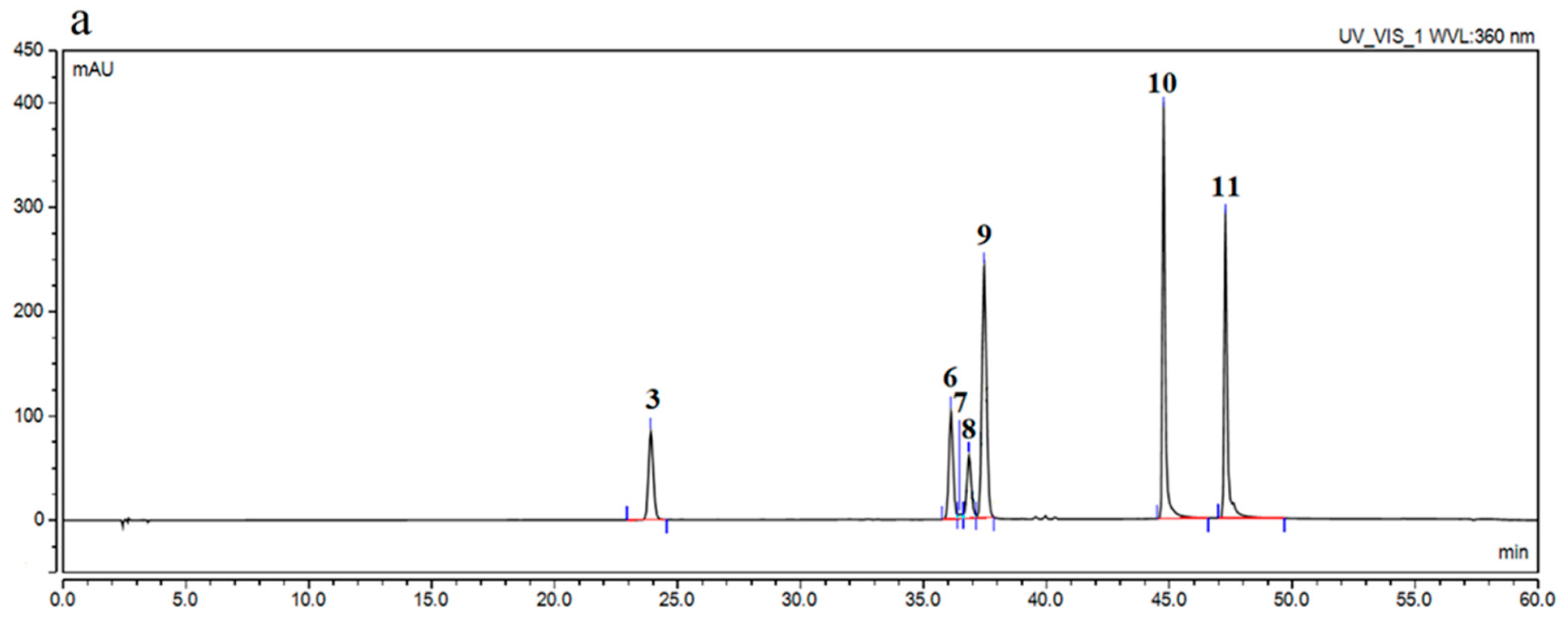
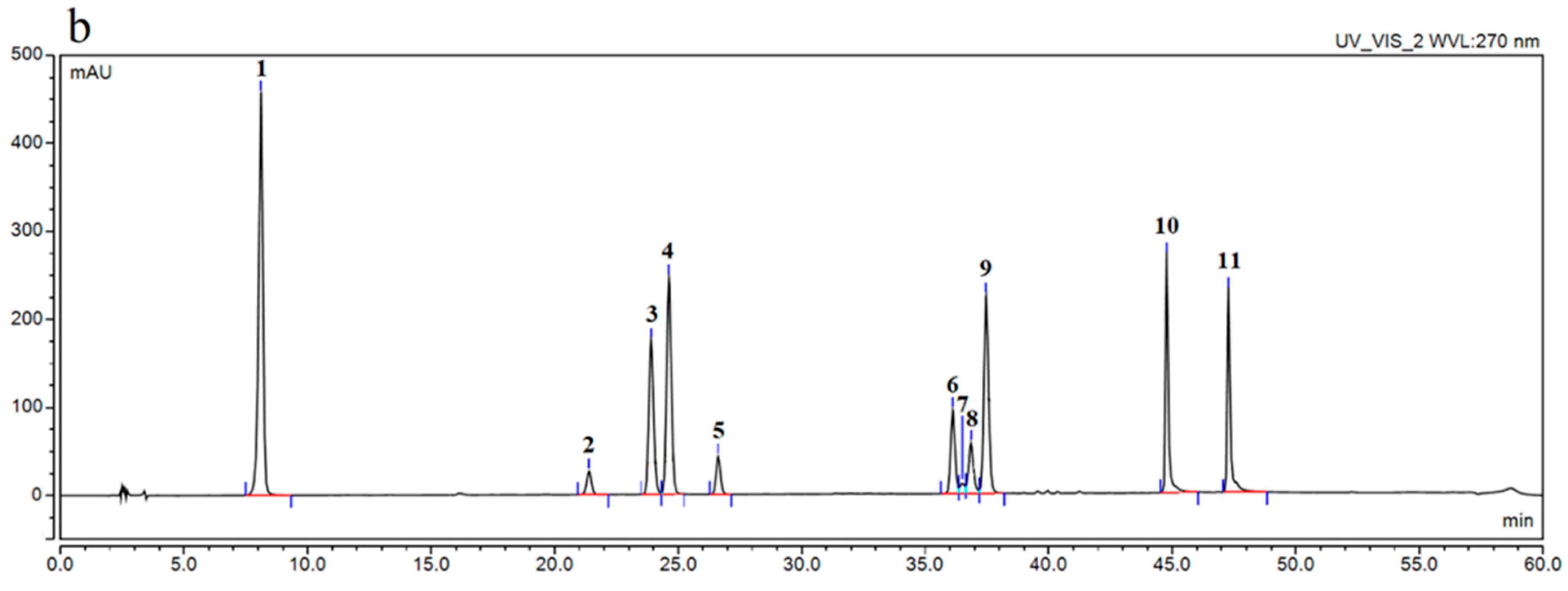
| Compound Name | |
|---|---|
| Phenolic Acids | Neo-chlorogenic acid Caffeic acid Gallic acid p-coumaric acid Ellagic acid |
| Flavonols | Quercetin Quercetin-3-O-galactoside, hyperoside Quercetin-3-O-glucuronide, miquelianin Kaempferol |
| Flavan-3-ols | Catechin Epicatechin Epicatechin gallate methyl gallate |
| Ellagitannins | Sanguiin H-6 /Lambertianin C Casuarinin |
| Anthocyanins | Cyanidin-3-O-glucoside |
| Varieties | Chester | Loch Ness | Loch Tay | Ruczaj | |||||
|---|---|---|---|---|---|---|---|---|---|
| Water Extract | Hydroalcoholic Extract | Water Extract | Hydroalcoholic Extract | Water Extract | Hydroalcoholic Extract | Water Extract | Hydroalcoholic Extract | ||
| Phenolic Compound | Content (µg/g Plant Material) | ||||||||
| Phenolic acids | |||||||||
| caffeic acid | 18.35 ± 1.57 | 17.12 ± 0.81 | 20.65 ± 0.46 | 1.77 ± 0.03 | 606.62 ± 4.22 | 255.25 ± 6.62 | 85.89 ± 5.15 | 55.86 ± 6.59 | |
| ellagic acid | 93.65 ± 9.48 | 515.30 ± 10.69 | 338.29 ± 10.89 | 703.78 ± 13.97 | 468.33 ± 5.17 | 783.06 ± 21.08 | 650.65 ± 11.17 | 876.82 ± 18.97 | |
| Flavonols | |||||||||
| quercetin | 9.26 ± 0.42 | 23.61 ± 0.86 | 1.10 ± 0.24 | 15.98 ± 0.41 | 40.25 ± 0.89 | 45.79 ± 0.18 | 10.91 ± 0.78 | 30.25 ± 0.24 | |
| kaempferol | 0.12 ± 0.05 | 0.72 ± 0.21 | 1.67 ± 0.29 | 2.12 ± 0.08 | 3.49 ± 0.27 | 4.46 ± 0.16 | 0.37 ± 0.07 | 1.69 ± 0.25 | |
| rutin | 113.21 ± 1.46 | 117.09 ± 7.15 | 9.56 ± 0.80 | 28.69 ± 2.40 | 179.01 ± 11.03 | 204.12 ± 6.55 | 162.70 ± 9.52 | 445.21 ± 32.02 | |
| hyperoside | 4723.72 ± 5.44 | 7094.32 ± 9.93 | 2234.92 ± 6.21 | 3775.87 ± 11.25 | 29,990.78 ± 14.07 | 30,854.28 ± 96.37 | 5969.87 ± 29.60 | 8047.17 ± 14.39 | |
| Flavon-3-ols | |||||||||
| epicatechin | 35.07 ± 0.99 | 598.91 ± 16.76 | 4.67 ± 1.30 | 157.14 ± 8.94 | 416.04 ± 19.19 | 703.96 ± 4.28 | 77.03 ± 7.88 | 961.14 ± 29.40 | |
| content (% of dry weight) | |||||||||
| Phenolic acids | |||||||||
| ellagic acid | 0.01 ± 0.01 | 0.05 ± 0.01 | 0.03 ± 0.01 | 0.07 ± 0.01 | 0.05 ± 0.01 | 0.08 ± 0.01 | 0.07 ± 0.01 | 0.09 ± 0.01 | |
| Flavonols | |||||||||
| rutin | 0.01 ± 0.01 | 0.01 ± 0.01 | >0.01 | >0.01 | 0.02 ± 0.01 | 0.02 ± 0.01 | 0.02 ± 0.01 | 0.04 ± 0.01 | |
| hyperoside | 0.47 ± 0.01 | 0.71 ± 0.01 | 0.22 ± 0.01 | 0.38 ± 0.01 | 3.00 ± 0.01 | 3.09 ± 0.01 | 0.60 ± 0.01 | 0.80 ± 0.01 | |
| Flavon-3-ols | |||||||||
| epicatechin | >0.01 | 0.06 ± 0.01 | >0.01 | 0.02 ± 0.01 | 0.04 ± 0.01 | 0.07 ± 0.01 | >0.01 | 0.10 ± 0.01 | |
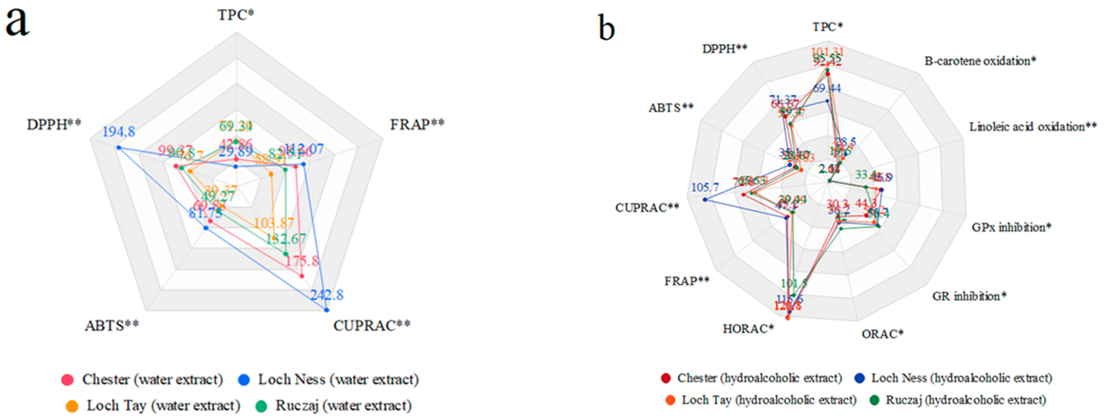
| TPC (mg GAE/g) * | DPPH IC50 (μg/mL) | ABTS IC50 (μg/mL) | CUPRAC IC0.5 (μg/mL) | FRAP IC0.5 (μg/mL) | HORAC Equivalent Gallic Acid Concentration (μg/cm3) | ORAC Equivalent Trolox Concentration (μM) | |
|---|---|---|---|---|---|---|---|
| Water extracts | |||||||
| Chester | 42.86 ± 0.71 | 99.37 ± 2.47 | 69.83 ± 1.15 | 175.80 ± 9.23 | 98.66 ± 2.60 | n/a | n/a |
| Loch Ness | 29.89 ± 0.19 | 194.8 ± 5.59 | 81.73 ± 2.31 | 242.80 ± 3.12 | 112.07 ±3.49 | n/a | n/a |
| Loch Tay | 71.29 ± 2.67 | 76.70 ± 3.92 | 39.37 ± 1.82 | 103.87 ± 11.94 | 58.91 ± 1.80 | n/a | n/a |
| Ruczaj | 69.34 ± 1.89 | 90.80 ± 1.31 | 49.27 ± 1.21 | 132.67 ± 7.94 | 82.51 ± 4.20 | n/a | n/a |
| Hydroalcoholic extracts | |||||||
| Chester | 92.42 ± 1.14 | 66.67 ± 1.27 | 30.40 ± 2.15 | 72.80 ± 3.16 | 45.20 ± 0.63 | 120.80 ± 8.20 | 30.40 ± 3.10 |
| Loch Ness | 69.44 ± 3.80 | 71.37 ± 2.19 | 35.10 ± 1.32 | 105.7 ± 3.89 | 47.10 ± 3.10 | 115.60 ± 10.3 | 36.20 ± 1.00 |
| Loch Tay | 101.31 ± 0.11 | 57.37 ± 3.61 | 24.83 ± 0.23 | 62.73 ± 3.89 | 39.99 ± 0.58 | 121.10 ± 11.2 | 34.70 ± 2.20 |
| Ruczaj | 95.55 ± 2.09 | 59.4 ± 0.87 | 28.67 ± 1.72 | 65.63 ± 2.91 | 39.44 ± 1.61 | 101.50 ± 4.20 | 42.00 ± 2.40 |
| Effect on SOD Activity | Effect on GR and GPx Activity | Linoleic Acid Oxidation | β-Carotene Oxidation | ||||
|---|---|---|---|---|---|---|---|
| Enzyme Inhibition (%) | GR Inhibition (%) | GR Inhibitory Activity (μmol Consumed NADPH/min Incubation) | GPx Inhibition (%) | GPx Inhibitory Activity (nmol Consumed NADPH/min Incubation) | Equivalent Ascorbic Acid Concentration (mg/mL) | Equivalent Ascorbic Acid Concentration (µg/mL) | |
| Hydroalcoholic extracts | |||||||
| Chester | 18.3 ± 1.5 | 44.3 ± 3.2 | 1.7 ± 0.2 | 45.8 ± 3.2 | 91.3 ± 4.6 | 2.04 ± 0.12 | 17.0 ± 3.2 |
| Loch Ness | 30.2 ± 2.5 | 56.7 ± 2.1 | 2.2 ± 0.1 | 46.9 ± 2.4 | 93.5 ± 3.5 | 2.51 ± 0.04 | 28.5 ± 1.7 |
| Loch Tay | 27.6 ± 2.0 | 53.2 ± 3.1 | 2.1 ± 0.3 | 42.0 ± 3.3 | 83.7 ± 4.7 | 2.18 ± 0.17 | 24.4 ± 2.1 |
| Ruczaj | 16.4 ± 4.5 | 58.4 ± 1.8 | 2.3 ± 0.3 | 33.4 ± 1.2 | 66.6 ± 2.1 | 2.11 ± 0.06 | 19.6 ± 1.8 |
| Sample | Equivalent Reference Concentration (μg/mL) | ||||
|---|---|---|---|---|---|
| Neostigmine | Magniflorine | Donepezil | Eserine | Rivastigmine | |
| Hydroalcoholic extract | |||||
| Loch Ness | 2.2 ± 0.1 | 6.9 ± 0.1 | 1.2 ± 0.0 | 1.4 ± 0.1 | 11.2 ± 0.1 |
| Anti-Hyaluronidase Activity | Effect on COX-2 Activity | |||
|---|---|---|---|---|
| IC50 (μg/mL) | Equivalent Acetylsalicylic Acid Concentration (mg/cm3) | COX-2 Inhibition (%) | ||
| Water extracts | Hydroalcoholic extract | |||
| Chester | 160.69 ± 15.20 | Chester | 3.23 ± 0.1 | 84.6 ± 3.5 |
| Loch Ness | 180.09 ± 9.14 | Loch Ness | 3.22 ± 0.1 | 82.1 ± 3.2 |
| Loch Tay | 129.30 ± 3.27 | Loch Tay | 3.22 ± 0.0 | 82.1 ± 2.0 |
| Ruczaj | 127.36 ± 4.13 | Ruczaj | 3.23 ± 0.0 | 84.6 ± 1.6 |
Publisher’s Note: MDPI stays neutral with regard to jurisdictional claims in published maps and institutional affiliations. |
© 2021 by the authors. Licensee MDPI, Basel, Switzerland. This article is an open access article distributed under the terms and conditions of the Creative Commons Attribution (CC BY) license (https://creativecommons.org/licenses/by/4.0/).
Share and Cite
Paczkowska-Walendowska, M.; Gościniak, A.; Szymanowska, D.; Szwajgier, D.; Baranowska-Wójcik, E.; Szulc, P.; Dreczka, D.; Simon, M.; Cielecka-Piontek, J. Blackberry Leaves as New Functional Food? Screening Antioxidant, Anti-Inflammatory and Microbiological Activities in Correlation with Phytochemical Analysis. Antioxidants 2021, 10, 1945. https://doi.org/10.3390/antiox10121945
Paczkowska-Walendowska M, Gościniak A, Szymanowska D, Szwajgier D, Baranowska-Wójcik E, Szulc P, Dreczka D, Simon M, Cielecka-Piontek J. Blackberry Leaves as New Functional Food? Screening Antioxidant, Anti-Inflammatory and Microbiological Activities in Correlation with Phytochemical Analysis. Antioxidants. 2021; 10(12):1945. https://doi.org/10.3390/antiox10121945
Chicago/Turabian StylePaczkowska-Walendowska, Magdalena, Anna Gościniak, Daria Szymanowska, Dominik Szwajgier, Ewa Baranowska-Wójcik, Piotr Szulc, Dagna Dreczka, Marek Simon, and Judyta Cielecka-Piontek. 2021. "Blackberry Leaves as New Functional Food? Screening Antioxidant, Anti-Inflammatory and Microbiological Activities in Correlation with Phytochemical Analysis" Antioxidants 10, no. 12: 1945. https://doi.org/10.3390/antiox10121945