Article

Effects of Dietary Chenodeoxycholic Acid Supplementation in a Low Fishmeal Diet Containing Clostridium autoethanogenum Protein on Growth, Lipid and Cholesterol Metabolism, and Hepatopancreas Health of Litopenaeus vannamei

1
Laboratory of Aquatic Nutrition and Feed, College of Fisheries, Guangdong Ocean University, Zhanjiang 524088, China
2
Aquatic Animals Precision Nutrition and High-Efficiency Feed Engineering Research Centre of Guangdong Province, Zhanjiang 524088, China
3
Key Laboratory of Aquatic, Livestock and Poultry Feed Science and Technology in South China, Ministry of Agriculture, Zhanjiang 524088, China
4
Guangdong Provincial Key Lab of Aquatic Animals Disease Control and Healthy Culture, Zhanjiang 524088, China
*
Author to whom correspondence should be addressed.
These authors contributed equally to this work.
Animals 2023, 13(13), 2109; https://doi.org/10.3390/ani13132109
Submission received: 19 May 2023 / Revised: 16 June 2023 / Accepted: 21 June 2023 / Published: 26 June 2023
(This article belongs to the Section Animal Nutrition)

Abstract

:

Simple Summary

Due to the scarcity of fishmeal resources, alternatives that provide the necessary nutrients for Litopenaeus vannamei are being sought. While fishmeal has been the main nutrition source of Litopenaeus vannamei due to its rich nutrients, the search for protein sources to replace it is becoming increasingly important. The protein derived from Clostridium autoethanogenum protein shows great potential as a substitute for fishmeal owing to its exceptional nutritional value. However, previous studies have suggested that the growth of Litopenaeus vannamei may be adversely affected by the fishmeal replacement of Clostridium autoethanogenum protein. Research has demonstrated that chenodeoxycholic acid can improve the growth and intestinal health of Litopenaeus vannamei. Five diets were formulated with varying concentrations of chenodeoxycholic acid to feed Litopenaeus vannamei. The results revealed that replacing partial fishmeal with Clostridium autoethanogenum protein and adding 0.06% chenodeoxycholic acid led to significant improvements in the growth performance, lipid metabolism, and sterol metabolism. Additionally, the hepatopancreas health of the Litopenaeus vannamei was also improved. The conducted experiment has set the groundwork for further investigation into determining the most effective quantity of chenodeoxycholic acid to be utilized in low fishmeal diets.

Abstract

The study aimed to assess the impact of adding chenodeoxycholic acid (CDCA) to the diet of Litopenaeus vannamei on their growth performance, lipid and cholesterol metabolism, and hepatopancreas health while being fed a low fishmeal diet. Five diets were formulated, one of which contained 25% fishmeal (PC); fishmeal was partially replaced with Clostridium autoethanogenum protein in the remaining four diets and supplemented with 0, 0.03, 0.06, and 0.09% CDCA (NC, BA1, BA2, and BA3, respectively). In this study, four replicates of each diet were assigned and each replicate consisted of 30 shrimp with an average weight of (0.25 ± 0.03 g). The shrimp were fed four times a day for a period of 56 days. The results of this study indicate that the inclusion of CDCA in the diet had a positive impact on the growth performance of the shrimp. The final body weight (FBW), weight gain (WG), and specific growth rate (SGR) of the shrimp in the PC group were similar to those in the BA2 group, and significantly higher than those in the other three groups. The survival rate (SR) was similar among all groups. In comparison to the PC group, the low fishmeal groups exhibited a significant decrease in the crude lipid content of the whole shrimp, as well as the Total cholesterol (T-CHOL), Low-density lipoprotein (LDL-C), and High-density lipoprotein (HDL-C) levels in the hemolymph. Regarding the sterol metabolism, the dietary supplementation of CDCA up-regulated the mRNA expression of intracellular cholesterol transporter 1-like (npc1), 7-dehydrocholesterol reductase (7dhcr), Delta (24) sterol reductase (Δ24), HMG-CoA reductase membrane form (hmgcr), and sterol carrier protein 2 (scp). In the lipid metabolism, the mRNA expression of sterol-regulatory element binding protein (srebp) was significantly down-regulated in the shrimp fed the BA1 diet and the expression of AMP-activated protein kinase (ampk) was significantly up-regulated in the shrimp fed the BA1 and BA3 diets compared to the PC group. The mRNA expression of triacylglycerol lipase (tgl) was significantly up-regulated in the shrimp fed the BA2 diet compared to the NC group. Compared with the shrimp fed the PC diets, the dietary supplementation of CDCA significantly down-regulated the protein expression of SREBP1. The lumen damage in the BA1 group was significantly less severe than those in the NC group. The addition of 0.06% CDCA to low fishmeal diets can improve the growth performance, lipid and cholesterol metabolism, and hepatopancreas health of L. vannamei.

1. Introduction

At present, L. vannamei a highly sought after aquatic product globally due to its abundance of high-quality protein [1,2,3]. The special palatability, high nutritional value, and protein content of fishmeal make it the primary protein source of aquatic organisms [4]. According to the current demand for fishmeal used in aquatic feed, the price of fishmeal is likely to go up by 30% in 2030 [5]. Thus, it is pressing to find alternative protein sources for fishmeal.
Previous studies have confirmed that concentrated cotton meal protein [6], soy protein concentrate [7], soybean meal [8], high-protein cottonseed meal [9], corn protein concentrate [10], methanotroph [11], black soldier fly [12], and yeast cultures [13] can be used as partial substitutes for fishmeal. Plant proteins are the main alternative to fishmeal due to their high protein yield and low cost [14]. Despite being a cost-effective and high-yielding alternative to fishmeal, plant proteins are not without their limitations. One of the major drawbacks of plant proteins is their high content of anti-nutritional factors, which can negatively impact their nutritional value. Additionally, plant proteins are often less palatable than fishmeal, which can affect their overall acceptability as a feed ingredient [15]. An increasing number of studies are proving that some plant proteins can hurt the intestine of shrimp [1,16]. Bacterial proteins do not have the same disadvantages as plant proteins and have been used, with good results, as an alternative to fishmeal. The advantages of bacterial proteins are their high nutritional value and easy cultivation [17]. As a result, increasing studies are focusing on bacterial proteins. Clostridium autoethanogenum proteins (CAP) are a novel type of bacterial protein, which is a by-product of the fermentation and combustion of ethanol in the steelmaking industry. Carbon monoxide and ammonia are the main raw materials for CAP production [18]. Compared with plant proteins, there is a higher crude protein component and lower anti-nutritional factors in CAP [18]. Previous studies have demonstrated that replacing 30% of fishmeal with CAP does not adversely affect the growth and muscle quality of L. vannamei [18]. Similarly, other studies have shown that CAP substituting 40% fishmeal did not adversely affect L. vannamei [19,20]. Moreover, the partial substitution of fishmeal with CAP has no adverse effects on yellow croaker (Larimichthys crocea) [21] and largemouth bass (Micropterus salmoides) [22].
However, the growth of aquatic animals would be impacted if an excessive quantity of fishmeal was replaced by CAP. When CAP replaced fishmeal by more than 30%, the growth, flesh quality, and immune function of L. vannamei were harmed to varying degrees [17,23]. Replacing 40% of fishmeal with CAP also had an effect on the lipid metabolism of L. vannamei, and excessive CAP may have negative effects on largemouth bass (Micropterus salmoides) [24,25]. Dietary supplementation with functional feed additives can reduce the adverse effects on shrimp. Chenodeoxycholic acid (CDCA) is a primary bile acid, which can also form secondary bile acids (lithocholic acid, deoxycholic acid, ursodeoxycholic acid) with glycine or taurine for the intestinal–hepatic cycle. Intestinal L-cells stimulate the secretion of cholecystokinins, which are then released from the gallbladder into the intestine via the common bile duct to aid in the absorption of lipids and fat-soluble vitamins [1]. Bile acids are catabolic products of cholesterol, which is then secreted by the bile ducts and digested by the intestines to be absorbed as a fat-soluble substance [26]. Due to the amphoteric structure of bile acids, they are essential for promoting the intestinal metabolism, as well as the further digestion of fat-soluble vitamins, cholesterol, and lipids [27,28]. According to earlier research, supplementing bile acids can enhance the growth performance and lipid metabolism of L. vannamei [27]. In addition, many studies have indicated that bile acids can enhance immune function and regulate the intestinal health of aquatic animals [29]. Early research has demonstrated that CDCA can boost the immune response and improve intestinal health in aquatic animals [30]. Prior studies have demonstrated that the addition of CDCA to plant protein feed can enhance the growth performance and intestinal health of shrimp [1,14]. However, limited research has been conducted on the impact of CDCA supplementation in low fishmeal diets on the lipid metabolism and immune response of L. vannamei.
The aim of this study was to examine the effects of adding CDCA to a low fishmeal diet on the growth performance, body composition, lipid metabolism, and intestinal health of L. vannamei.

2. Materials and Methods

2.1. Experimental Diets

In this study, five iso-nitrogenous and iso-lipid diets were formulated, including a diet containing 25% fishmeal (PC) and four diets containing 12.5% fishmeal and supplemented with 0, 0.03, 0.06, and 0.09% CDCA (NC, BA1, BA2, and BA3, respectively). Fishmeal was substituted with 10% CAP and supplemented with fish oil. Table 1 displays the proximate composition analyses and formulation details of the diets utilized in this study.

2.2. Shrimp and Experimental Conditions

Shrimp were purchased from the Guangdong Yuehai Seed Co., Ltd. (Zhanjiang, China). The shrimp were given commercial feed (48.0% crude protein and 8.0% crude lipid) for one week prior to the experiment. After 24 h of starvation, 600 healthy shrimp (0.25 ± 0.03 g) were selected and randomly allocated into 5 groups. Each group consisted of 4 replicates, with each replicate containing 30 shrimp. They were fed at 07:15, 12:15, 17:15, and 22:15 every day for 56 days. Water temperatures were 27–30 °C, the pH was 7.7–8.1, and the salinities were around 31‰.

2.3. Sampling and Chemical Analysis

Following a feeding period of 56 days, the shrimp in each tank were subjected to 24 h fasting. The weight of the shrimp was measured and the survival rate was determined through calculations. To determine the moisture content, crude lipid, and crude protein levels, a random sample of five shrimp was chosen from a tank. The hemolymph of five shrimp per tank was collected and subsequently centrifuged at 4 °C and 3500 rpm for 15 min. After the hemolymph supernatant was obtained, it was carefully collected and immediately preserved at a temperature of −80 °C. Hepatopancreas of four shrimp were taken, washed in normal saline (0.9% NaCl solution), then transferred to liquid nitrogen, and then stored at −80 °C for subsequent analysis. The intestine and hepatopancreas were collected from four shrimp, kept in RNA later reagent (Ambion®, ThermoFisher, Waltham, MA, USA), and stored at −80 °C.
The whole shrimps underwent a drying process in a 105 °C oven, which was carried out with the aim of assessing their moisture content. The determination of the crude protein content was carried out using the Dumas Nitrogen method with a Primacs100 analyzer (Skalar, Breda, Dutch). For the determination of the crude lipid, the XT15 extractor (Ankom, NY, USA) was utilized. Additionally, the amino acid content was analyzed in accordance with the standard GB/T 18246-2019.

2.4. Enzyme Activity Assays

The contents of triglyceride (TG), Total cholesterol (T-CHOL), High-density lipoprotein (HDL-C), Low-density lipoprotein (LDL-C), and the activities of alanine aminotransferase (ALT) and aspartate aminotransferase (AST) in the hemolymph were analyzed using the commercial Kit of Nanjing Jiancheng (Nanjing Jiancheng Institute Biological Engineering, China). The total cholesterol (T-CHOL) content and the activity of lipase in the hepatopancreas were determined according to the commercial Kit of Nanjing Jiancheng (Nanjing Jiancheng Institute Biological Engineering, Nanjing, China).

2.5. Quantitative Real-Time PCR Analysis

Total RNA was extracted from the hepatopancreas and intestine according to Trizol Reagent (Transgen Biotech, Beijing, China). The extracted RNA was reverse transcribed into cDNA using the QuantiTect Reverse Transcription Kit (Accurate Biotechnology, Changsha, China). The primers used in the experiment were synthesized by the Sangon Biotech (Guangdong, China) Co., Ltd. The sequence is shown in Table 2. Subsequently, real-time fluorescence quantitative PCR was performed using Light Cycler 480 (Roche Applied Science) to detect gene expression.

2.6. Morphological Analysis of Hepatopancreas

Hepatopancreas samples were soaked in 4% formaldehyde for 24 h before being transferred to Bonn solution and 70% ethanol. After that, the samples were dehydrated with different concentrations of ethanol, followed by embedding in paraffin. The sections were stained with hematoxylin-eosin and observed under microscope (Nikon Ni-U, Tokyo, Japan).

2.7. Western Blot Analyses

According to the method of Xu et al. [31], 30 mg of hepatopancreatic tissue was dissected, PBS, cell lysate, protease inhibitor, and phosphorylase inhibitor were added, and finally, PMSF was added for disruption. The concentration of hepatopancreatic protein was measured using the BCA method and subsequently adjusted to a concentration of 4 mg/mL with PBS and loading buffer. Afterwards, the protein samples underwent denaturation in boiling water for a duration of 10 min before being utilized for the separation of proteins (36 mg of total protein per gel hole) through SDS-PAGE. After electrophoresis, they were transferred to membrane and block with rapid blocking solution for 25 min at room temperature. After development with ECL reagents, bands were quantified using Image J (version 1.43, National Institutes of Health). In this study, the following antibodies were used: antibodies against SREBP1(1:800, ab28481, Abcam), FXR (1:1500, bs-12867R, Bioss), GAPDH (1:1000, 2118 S, Cell Signaling Technology).

2.8. Calculations and Statistical Analysis

The parameters were calculated as follows:
Weight gain (WG, %) = 100 × (m1 − m0)/m0
Specific growth rate (SGR, % day − 1) = 100 × (Ln m1 − Ln m0)/t
Survival rate (SR, %) = 100 × (n1)/(n0)
Feed conversion rate (FCR) = feed consumed (g)/(m1 − m0)
Feeding intake (FI) = feed consumed/n1
Protein efficiency rate (PER) = (m1 − m0)/protein intake
where m0, initial body weight; m1, final body weight; n0, initial amount of shrimp; n1, final amount of shrimp.
Prior to conducting a one-way analysis of variance (ANOVA), the data underwent normality distribution testing using the Kolmogorov-Smirnov test and homogeneity of variances testing using Levene’s test. The data were then analyzed using one-way ANOVA followed by Tukey’s multiple range test to identify significant differences among treatments. The statistical software used was SPSS 21.0 (SPSS, Chicago, IL, USA). A probability value of p < 0.05 was considered statistically significant.

3. Results

3.1. Growth Performance

Based on the data presented in Table 3, the final body weight (FBW), weight gain (WG), and specific growth rate (SGR) of the shrimp in the PC group were similar to those in the BA2 group, and significantly higher than those in the other three groups (p < 0.05). The survival rate (SR) was similar among all groups (p > 0.05). The feed conversion rate (FCR) and feeding (FI) of the five experimental groups had no differences (p > 0.05). The protein efficiency rate (PER) of the shrimp in the PC and BA1 groups was significantly higher than that of the NC group (p < 0.05).

3.2. Proximate Composition of Whole Shrimp

As indicated in Table 4, no noticeable differences in the moisture content were observed among the five groups. (p > 0.05). The NC group exhibited a significantly lower crude protein content in comparison to the BA1 and BA3 groups (p < 0.05). The crude lipid content of the shrimp was significantly decreased when CDCA was added into their diet (p < 0.05).

3.3. Hemolymph and Hepatopancreas Biochemical Indicators

According to Table 5, there was no significant difference in the content of TG in the hemolymph (p > 0.05). The T-CHOL content in the PC group was significantly higher than the other four groups (p < 0.05). The HDL-C content of the shrimp fed the BA2 diet was significantly higher than those fed the NC and BA1 diet (p < 0.05). The LDL-C and ALT content significantly increased with the increase in the CDCA content (p < 0.05). The AST content of the shrimp fed the BA3 diet was significantly higher than those fed the NC diet (p < 0.05). The hepatopancreas T-CHOL content in the shrimp fed the PC diet was significantly higher than those fed the other diets, except for the BA3 diet (p < 0.05). The lipase content in the hepatopancreas was significantly lower in the PC diet than that in the BA1 diet (p < 0.05).

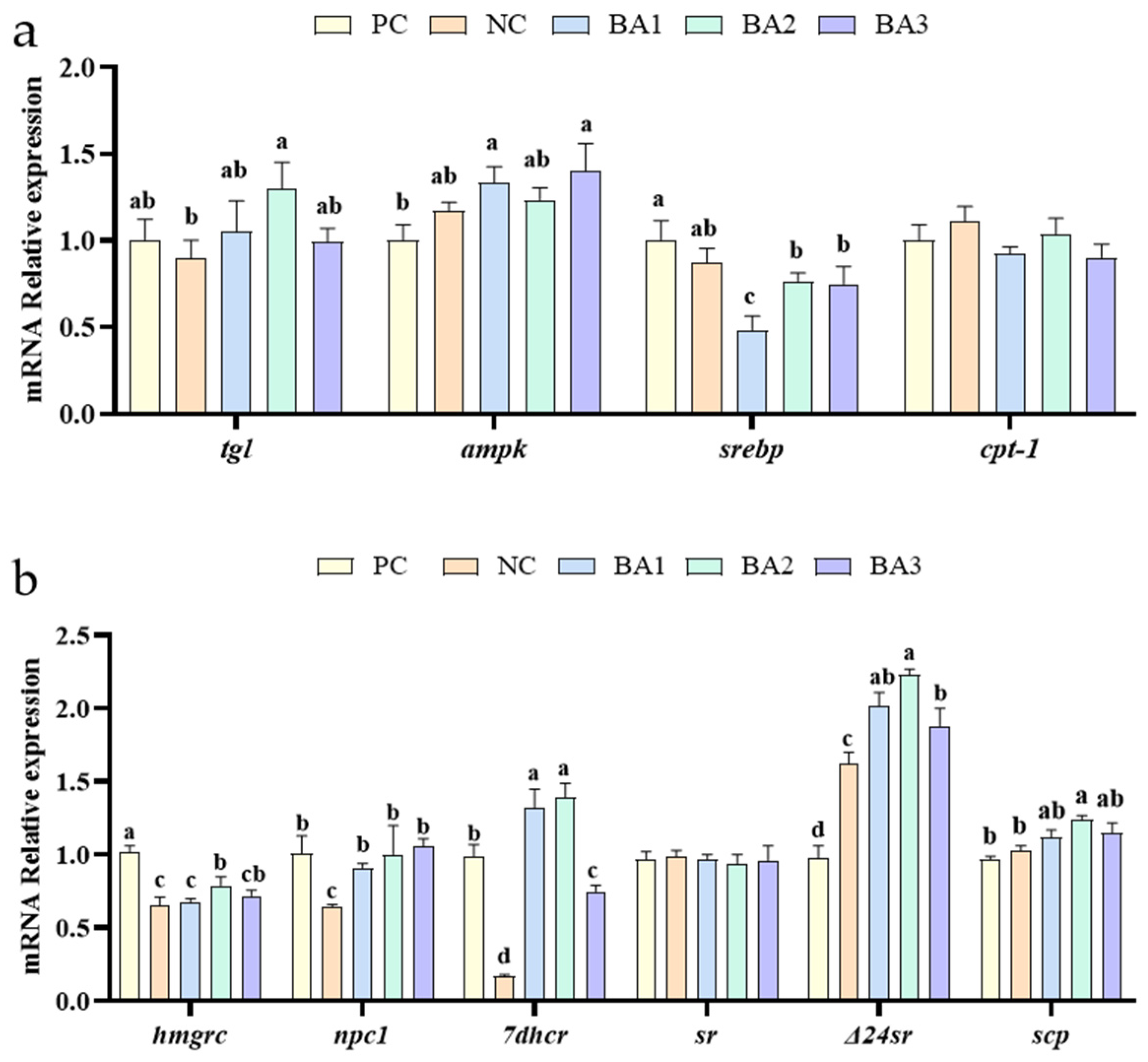
3.4. Lipid and Sterol Metabolism-Related Genes Expression

The relative expression of lipid metabolism-related genes in the hepatopancreas is shown in Figure 1. Among the genes related to lipid metabolism, the mRNA expression of tgl in the BA2 group was significantly higher than that in the NC group, and the mRNA expression of ampk in the BA1 and BA3 groups were significantly higher than that in the PC group (p < 0.05). Compared with the shrimp fed the PC diet, the supplementation of CDCA in the diets down-regulated the mRNA expression of srebp (p < 0.05).
As shown in Figure 1, adding CDCA to low fishmeal diets affected the expression levels of sterol metabolism-related genes in the hepatopancreas of L. vannamei. Compared with the shrimp fed the NC diets, the dietary supplementation of CDCA up-regulated the mRNA expression of npc1, 7dhcr, and Δ24 (p < 0.05). The mRNA expression of hmgcr significantly increased in the BA2 and BA3 diets compared to the NC diet (p < 0.05). In all groups, the mRNA expression of Steroid reductase (sr) in the hepatopancreas was similar (p > 0.05). The shrimp fed the BA2 diet achieved the highest scp expressions compared to the other groups (p < 0.05).

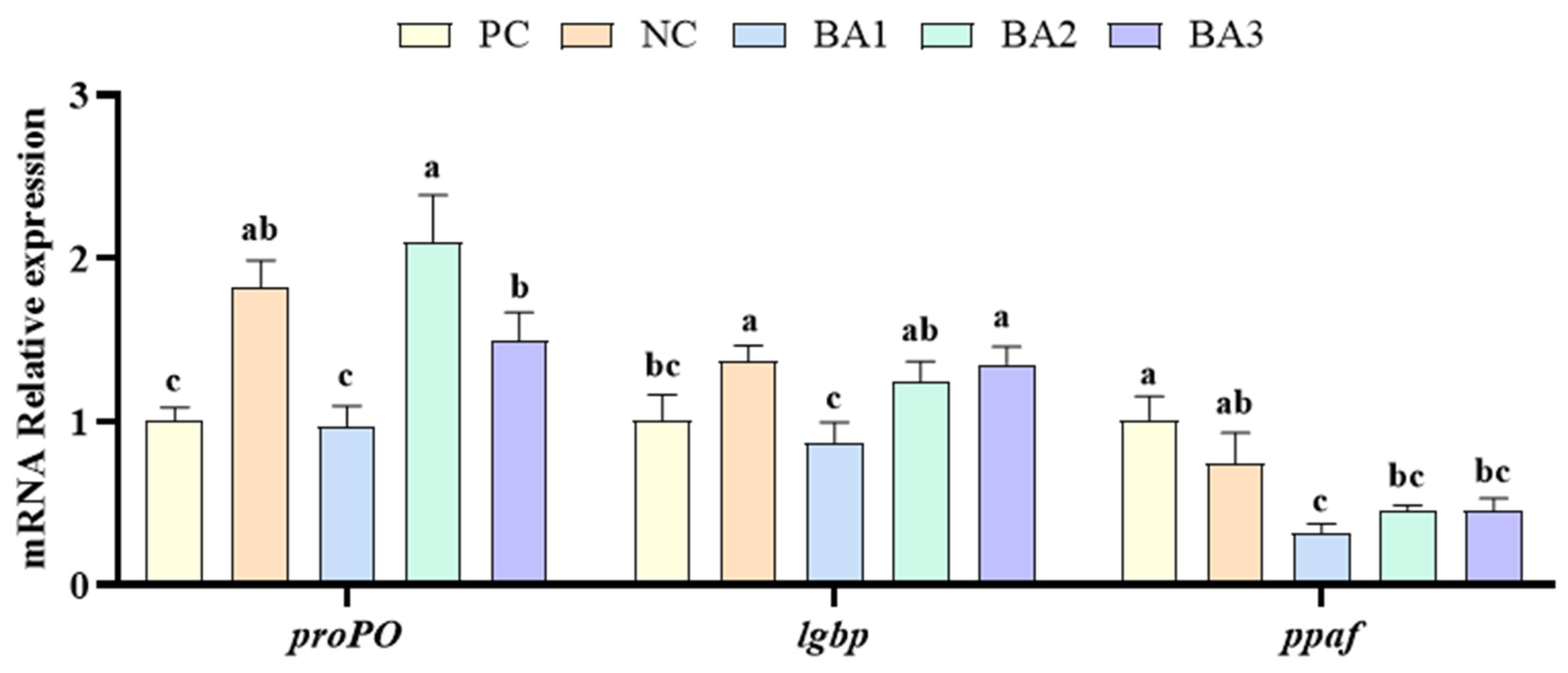
3.5. Phenol Oxidase-Related Genes Expression

The relative expression of phenol oxidase-related genes in the hepatopancreas is shown in Figure 2. Among the genes related to the phenol oxidase system, the mRNA expression of prophenoloxidas (proPO) and lipopolysaccharide and beta-1,3-glucan binding protein (lgbp) in the NC group was significantly higher than that in the PC group; the mRNA expression of proPO, lgbp, and prophenoloxidase activating factor (ppaf) showed a decreasing and then increasing trend with of the increase in CDCA levels (p < 0.05).

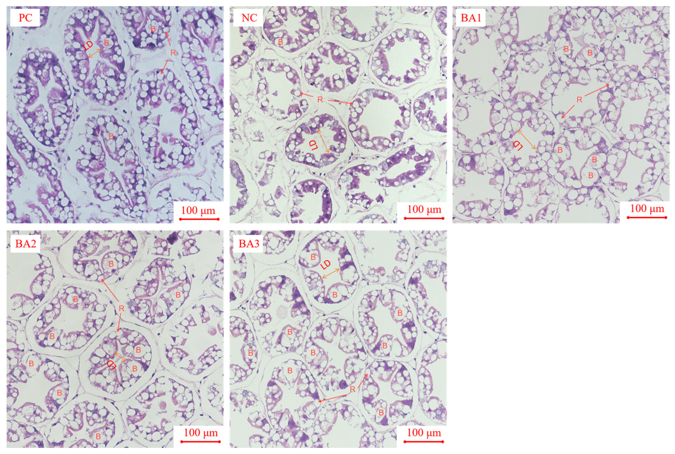
3.6. Morphological Analysis of Hepatopancreas

The morphology of the hepatopancreas is shown in Figure 3. The hepatopancreas of L. vannamei is composed of multiple hepatic tubules. The large vacuoles in the hepatic tubules are B cells (Blasenzellen cells), the small lipid vacuoles are R cells (Restzellen cells), and the area with a five- or four-pointed star is the lumen of the hepatopancreatic tubules (Lumen of hepatopancreatic tubules). The HE-stained hepatopancreas showed that with the increase in CDCA addition, the number of B cells in the hepatic tubule gradually increased, and the number of R cells gradually decreased. The lumen of the shrimp in the NC group was dilated or even ruptured. The lumen of the shrimp in the BA1 group was slightly damaged, and the lumen structure of the shrimp in the other groups was normal.

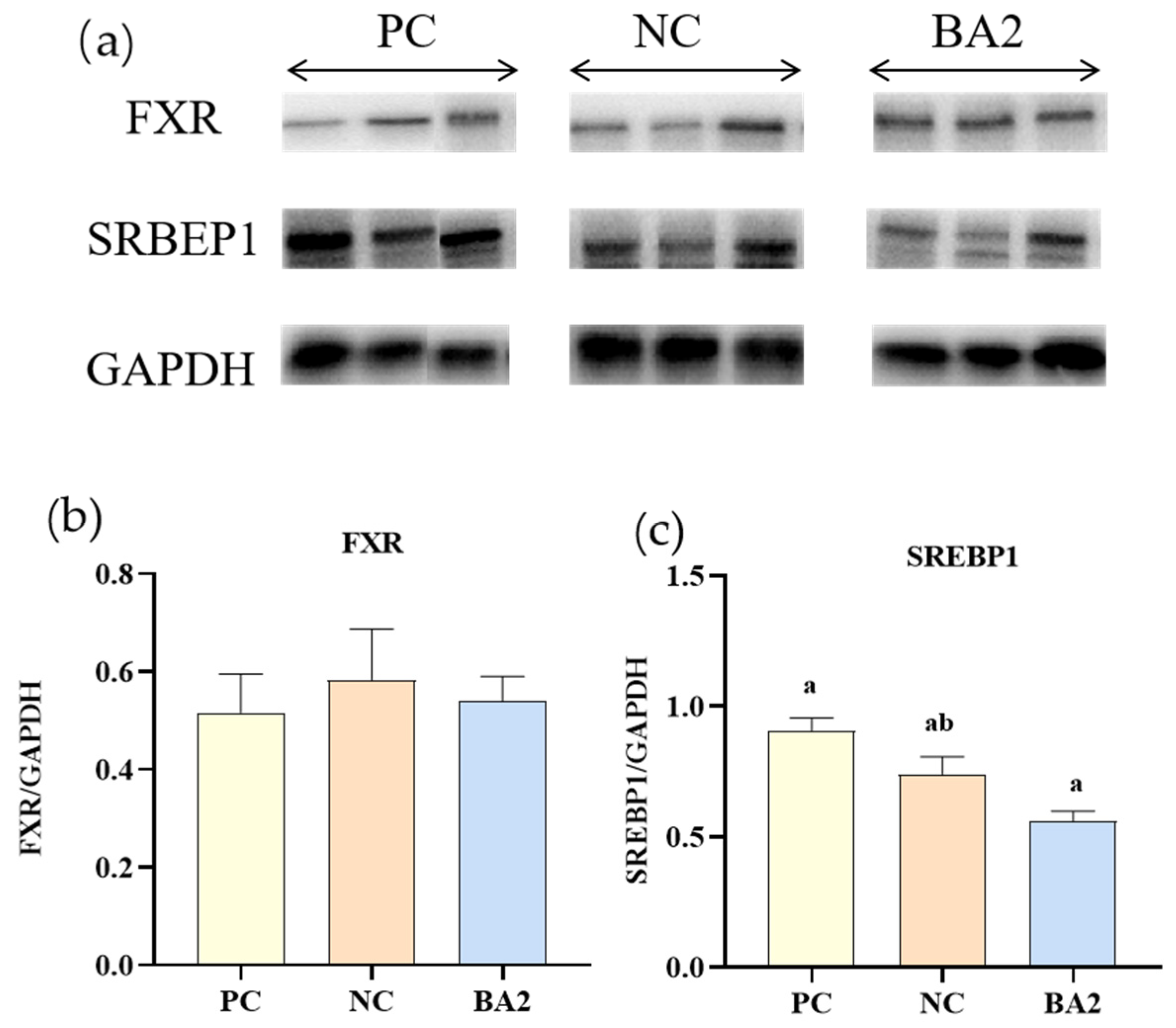
3.7. Western Blot Analysis of Bile Acid Metabolism Related Proteins Expression

As shown in Figure 4, compared with the shrimp fed the PC diet, the dietary supplementation of 0.06% CDCA significantly down-regulated the protein expression of SREBP1 (p < 0.05). In all of the groups, the protein expression of FXR in the hepatopancreas was similar (p > 0.05).

4. Discussion

Although many studies have proven that excessive dietary CAP replacement for fishmeal would cause some adverse effects, this study revealed that substituting partial fishmeal with CAP could still significantly enhance the growth performance of L. vannamei by adding appropriate CDCA. The addition of 0.06% CDCA can effectively mitigate this negative effect. The study revealed that the partial replacement of fishmeal with CAP significantly reduced the growth of L. vannamei [17]. According to previous research, the growth of L. vannamei significantly decreased when fishmeal was substituted with more than 30% CAP [23].
Similar to our results, a low fishmeal diet supplemented with CDCA can improve the growth performance and intestinal health of L. vannamei [32], and dietary supplementation of CDCA can also improve the hepatopancreas health of L. vannamei [1]. The growth of L. vannamei can be stimulated through dietary supplementation of 0.02–0.03% bile acid, as reported by Su et al. [27]. Iwashita et al. [33] demonstrated that supplementing plant protein-based feeds with choline taurine (a type of bile acid) resulted in the improved growth of rainbow trout (Oncorhynchus mykiss). Adding CDCA to feeds can enhance the growth performance of shrimps, probably due to their ability to promote the emulsification and digestion of feeds [34]. In this study, the growth performance of shrimp was not further improved with the increase in the bile acid content, indicating that the addition of excessive CDCA will cause negative effects. This is similar to the findings of Su et al. [24]. In fact, the excessive addition of CDCA had a negative impact on shrimp growth. Jiang et al. [35] found that a whole plant protein diet supplemented with 0.015–0.045% bile acid significantly improved the growth of gefilte fish (Oreochromis niloticus), but when the level of bile acid addition was increased to 0.135%, it hindered the growth. In terms of feed utilization, the FCR and FI were similar among all the groups. The addition of 0.03% CDCA to low fishmeal feeds can significantly improve protein efficiency. Studies conducted previously have shown that the addition of 0.06% bile acid to the diet of grass carp (Ctenopharyngodon idella) can mitigate the reduction in protein efficiency associated with plant-based raw materials [36].
Adding CDCA at a concentration range of 0.03%~0.09% has been found to enhance the crude protein content of whole shrimp. Another study reported that adding 0.006% bile acid significantly improved the whole fish crude protein of grass carp, and it was speculated that the muscle glutamate dehydrogenase involved in protein catabolism was downregulated by adding bile acid, thus promoting the protein synthesis required for somatic cell growth [36]. This experiment has also shown that either replacing fishmeal with CAP or adding CDCA to fishmeal could significantly reduce the crude lipid composition of whole shrimp. Studies of grass carp [35,37] showed that a low dose of dietary bile acid could reduce the accumulation of body lipid. This may be due to the enhancement of triglycerides breakdown promoting the lipids’ decomposition. Bile acid is an essential component in the process of lipid emulsification as it plays a critical role in breaking down large lipid molecules into smaller ones, which in turn increases the surface area for lipase reaction, which is crucial for the digestion and absorption of lipids [38]. Both the replacement of fishmeal with CAP and the supplementation of CDCA can play a role in enhancing lipase activity in the hepatopancreas, which corresponds to the trend of crude lipid in whole shrimp. Replacing fishmeal with CAP significantly reduced the content of T-CHOL and HDL-C in the hemolymph, as well as the content of T-CHOL in the hepatopancreas, which are closely related to lipid metabolism [39,40]. However, the supplementation of CDCA could effectively reverse this trend. These findings indicate that the addition of CDCA is crucial for regulating the lipid metabolism in shrimp. The main way in which HDL-C regulates lipid metabolism is through mediating reverse cholesterol transport, which refers to the conversion of cholesterol from the peripheral hepatopancreatic Cholesterol from peripheral tissues, such as lipid and muscle, being taken into the hepatopancreas and converted to bile acids before entering the next level of the metabolic cycle. This process is accompanied by the formation of HDL-C. In addition, Chang Yumei et al. suggested that a decrease in serum T-CHOL may be an indication of liver injury.
The tgl—as a regulatory transcription factor for lipolysis—and its translated products can hydrolyze triglycerides to convert lipids into glycerol and free fatty acids [41]. In general, higher tgl expression indicates a higher degree of triglyceride hydrolysis. The ampk is an important cellular energy sensor, and the activation of the ampk stimulates the catabolic pathway of adenosine triphosphate (ATP) breakdown, leading to accelerated fatty acid oxidation and glycolysis, while limiting the production of fatty acids, protein, and glycogen. Polakof et al. [42] showed that the ampk limited a reduction in fatty acid synthesis in rainbow trout (Oncorhynchus mykiss). In the regulation of lipid synthesis, srebp is one of the most important transcription factors [43]. This study found that supplementing shrimp diets with CDCA led to an increase in the expression of the genes related to energy metabolism and lipid oxidation, while inhibiting the genes associated with lipid synthesis, thereby significantly reducing the body lipid in shrimp. The Western blot results showed that dietary supplementation with CDCA significantly reduced SREBP1 expression, indicating reduced lipid synthesis, which was consistent with the molecular results.
Sterol synthesis has an important impact on lipid metabolism [44]. Hmgrc plays a crucial role in regulating the biosynthesis of cholesterol in vertebrates by acting as the rate-limiting enzyme in the process [45]. The results of this experiment revealed that a low fishmeal diet dramatically reduced hmgrc mRNA expression, but CDCA supplementation increased hmgrc expression. The results obtained in this study are consistent with the findings reported by Rozner et al. [46]. 7dhcr is a critical enzyme in the process of cholesterol synthesis, playing a pivotal role in promoting cholesterol production [47]. In this experiment, low fishmeal diets down-regulated 7dhcr expression and supplementation with CDCA up-regulated 7dhcr expression. Sterols are converted to cholesterol through the action of Δ24sr and a number of other enzymes [25,48]. Sterols carrier proteins are essential for cholesterol absorption and storage. The addition of CDCA to the diet led to a significant increase in the mRNA expression of scp in the BA2 group, implying that it can promote the absorption and utilization of sterols. Npc1 and npc2 are crucial proteins involved in intracellular lipid transport. Cholesterol initially binds to npc2, and subsequently to npc1, which accelerates the transport of cholesterol [49]. In this experiment, the low fishmeal diet down-regulated the expression of npc1, while the supplementation of CDCA could up-regulated the expression of npc1, which indicated that the addition of CDCA could accelerate intracellular cholesterol transport in shrimp.
ALT and AST are considered to be markers of hepatopancreas damage. In general, the activity of transaminase in the hemolymph is low. However, in cases of hepatopancreatic disease, increased cell membrane permeability can lead to the release of transaminase into the hemolymph [50,51]. According to the findings of this study, the NC diet significantly increased the activities of these two transaminases in the hemolymph, which seemed to indicate that the shrimp hepatopancreas was damaged, and the addition of 0.03–0.09% CDCA could significantly alleviate this damage. According to the study conducted by Su et al. [27], it was observed that the supplementation of bile acid below 0.05% did not affect the hemolymph ALT and AST content of L. vannamei, but adding more than 0.05% caused hepatopancreatic toxicity. The difference in these results might be due to the different types of bile acids. On the level of gene expression, the proPO and lgbp expression levels were significantly increased in the NC, BA2, and BA3 groups. This indicated that the CAP replacement of fishmeal and the supplementation of 0.06–0.09% CDCA have the potential to activate the proPO immune system.
In crustaceans, the hepatopancreas are the main organs for absorbing and storing substances, and their morphology indicates the nutritional status of crustaceans [52,53]. The results of Xu et al. [54] showed that R cells are the main site of lipid droplets and glycogen nutrient reserves, and their research also showed that the lower the lipid level or the higher the energy metabolism of the organism, the smaller the number of R cells. B cells are the primary location where the complex process of synthesizing, secreting, and breaking down critical molecules occurs, making them an integral component of the immune system. The more vigorous the synthesis of digestive enzymes, the greater the number of B cells [55]. However, studies have shown that B cell hypertrophy may also be associated with fat oxidative damage [56]. Studies have shown that bile acids were potentially cytotoxic, with previous results confirming that high concentrations (0.135%) of dietary bile acids damaged tilapia liver cells [35,57]. The study’s findings indicate that increased dietary CDCA levels are positively correlated with the number of hepatopancreas B cells, while a negative correlation was observed between the CDCA levels and the number of R cells. The findings indicate that CDCA being added to fishmeal can effectively promote shrimp growth. In addition, the dilated and irregular lumen of the central hepatic tubule is also a sign of hepatopancreas damage. The current research findings demonstrate that the hepatic canalicular lumen of the shrimp in the NC group showed obvious dilatation, but there was no obvious abnormality in the lumen of the shrimp in the BA2 and BA3 groups, indicating that dietary CDCA supplementation can alleviate the hepatopancreas injury of shrimp.

5. Conclusions

This study aimed to assess the impact of CDCA supplementation on L. vannamei when fed a low fishmeal diet with CAP replacing fishmeal. The study revealed that the addition of 0.06% CDCA could alleviate the negative effects on the growth performance, lipid and cholesterol metabolism, and hepatopancreas health of L. vannamei.

Author Contributions

M.S.: Conceptualization, Formal analysis, Investigation, Writing-original draft. C.Z.: Conceptualization, Methodology, Investigation. X.L. and G.H.: Methodology, Resources, Supervision. J.C. and B.T.: Methodology, Investigation. Y.S., S.X.: Methodology, Supervision, Writing-review and editing, Project administration, Funding acquisition. All authors have read and agreed to the published version of the manuscript.

Funding

This study was supported by the fund of the National Natural Science Foundation of China (32002402) and the Guangdong Basic and Applied Basic Research Foundation (2021A1515010428, 2023A1515030007), Characteristic innovation projects of ordinary universities in Guangdong (2022KTSCX049), ModernSeed Industry Park for Whiteleg Shrimp of Guangdong Province, program for scientific research start-up funds of Guangdong Ocean University, Science and Technology Bureau of Zhanjiang (2020A05003, 2022A 01002).

Institutional Review Board Statement

The animal protocol utilized in this study was authorized by the Institutional Animal Care and Use Committee (IACUC), Fisheries College, Guangdong Ocean University (protocol code GDOU-IACUC-2021-A2313 and date of May 2021). All experimental procedures involving animals were conducted in compliance with the National Center for the Replacement Refinement & Reduction of Animals in Research, as well as the ARRIVE guidelines 2.0 and other relevant regulations.

Informed Consent Statement

The study did not involve client-owned animals.

Data Availability Statement

The corresponding authors can provide data on request to support the findings of this study. It is important to note that the data will not be made public for privacy or ethical reasons.

Conflicts of Interest

The authors declare that they have no known competing financial interest or personal relationships that could have appeared to influence the work reported in this paper.

References

  1. Li, X.; Yao, X.; Zhang, X.; Dong, X.; Chi, S.; Tan, B.; Zhang, S.; Xie, S. Effects of dietary chenodeoxycholic acid supplementation in a low fishmeal diet on growth performance, lipid metabolism, autophagy and intestinal health of Pacific white shrimp. Litopenaeus vannamei. Fish Shellfish Immunol. 2022, 127, 1088–1099. [Google Scholar] [CrossRef]
  2. Wu, Y.; Li, R.; Shen, G.; Huang, F.; Yang, Q.; Tan, B.; Chi, S. Effects of dietary small peptides on growth, antioxidant capacity, nonspecific immunity and ingut microflorastructure of Litopenaeus vannamei. J. Guangdong Ocean Univ. 2021, 41, 1–9. [Google Scholar] [CrossRef]
  3. Huang, B.; Zhang, S.; Dong, X.; Chi, S.; Yang, Q.; Liu, H.; Tan, B.; Xie, S. Effects of fishmeal replacement by black soldier fly on growth performance, digestive enzyme activity, intestine morphology, intestinal flora and immune response of pearl gentian grouper (Epinephelus fuscoguttatus ♀ × Epinephelus lanceolatus ♂). Fish Shellfish. Immunol. 2022, 120, 497–506. [Google Scholar] [CrossRef]
  4. Xu, J.; Sheng, Z.; Chen, N.; Xie, R.; Zhang, H.; Li, S. Effect of dietary fish meal replacement with spray dried chicken plasma on growth, feed utilization and antioxidant capacity of largemouth bass (Micropterus salmoides). Aquac. Rep. 2022, 24, 101112. [Google Scholar] [CrossRef]
  5. Food and Agriculture Organization (F.A.O.). The State of World Fisheries and Aquaculture 2020; FAO Yearbook: Rome, Italy, 2020. [Google Scholar]
  6. Jiang, S.; Chen, Z.; Zhou, F.; Yang, Q.; Huang, J.; Yang, L.; Jiang, S. Study on partial replacement of fish meal with concentrated dephenolized cottonseed protein in feed of Penaeus monodon. Aquac. Res. 2020, 52, 3871–3881. [Google Scholar] [CrossRef]
  7. Ray, G.W.; Liang, D.; Yang, Q.; Tan, B.; Dong, X.; Chi, S.; Liu, H.; Zhang, S.; Rimei, L. Effects of replacing fishmeal with dietary soybean protein concentrate (SPC) on growth, serum biochemical indices, and antioxidative functions for juvenile shrimp Litopenaeus vannamei. Aquaculture 2020, 516, 734630. [Google Scholar] [CrossRef]
  8. Zhou, H.; Chen, G.; Ji, D.; Wang, S.; Zhang, J. The effects of partial rReplacement of fish meal by three protein sources on growth performance approximate compositions and energy budget of Juvenile Brown-marbled Grouper (Epinephelus fuscoguttatus). J. Guangdong Ocean Univ. 2012, 32, 10–16. [Google Scholar]
  9. Zhang, H.; Pu, X.; Yang, Q.; Tan, B.; Dong, X.H.; Chi, S.; Liu, H.Y.; Zhang, S. Effects of replacing fish meal with high-protein cottonseed meal on growth performance, non-specific immune index and disease resistance for Litopenaeus vannamei. J. Guangdong Ocean Univ. 2018, 38, 20–26. [Google Scholar] [CrossRef]
  10. Tu, G.; Chen, G.; Zhou, H.; Wang, S.; Ji, D.; Zhng, J.; Ye, F. Effects of partial replacement dietary fish meal by three kinds of protein sources on haematological indices of Juvenile Epinephelus fuscoguttatus. J. Guangdong Ocean Univ. 2012, 32, 12–19. [Google Scholar]
  11. Chen, Y.; Chi, S.; Zhang, S.; Dong, X.; Yang, Q.; Liu, H.; Tan, B.; Xie, S. Evaluation of Methanotroph (Methylococcus capsulatus, Bath) bacteria meal on body composition, lipid metabolism, protein synthesis and muscle metabolites of Pacific white shrimp (Litopenaeus vannamei). Aquaculture 2022, 547, 737517. [Google Scholar] [CrossRef]
  12. Xie, Y.; Peng, K.; Hu, J.; Wang, G. Review on application of black soldier fly (Hermetia illucens Linnaeus) in aquatic feed. J. Guangdong Ocean Univ. 2022, 42, 144–150. [Google Scholar] [CrossRef]
  13. Ayiku, S.; Shen, J.; Tan, B.P.; Dong, X.H.; Liu, H.Y. Effects of reducing dietary fishmeal with yeast supplementations on Litopenaeus vannamei growth, immune response and disease resistance against Vibrio harveyi. Microbiol. Res. 2020, 239, 126554. [Google Scholar] [CrossRef]
  14. Xie, S.; Wei, D.; Tian, L.; Liu, Y. Dietary supplementation of chenodeoxycholic acid improved the growth performance, immune response and intestinal health of juvenile Penaeus monodon fed a low fish-meal diet. Aquac. Rep. 2021, 20, 100773. [Google Scholar] [CrossRef]
  15. Zhang, W.; Tan, B.; Pang, A.; Deng, J.; Yang, Q.; Zhang, H. Screening of potential biomarkers for soybean meal induced enteritis in pearl gentian Grouper (Epinephelus fuscoguttatus♀ × Epinephelus lanceolatus♂). J. Guangdong Ocean Univ. 2022, 42, 1–12. [Google Scholar] [CrossRef]
  16. Chen, G.; Liu, B.; Chen, J.; Liu, H.; Tan, B.; Dong, X.; Yang, Q.; Chi, S.; Zhang, S.; Yao, M. Supplementing sulfate-based alginate polysaccharide improves Pacific white shrimp (Litopenaeus vannamei) fed fishmeal replacement with cottonseed protein concrntrate: Effects on growth, intestinal health, and disease resistance. Aquac. Nutr. 2022, 21, 7132362. [Google Scholar] [CrossRef]
  17. Jiang, X.; Yao, W.; Yang, H.; Tan, S.; Leng, X.; Li, X. Dietary effects of Clostridium autoethanogenum protein substituting fish meal on growth, intestinal histology and immunity of Pacific white shrimp (Litopenaeus vannamei) based on transcriptome analysis. Fish Shellfish. Immunol. 2021, 119, 635–644. [Google Scholar] [CrossRef]
  18. Yao, W.; Yang, P.; Zhang, X.; Xu, X.; Zhang, C.; Li, X.; Leng, X. Effects of replacing dietary fish meal with Clostridium autoethanogenum protein on growth and flesh quality of Pacific white shrimp (Litopenaeus vannamei). Aquaculture 2022, 549, 737770. [Google Scholar] [CrossRef]
  19. Zheng, C.; Cao, J.; Chi, S.; Dong, X.; Yang, Q.; Liu, H.; Zhang, S.; Xie, S.; Tan, B. Dietary phosphorus supplementation in the diet of Pacific white shrimp (Litopenaeus vannamei) alleviated the adverse impacts caused by high Clostridium autoethanogenum protein. Fish Shellfish. Immunol. 2022, 131, 137–149. [Google Scholar] [CrossRef]
  20. Zheng, C.; Xie, S.; Tan, B.; Cao, J.; Chi, S.; Dong, X.; Yang, Q.; Liu, Y.H.; Zhang, S. Effects of dietary lipid sources on alleviating the negative impacts induced by the fishmeal replacement with Clostridium autoethanogenum protein in the diet of pacific white shrimp (Litopenaeus vannamei). Front. Mar. Sci. 2022, 9, 879364. [Google Scholar] [CrossRef]
  21. Wu, Y.; Tian, S.; Yuan, J.; Zhang, Z.; Zhou, H.; Gao, W.; Zhang, W.; Mai, K. Effects of Clostridium autoethanogenum protein as substitute for dietary fishmeal on the growth, feed utilization, intestinal health and muscle quality of large yellow croaker Larimichthys crocea. Aquaculture 2022, 561, 738591. [Google Scholar] [CrossRef]
  22. Ma, S.; Liang, X.; Chen, P.; Wang, J.; Gu, X.; Qin, Y.; Blecker, C.; Xue, M. A new single-cell protein from Clostridium autoethanogenum as afunctional protein for largemouth bass (Micropterus salmoides). Animal Nutr. 2022, 10, 99–110. [Google Scholar] [CrossRef]
  23. Yao, W. Dietary Effects of Replacing Fish Meal with Clostridium autoethanogenum Protein on Growth and Flesh Quality of Pacific White Shrimp (Litopenaeus vannamei) and Nutritional Improvement Strategy; Shanghai Ocean University: Shanghai, China, 2022. [Google Scholar]
  24. Lu, Q.; Xi, L.; Liu, Y.; Gong, Y.; Su, J.; Han, D.; Yang, Y.; Jin, J.; Liu, H.; Zhu, X. Effects of dietary inclusion of Clostridium autoethanogenum protein on the growth performance and liver health of largemouth bass (Micropterus salmoides). Front. Mar. Sci. 2021, 8, 764964. [Google Scholar] [CrossRef]
  25. Chen, Y.; Zheng, C.; Zhang, X.; Li, X.; Yao, X.; He, G.; Cao, J.; Chi, S.; Zhang, S.; Tan, B. Evaluation of ratios of fish-to-soybean oils on growth, lipid and cholesterol metabolism and muscle metabolites of Pacific white shrimp (Litopenaeus vannamei) fed low fishmeal diets containing Clostridium autoethanogenum protein. Aquac. Rep. 2022, 27, 101417. [Google Scholar] [CrossRef]
  26. Hylemon, P.B.; Takabe, K.; Dozmorov, M.; Nagahashi, M.; Zhou, H. Bile acids as global regulators of hepatic nutrient metabolism. Liver Res. 2017, 1, 10–16. [Google Scholar] [CrossRef]
  27. Su, C.; Liu, X.; Li, J.; Zhang, M.; Pan, L.; Lu, Y.; Wang, Y.; Ding, Y. Effects of bile acids on the growth performance, lipid metabolism, non-specific immunity and intestinal microbiota of Pacific white shrimp (Litopenaeus vannamei). Aquac. Nutr. 2021, 27, 2029–2041. [Google Scholar] [CrossRef]
  28. Liao, M.; Chen, S.; Wang, W.; Zhong, S.; Yv, S.; Liao, S. Cholesterol-lowering activity of Sargassum zhangii polysaccharide in vitro. J. Guangdong Ocean Univ. 2017, 37, 80–85. [Google Scholar] [CrossRef]
  29. Li, X.; Shi, M.; Chen, L.; Zhang, S.; Chi, S.; Dong, X.; Deng, J.; Tan, B.; Xie, S. Effects of bile acids supplemented into low fishmeal diet on growth, molting, and intestinal health of Pacific white shrimp, Litopenaeus vannamei. Aquac. Rep. 2023, 29, 101491. [Google Scholar] [CrossRef]
  30. Du, J.; Xu, H.; Li, S.; Cai, Z.; Mai, K.; Ai, Q. Effects of dietary chenodeoxycholic acid on growth performance, body composition and related gene expression in large yellow croaker (Larimichthys crocea) fed diets with high replacement of fish oil with soybean oil. Aquaculture 2017, 479, 584–590. [Google Scholar] [CrossRef]
  31. Xu, J.; He, G.; Chen, L.; Xie, S.; Chi, S.; Zhang, S.; Cao, J.; Tan, B. Farnesoid X receptor (FXR) and G protein-coupled bile acid receptor 1 (TGR5) signaling pathways improved the hepatic lipid metabolism in hybrid grouper. Aquac. Rep. 2022, 22, 100997. [Google Scholar] [CrossRef]
  32. Li, X.; Li, H.; Qu, K.; Liu, Y.; Chi, S.; Yang, Q.; Tan, B.; Zhang, S.; Xie, S. Dietary bile acids promote sterol metabolism, bile acids enterohepatic circulation, and apoptosis in juvenile Pacific white shrimp (Litopenaeus vannamei). Anim. Feed. Sci. Technol. 2023, 115710. [Google Scholar] [CrossRef]
  33. Iwashita, Y.; Suzuki, N.; Yamamoto, T.; Shibata, J.-i.; Isokawa, K.; Soon, A.H.; Ikehata, Y.; Furuita, H.; Sugita, T.; Goto, T. Supplemental effect of cholyltaurine and soybean lecithin to a soybean meal-based fish meal-free diet on hepatic and intestinal morphology of rainbow trout Oncorhynchus mykiss. Fish. Sci. 2008, 74, 1083–1095. [Google Scholar] [CrossRef]
  34. Reschly, E.J.; Ai, N.; Ekins, S.; Welsh, W.J.; Hagey, L.R.; Hofmann, A.F.; Krasowski, M.D. Evolution of the bile salt nuclear receptor FXR in vertebrates *s. J. Lipid Res. 2008, 49, 1577–1587. [Google Scholar] [CrossRef] [Green Version]
  35. Jiang, M.; Wen, H.; Gou, G.; Liu, T.; Lu, X.; Deng, D. Preliminary study to evaluate the effects of dietary bile acids on growth performance and lipid metabolism of juvenile genetically improved farmed tilapia (Oreochromis niloticus) fed plant ingredient-based diets. Aquac. Nutr. 2018, 24, 1175–1183. [Google Scholar] [CrossRef]
  36. Zhou, J.; Chen, H.; Ji, H.; Shi, X.; Li, X.; Chen, L.; Du, Z.; Yu, H. Effect of dietary bile acids on growth, body composition, lipid metabolism and microbiota in grass carp (Ctenopharyngodon idella). Aquac. Nutr. 2018, 24, 802–813. [Google Scholar] [CrossRef]
  37. Cao, B.; Liao, Z.; Xiang, X.; He, W.; Cen, Y.; He, S. Effect of bile acids on growth performance, muscle nutrient composition and digestive enzyme activity in grass carp (Ctenopharyngodon idellus). Prog. Fish. Sci. 2017, 38, 99–106. [Google Scholar] [CrossRef]
  38. Ogata, Y.; Nishi, M.; Nakayama, H.; Kuwahara, T.; Ohnishi, Y.; Tashiro, S. Role of bile in intestinal barrier function and its inhibitory effect on bacterial translocation in obstructive jaundice in rats. J. Surg. Res. 2003, 115, 18–23. [Google Scholar] [CrossRef]
  39. Chen, Q.; Liu, H.; Tan, B.; Dong, X.; Chi, S.; Yang, Q.; Zhang, S. Effects of dietary cholesterol level on growth performance, blood biochemical parameters and lipid metabolism of juvenile Cobia (Rachycentron canadum). J. Guangdong Ocean Univ. 2016, 36, 35–43. [Google Scholar] [CrossRef]
  40. Shao, Y.; Chen, C.; Zhang, T.; Li, Y.; Zhang, M.; Li, W. Influence of high temperature stress on survival rate and serum biochemical indexes of 2 Epinephelus Hybrids. J. Guangdong Ocean Univ. 2017, 37, 89–95. [Google Scholar] [CrossRef]
  41. Rivera-Pérez, C.; García-Carreño, F. Purification and characterization of an intracellular lipase from pleopods of whiteleg shrimp (Litopenaeus vannamei). Comp. Biochem. Physiol. Part B Biochem. Mol. Biol. 2011, 158, 99–105. [Google Scholar] [CrossRef]
  42. Polakof, S.; Panserat, S.; Craig, P.; Martyres, D.; Plagnes-Juan, E. The Metabolic Consequences of Hepatic AMP-Kinase Phosphorylation in Rainbow Trout. PLoS ONE 2011, 6, e20228. [Google Scholar] [CrossRef] [Green Version]
  43. Jump, D.B. Dietary polyunsaturated fatty acids and regulation of gene transcription. Curr. Opin. Lipidol. 2002, 13, 155–164. [Google Scholar] [CrossRef]
  44. Yamauchi, Y.; Rogers, M.A. Sterol metabolism and transport in atherosclerosis and cancer. Front. Endocrinol. 2018, 9, 509. [Google Scholar] [CrossRef] [Green Version]
  45. Endo, A. The discovery and development of HMG-CoA reductase inhibitors. J. Lipid Res. 1992, 33, 1569–1582. [Google Scholar] [CrossRef]
  46. Rozner, S.; Garti, N. The activity and absorption relationship of cholesterol and phytosterols. Colloids Surf. A Physicochem. Eng. Asp. 2006, 282, 435–456. [Google Scholar] [CrossRef]
  47. Liland, N.S.; Espe, M.; Rosenlund, G.; Waagbø, R.; Hjelle, J.I.; Lie, Ø.; Fontanillas, R.; Torstensen, B.E. High levels of dietary phytosterols affect lipid metabolism and increase liver and plasma TAG in Atlantic salmon (Salmo salar L.). Br. J. Nutr. 2013, 110, 1958–1967. [Google Scholar] [CrossRef] [Green Version]
  48. Fernández, C.; Suárez, Y.; Ferruelo, A.J.; Gómez-Coronado, D.; Lasunción, M.A. Inhibition of cholesterol biosynthesis by Δ22-unsaturated phytosterols via competitive inhibition of sterol Δ24-reductase in mammalian cells. Biochem. J. 2002, 366, 109–119. [Google Scholar] [CrossRef] [Green Version]
  49. Pfeffer, S.R. NPC intracellular cholesterol transporter 1 (NPC1)-mediated cholesterol export from lysosomes. J. Biol. Chem. 2019, 294, 1706–1709. [Google Scholar] [CrossRef] [Green Version]
  50. Cao, J.; Yan, J.; Wang, G.; Huang, Y.; Zhang, R.; Zhou, T.; Liu, Q.; Sun, Z. Effects of replacement of fish meal with housefly maggot meal on digestive enzymes, transaminases activities and hepatopancreas histological structure of Litopenaeus vannamei. South China Fish. Sci. 2012, 8, 72–79. [Google Scholar] [CrossRef]
  51. Xv, D.; Wu, J.; Sun, L.; Qin, X.; Fan, X. Physiological rResponse and metabolicregulation of Litopenaeus vannamei exposed to combination stress of acute cold exposure and chronic waterless duration. J. Guangdong Ocean Univ. 2022, 42, 20–28. [Google Scholar] [CrossRef]
  52. DJ, J.; CG, A.; Yellowlees, D. Epithelial cytology and function in the digestive gland of Thenus orientalis (Decapoda: Scyllaridae). J. Crustac. Biol. 1998, 18, 271–278. [Google Scholar] [CrossRef]
  53. Deng, S.; Ye, M.; Zhu, C.; Chen, H.; Li, G. Influence of eyestalk ablation on biochemical composition of ovary, hepatopancreas and serum in pacific white shrimp, Litopenaeus vannamei broodstock. J. Guangdong Ocean Univ. 2016, 36, 29–34. [Google Scholar] [CrossRef]
  54. Xu, C.; Li, E.; Liu, Y.; Wang, S.; Wang, X.; Chen, K.; Qin, J.; Chen, L. Effect of dietary lipid level on growth, lipid metabolism and health status of the Pacific white shrimp Litopenaeus vannamei at two salinities. Aquac. Nutr. 2018, 24, 204–214. [Google Scholar] [CrossRef]
  55. Romano, N.; Koh, C.-B.; Ng, W.-K. Dietary microencapsulated organic acids blend enhances growth, phosphorus utilization, immune response, hepatopancreatic integrity and resistance against Vibrio harveyi in white shrimp, Litopenaeus vannamei. Aquaculture 2015, 435, 228–236. [Google Scholar] [CrossRef]
  56. Chen, W.W.; Romano, N.; Ebrahimi, M.; Natrah, I. The effects of dietary fructooligosaccharide on growth, intestinal short chain fatty acids level and hepatopancreatic condition of the giant freshwater prawn (Macrobrachium rosenbergii) post-larvae. Aquaculture 2017, 469, 95–101. [Google Scholar] [CrossRef]
  57. Poupon, R. Ursodeoxycholic acid and bile-acid mimetics as therapeutic agents for cholestatic liver diseases: An overview of their mechanisms of action. Clin. Res. Hepatol. Gastroenterol. 2012, 36, S3–S12. [Google Scholar] [CrossRef]
Figure 1. Lipid metabolism and cholesterol metabolism-related gene mRNA relative expression. (a) the expression of lipid metabolism-related genes in the hepatopancreas. (b) the expression of cholesterol metabolism-related genes in the hepatopancreas. tgl: triacylglycerol lipase; cpt-1: carnitine palmitoyl transferase-1; ampk: AMP-activated protein kinase; srebp: sterol-regulatory element binding protein; hmgcr: HMG-CoA reductase membrane form; scp: sterol carrier protein 2; Δ24sr: Delta (24) sterol reductase; sr: Steroid reductase; 7dhcr: 7-dehydrocholesterol reductase; npc1: NPC1 intracellular cholesterol transporter 1-like. Data represent mean ± SD (n = 4). Values in the same row with different letters are significantly different (p < 0.05) based on Tukey’s multiple-test. PC, 25% fishmeal; NC, 12.5% fishmeal; BA1, NC + 0.03 g kg−1 chenodeoxycholic acid; BA2, NC + 0.06 g kg−1 chenodeoxycholic acid; BA3, NC + 0.03 g kg−1 chenodeoxycholic acid.
Animals 13 02109 g001
Figure 2. The relative expression of phenol oxidase -related gene. proPO: prophenoloxidas, lgbp: lipopolysaccharide and beta-1,3-glucan binding protein; ppaf: prophenoloxidase activating factor. Data represent mean ± SD (n = 4). Values in the same row with different letters are significantly different (p < 0.05) based on Tukey’s multiple-test. PC, 25% fishmeal; NC, 12.5% fishmeal; BA1, NC + 0.03 g kg−1 chenodeoxycholic acid; BA2, NC + 0.06 g kg−1 chenodeoxycholic acid; BA3, NC + 0.03 g kg−1 chenodeoxycholic acid.
Animals 13 02109 g002
Figure 3. The effects of CDCA addition to low fishmeal diets on hepatopancreas morphology of the shrimp. The letter B indicates B cells (Blasenzellen cells), the letter R indicates R cells (Restzellen cells), and the LDindicates lumen diameter. PC, 25% fishmeal; NC, 12.5% fishmeal; BA1, NC + 0.03 g kg−1 chenodeoxycholic acid; BA2, NC + 0.06 g kg−1 chenodeoxycholic acid; BA3, NC + 0.00 g kg−1 chenodeoxycholic acid.
Animals 13 02109 g003
Figure 4. The impact of supplementing low fishmeal diets with bile acids on the protein deposition of L. vannamei. (a) The western blot analysis of FXR (farnesoid X receptor), SREBP1 (sterol regulatory element binding proteins 1), and GAPDH in the hepatopancreas. (b,c) The relative quantification of protein levels of FXR and SREBP1 normalized to the GAPDH. Data represent mean ± SD (n = 4). Values in the same row with different letters are significantly different (p < 0.05) based on Tukey’s multiple-test. PC, 25% fishmeal; NC, 12.5% fishmeal; BA1, NC + 0.03 g kg−1 chenodeoxycholic acid; BA2, NC + 0.06 g kg−1 chenodeoxycholic acid; BA3, NC + 0.09 g kg−1 chenodeoxycholic acid.
Animals 13 02109 g004
Table 1. Formulation and proximate of experimental diets (% dry matter).
IngredientsDiets
PCNCBA1BA2BA3
Fishmeal 125.00 12.50 12.50 12.50 12.50
Clostridium autoethanogenum protein 20.00 10.00 10.00 10.00 10.00
Fish oil1.50 2.70 2.70 2.70 2.70
Soybean oil2.50 2.50 2.50 2.50 2.50
Soybean lecithin1.00 1.00 1.00 1.00 1.00
Soybean meal25.00 25.00 25.00 25.00 25.00
Peanut meal10.00 10.00 10.00 10.00 10.00
Shrimp shell meal5.00 5.00 5.00 5.00 5.00
Beer yeast3.00 3.00 3.00 3.00 3.00
Wheat flour23.55 23.55 23.55 23.55 23.55
Vitamin C0.10 0.10 0.10 0.10 0.10
Choline chloride0.30 0.30 0.30 0.30 0.30
Vitamin and Mineral Premix 31.00 1.00 1.00 1.00 1.00
Ethoxyquin0.05 0.05 0.05 0.05 0.05
Cellulose microcrystalline1.00 0.80 0.77 0.74 0.71
CaH2PO41.00 2.50 2.50 2.50 2.50
Chenodeoxycholic acid0.00 0.00 0.03 0.06 0.09
Proximate composition
Dry matter 89.7289.8489.9 89.94 89.86
Crude protein 44.15 43.69 44.10 44.09 43.71
Crude lipid 8.54 8.48 8.66 8.65 8.31
Gross energy (KI/g)20.0420.3320.3720.3520.29
PC, 25% fishmeal; NC, 12.5% fishmeal; BA1, NC + 0.03 g kg−1 chenodeoxycholic acid; BA2, NC + 0.06 g kg−1 chenodeoxycholic acid; BA3, NC + 0.09 g kg−1 chenodeoxycholic acid. 1 Fishmeal: Peruvian fishmeal, 68.21% crude protein, 9.00% crude lipids, provided by Technology de Alimentos S.A., Callao, Peru. 2 CAP, Clostridium autoethanogenum protein; 84.20% crude protein, 0.2% crude lipids, provided by Hebei Shoulang New Energy Technology Co. Ltd., Tangshan, China. 3 Vitamin and Mineral Premix (kg−1 of diet): were provided by Beijing Enhalor International Tech Co., Ltd., Beijing, China.
Table 2. Primers used for quantitative real-time PCR.
Gene Name Forward Primers (5′−3′)Reverse Primers (5′−3′) GenBank No.
β-actinCCACGAGACCACCTACAACAGCGAGGGCAGTGATTTCAF300705.2
proPOTCCATTCCGTCCGTCTGGGCTTCGCTCTGGTTAGGAY723296
lgbpCCATGTCCGGCGGTGGAAGTCATCGCCCTTCCAGTTGAY723297
ppafGAGAAGGAGCTGAACCTGTACAGCGCCTGAGTTGTAGTTAGJX644454.1
tglACAAGGTGGATAAGGAAGAGTAATCAGTAGTTGGCGAAGAXM_027361886.1
cpt-1CAACTTCTACGGCACTGATGTCGGTCCACCAATCTTCXM_027361886·1
ampkTCAGAGGAGGAGCAGGAACCCCGAGGTCTAATAGGCACKP272117·1
srebpACCATTGCCACTCCCCTAGTTGCGTTTCTCGCCTTIMG770374.1
hmgcrAGGTGCCCACAAAGACACTCTGATAGTTCCCCAGCCAGGAXM_027354586.1
scpTCAGAGGAAATGAACGGGGGTGGAAGCAGTACACACCCTTXM_027375905.1
Δ24srTGCTGATTGTGCTACCGCTTTGCTGATTGTGCTACCGCTTXM_027382756.1
srTGCTTGGACCATTCAAGGGGACCCGCATAGTCTCTTGTGCXM_027383297.1
7dhcrAGACCTGTTACGGCTGTTGAGGACTGGTCGGGACTCCAAAAXM_027377095.1
npc1CGAAGGGGAAAAGCCAGAGTTTGAGGAGGAAGGGAGCGTAXM_027363410.1
proPO: prophenoloxidas, lgbp: lipopolysaccharide and beta-1,3-glucan binding protein; ampk: AMP-activated protein kinase; tgl: triacylglycerol lipase; cpt-1: carnitine palmitoyl transferase-1; srebp: sterol-regulatory element binding protein; ppaf: prophenoloxidase activating factor; hmgcr: HMG-CoA reductase membrane form; scp: sterol carrier protein 2; Δ24sr: Delta (24) sterol reductase; sr: Steroid reductase; 7dhcr: 7-dehydrocholesterol reductase; npc1: NPC1 intracellular cholesterol transporter 1-like.
Table 3. Growth performance of L. vannamei fed different diets.
Diets
PCNCBA1BA2BA3
IBW (g)0.25 ± 0.030.25 ± 0.030.25 ± 0.030.25 ± 0.030.25 ± 0.03
FBW (g)5.15 ± 0.06 a4.79 ± 0.05 b4.89 ± 0.08 b4.96 ± 0.10 ab4.77 ± 0.09 b
WG (%)1927.18 ± 23.78 a1787.45 ± 18.08 b1823.38 ± 29.92 b1853.51 ± 38.18 ab1778.39 ± 36.95 b
SGR (%d−1)5.37 ± 0.02 a5.25 ± 0.02 b5.28 ± 0.03 b5.31 ± 0.04 ab5.24 ± 0.04 b
SR (%)95.56 ± 3.8595.56 ± 7.70100 ± 0.0095.56 ± 3.8594.44 ± 6.94
FCR1.17 ± 0.041.17 ± 0.041.16 ± 0.011.22 ± 0.031.22 ± 0.05
FI (g/shrimp)6.35 ± 0.186.14 ± 0.315.94 ± 0.126.35 ± 0.236.16 ± 0.18
PER1.77 ± 0.02 a1.67 ± 0.04 c1.75 ± 0.02 ab1.72 ± 0.01 abc1.69 ± 0.02 bc
Data represent mean ± SD (n = 4). Values in the same row with different letters are significantly different (p < 0.05) based on Tukey’s multiple-test. PC, 25% fishmeal; NC,12.5% fishmeal; BA1, NC + 0.03 g kg−1 chenodeoxycholic acid; BA2, NC + 0.06 g kg−1 chenodeoxycholic acid; BA3, NC + 0.09 g kg−1 chenodeoxycholic acid. IBW, initial body weight; FBW, final body weight; WG, weight gain; SGR, specific growth rate; SR, survival rate; FCR, feed conversion rate; FI, feed intake; PER, protein efficiency rate.
Table 4. Composition of L. vannamei fed different diets.
Diets
PCNCBA1BA2BA3
Moisture77.46 ± 0.2777.43 ± 0.3777.10 ± 0.2277.74 ± 0.2377.51 ± 0.35
Crude protein17.28 ± 0.21 ab17.01 ± 0.11 b17.87 ± 0.11 a17.30 ± 0.16 ab17.64 ± 0.38 a
Crude lipid1.85 ± 0.06 a1.64 ± 0.06 b1.49 ± 0.07 bc1.41 ± 0.04 c1.33 ± 0.08 c
Data represent mean ± SD (n = 4). Values in the same row with different letters are significantly different (p < 0.05) based on Tukey’s multiple-test. PC, 25% fishmeal; NC, 12.5% fishmeal; BA1, NC + 0.03 g kg−1 chenodeoxycholic acid; BA2, NC + 0.06 g kg−1 chenodeoxycholic acid; BA3, NC + 0.09 g kg−1 chenodeoxycholic acid.
Table 5. Biochemical indices of hemolymph and hepatopancreas of L. vannamei fed different diets.
Diets
PCNCBA1BA2BA3
Haemolymph
TG (mmol/L)3.67 ± 0.153.51 ± 0.363.63 ± 0.093.67 ± 0.263.57 ± 0.22
T-CHOL (mmol/L)3.16 ± 0.23 a1.62 ± 0.16 b1.50 ± 0.17 b1.73 ± 0.17 b1.87 ± 0.24 b
HDL-C(mmol/L)1.36 ± 0.29 abc0.81 ± 0.32 c0.84 ± 0.16 bc1.59 ± 0.32 a1.56 ± 0.22 ab
LDL-C(mmol/L)0.62 ± 0.04 a0.23 ± 0.03 c0.19 ± 0.02 c0.39 ± 0.04 b0.51 ± 0.09 ab
ALT (U/L)5.83 ± 1.35 b11.33 ± 2.24 a5.65 ± 1.04 b4.17 ± 1.25 b3.75 ± 0.08 b
AST (U/L)21.29 ± 1.85 ab23.94 ± 1.52 a18.94 ± 1.95 ab18.73 ± 3.03 ab16.87 ± 2.50 b
Hepatopancreas
T-CHOL (mmol/L)0.93 ± 0.13 a0.60 ± 0.05 cd0.51 ± 0.05 d0.72 ± 0.05 bc0.81 ± 0.04 ab
LPS (U/gprot)1.07 ± 0.28 b1.52 ± 0.50 ab2.49 ± 0.54 a2.04 ± 0.27 ab2.02 ± 0.31 ab
Data represent mean ± SD (n = 4). Values in the same row with different letters are significantly different (p < 0.05) based on Tukey’s multiple-test. PC, 25% fishmeal; NC, 12.5% fishmeal; BA1, NC + 0.03 g kg−1 chenodeoxycholic acid; BA2, NC + 0.06 g kg−1 chenodeoxycholic acid; BA3, NC + 0.09 g kg−1 chenodeoxycholic acid. TG, Triglyceride; T-CHOL, Total cholesterol; HDL-C, High-density lipoprotein; LDL-C, Low-density lipoprotein; ALT, Glutathione aminotransferase; AST, Glutathione transaminase; T-CHOL, Total cholesterol; LPS, lipas.
Disclaimer/Publisher’s Note: The statements, opinions and data contained in all publications are solely those of the individual author(s) and contributor(s) and not of MDPI and/or the editor(s). MDPI and/or the editor(s) disclaim responsibility for any injury to people or property resulting from any ideas, methods, instructions or products referred to in the content.

