Article

Characterization and Comparison of Enterococcus spp. Isolates from Feces of Healthy Dogs and Urine of Dogs with UTIs

by
Dagmara Stępień-Pyśniak
1,
Fabrizio Bertelloni
2,*,
Marta Dec
1,
Giulia Cagnoli
2,
Dorota Pietras-Ożga
3,
Renata Urban-Chmiel
1 and
Valentina Virginia Ebani
2
1
Department of Veterinary Prevention and Avian Diseases, Faculty of Veterinary Medicine, University of Life Sciences in Lublin, Głęboka 30, 20-612 Lublin, Poland
2
Department of Veterinary Science, University of Pisa, 56124 Pisa, Italy
3
Department of Epizootiology and Clinic of Infectious Diseases, Faculty of Veterinary Medicine, University of Life Sciences in Lublin, Głęboka 30, 20-612 Lublin, Poland
*
Author to whom correspondence should be addressed.
Animals 2021, 11(10), 2845; https://doi.org/10.3390/ani11102845
Submission received: 30 August 2021 / Revised: 21 September 2021 / Accepted: 27 September 2021 / Published: 29 September 2021
(This article belongs to the Special Issue Antimicrobial Resistance in Animals)

Abstract

:

Simple Summary

Infections caused by Enterococcus spp. represent a serious threat to human and animal health due to difficulties in treatment. Indeed, these bacteria are a very able “trafficker” of antimicrobial resistance genes and for this reason they are often resistant to many antimicrobials. In this study we explored the role of pet dogs as possible carriers and targets of antimicrobial resistant and virulent enterococci. Isolates collected from feces of healthy animals and urine of dogs suffering with UTIs were characterized and compared. Strains resulted as resistant to many of the antimicrobials tested and almost of them were multidrug-resistant. Diffuse resistance was recorded for compounds routinely employed in human and pet therapy. Genes responsible for antimicrobial resistance were widely detected. E. faecalis and E. faecium resulted as equally distributed in stool samples, while E. faecalis prevailed among UTI isolates; virulence genes were more often detected in bacteria belonging to this species. Our data confirm that enterococci inhabitant of the gut flora probably represent the main source of UTI in dogs. Furthermore, healthy and sick pet dogs could be spreaders of antimicrobial and virulent enterococci, representing a possible hazard for other animals and owners.

Abstract

Enterococcus spp. are opportunistic pathogens of both humans and animals characterized by high resistance to antimicrobials. Dogs could be intestinal carriers or suffer from Enterococcus infections, mainly urinary tract infections (UTIs). This study aimed to analyze and compare Enterococcus spp. isolated from healthy dog stools and sick dog urine. Overall, 51 isolates (29 from stools and 22 from UTI) were characterized at species level and tested for antimicrobial resistance, biofilm production and presence of resistance and virulence genes. E. faecium and E. faecalis resulted as equally distributed in stools samples, while E. faecalis predominated among UTI isolates. HLAR phenotype was detected in 47.1% isolates; 64.7% isolates were resistant to ampicillin (47.1% with a MIC ≥ 64 µg/mL). High levels of resistance were recorded for fluoroquinolones (enrofloxacin 74.5%, ciprofloxacin 66.7%), clindamycin (84.3%), tetracycline (78.4%) and quinupristin–dalfopristin (78.4%). No vancomycin resistant strains were detected. All but one isolate were multidrug-resistant. Most detected resistance genes were tetM (70.5%), pbp4 (52.9%) and aph(3′)-IIIa (39.2%). All isolates were able to produce biofilm, but isolates from UTIs and belonging to E. faecalis more frequently resulted in strong biofilm producers. Most detected virulence genes were asa1 (52.9%), gelE (41.2%), cylA (37.3%) and esp (35.3%); all of them resulted as more frequently associated to E. faecalis. No particular differences emerged between isolates from feces and UTI, considering all evaluated aspects. Our results confirm pet dogs as carriers of multidrug-resistant enterococci; stool microflora could be considered as the most probable source of enterococcal UTI and E. faecalis carried by dogs seems to be more virulent than E. faecium, justifying its more frequent involvement in urinary tract infections.

1. Introduction

Bacteria belonging to Enterococcus genus are Gram-positive, catalase-negative, facultative anaerobic and non-spore-forming. Enterococci are commensal bacteria living in the gastrointestinal tract of many animals, from invertebrates to humans. More than 50 species have been described, but Enterococcus faecium and Enterococcus faecalis are the most detected in both humans and animals [1]. These bacteria are very hardy organisms; indeed, they can survive for months in the environment, tolerate different adverse conditions and endure host defenses [1,2]. For these reasons, enterococci are considered opportunistic pathogens in humans as well in animals and they are often causes of nosocomial infections [3]. The main problem associated with enterococcal infections is antimicrobial resistance. On one hand enterococci are intrinsically resistant to some antimicrobials; on the other hand they are very able to acquire and transfer resistant genes from other bacteria via plasmids and/or transposons [2,4]. The selective pressure linked to widespread use of antimicrobials drives an accumulation process of resistance genes in these bacteria and the selection of multidrug-resistant strains. As consequence, antimicrobial therapy of Enterococcus infection in humans need to be constantly modulated and changed [5]. One of the main raised problems was the resistance to vancomycin, considered as last line therapy [1]. Another example is the acquisition of genes conferring high level resistance to streptomycin or gentamicin and the magnitude of minimum inhibitory concentrations for ampicillin that removed the possibility of taking advantage of the synergism between these two compounds for therapy [2,5,6].
Dogs can be affected by enterococci and can act as reservoirs of virulent and multidrug-resistant strains. Different authors stressed that healthy dogs could be asymptomatic carriers of Enterococcus spp. at the intestinal level and E. faecalis and E. faecium are the most abundant species detected [3,7,8,9]. Vancomycin Resistant Enterococci (VRE) are rarely detected in pet healthy dogs [4,9,10], however, resistant and multidrug-resistant strains are frequently isolated [8,11,12]. As well as in humans, enterococci could be responsible of nosocomial infections in dogs [13]; nevertheless, the most common pathological condition associated with Enterococcus is urinary tract infection (UTI). UTI occurs as consequence of local host defenses alteration and/or invasion by virulent microorganisms, generally bacteria [14]. Most UTIs are ascending infections caused by rectal, perineal and genital flora [15]. Although Escherichia coli is the main pathogen isolated from UTIs in dogs, about 25% of canine infections are caused by Gram-positive cocci [16] and recent investigations suggest that Enterococcus species are involved in a percentage of cases between 3 and 11% [17,18,19,20].
The aim of this study was to characterize and compare Enterococcus spp. isolated from healthy dog feces and from dogs suffering from UTI. In particular, species involved, phenotypic and genotypic antimicrobial resistance, as well as multidrug-resistance, biofilm production and virulence of the selected isolates were investigated.

2. Materials and Methods

2.1. Bacteria Strains Provenience and Identification

The study was carried out on 51 enterococci that were previously isolated and kept in biobank of Infectious Disease Laboratory of Department of Veterinary Sciences, University of Pisa. In particular, 22 isolates were cultured from urine of dogs suffering UTIs, while 29 were from healthy dog fecal samples subjected to the laboratory for routine controls. Urines and stools samples were collected during 2019 from dogs living in Tuscany, Central Italy. Enterococci were isolated on Kanamycin Aesculin Azide Agar (KAAA) (Oxoid Ltd., Basingstoke, United Kingdom) and confirmed to genus, and presumptively species level by API 20Strep® (Biomerieux, Marcy l’Etoile, France), during the routine clinical activities; all isolates were kept at −80 °C in Brain Heart Infusion (BHI) broth (Oxoid Ltd.) using glycerol as cryoprotectant.
Bacteria species was evaluated using an Ultraflextreme Matrix Assisted Laser Desorption Ionization Time of Flight Mass Spectrometry (MALDI-TOF MS) and MALDI-Biotyper 3.0 software (Bruker Daltonics, Bremen, Germany) as described previously [21]. The MALDI Biotyper output is a log (score) between 0 and 3.0, which is calculated from a comparison of the peak list from an unknown isolate with the reference main spectra in the database. According to the criteria proposed by the manufacturer, a log (score) below 1.700 does not allow for reliable identification; a log (score) between 1.700 and 1.999 allows identification to the genus level; a log (score) between 2.000 and 2.299 means highly probable identification at the genus level and probable identification at the species level and a log (score) higher than 2.300 (2.300–3.000) indicates highly probable identification at the species level.
Phenotypic identification was confirmed with genotypic method, using primers and protocols previously reported. Species-specific primers were selected for the identification of E. faecalis (E. faecalis-F: ATCAAGTACAGTTAGTCTTTATTAG; E. faecalis-R: ACGATTCAAAGCTAACTGAATCAGT), E. faecium (E. faecium-F: TTGAGGCAGACCAGATTGACG and E. faecium-R: TATGACAGCGACTCCGATTCC) and E. gallinarum (E. gallinarum-F: TTACTTGCTGATTTTGATTCG and E. gallinarum-R: TGAATTCTTCTTTGAAATCAG) [22,23]. From overnight culture, the genomic DNA was extracted using the commercial Genomic Mini kit (A&A Biotechnology, Poland); extracted DNA was kept at 4 °C and employed for all the molecular investigations described subsequently. Uniplex PCRs were conducted using a DNA Mastercycler (Eppendorf, Germany) at following conditions: an initial denaturation at 95 °C for 4 min, followed by 30 cycles of denaturation at 95 °C for 30 s, annealing at 54 °C (E. faecalis and E. faecium) or 55 °C (E. gallinarum) for 1 min, extension at 72 °C for 1 min; a final extension step at 72 °C for 8 min was added. The reaction mixture was composed of 1 µL (~20 ng) of DNA as a template, 12.5 µL of 2X Taq PCR Master Mix (Qiagen, Poland), 1 µL of each of the two primers (10 pmol/µL, Genomed, Poland) and RNAse-free water (Qiagen, Poland) to a final volume of 20 µL. PCR products were resolved by electrophoresis on a 1.5% agarose gel stained with GelView (Novazym, Poland), and then visualized using a gel imaging analysis system with Quantity One software (Bio-Rad Laboratories, USA). The product lengths for the expected species of enterococci were 941bp for E. faecalis, 658bp for E. faecium and 173bp for E. gallinarum. Positive/negative controls for PCR were E. faecalis ATCC 51299 (vanB+), E. faecium ATCC 51559 (vanA+) and E. gallinarum ATCC 49573 (vanC1+).

2.2. Phenotypic Resistance

Disk diffusion method was employed to test the resistance of selected isolates to 21 antimicrobials, following CLSI guidelines [24]. The tested antimicrobial disks (Oxoid Ltd., Hampshire, United Kingdom) were: amoxicillin–clavulanic acid (30 μg), ampicillin (10 μg), cefalotin (30 μg), chloramphenicol (30 μg), ciprofloxacin (5 μg), clindamycin (2 μg), enrofloxacin (5 μg), erythromycin (10 μg), gentamicin (10 μg), linezolid (30 μg), neomycin (10 μg), nitrofurantoin (300 μg), oxacillin (1 μg), quinupristin–dalfopristin (15 μg), rifampicin (30 μg), streptomycin (10 μg), teicoplanin (30 μg), tetracycline (30 μg), tigecycline (15 μg), trimethoprim (5 μg),and vancomycin (30 μg). Results were interpreted following CLSI and EUCAST breakpoint tables [25,26]. Isolates resistant to streptomycin or gentamicin in disk diffusion test were examined for high level aminoglycoside resistance (HLAR) following CLSI guidelines [26] and employing high concentration of gentamicin (500 μg/mL) and streptomycin (1000 μg/mL). Minimum Inhibitory Concentration (MIC) for vancomycin and ampicillin was evaluated in all disk diffusion test resistant and intermediate isolates [26,27].
On the basis of phenotypic resistance patterns, isolates were classified as multidrug-resistant (MDR), extensively drug-resistant (XDR) or pandrug-resistant (PDR) [28].

2.3. Genotypic Resistance

To detect resistance genes and Tn916/Tn1545-like transposon (integrase gene Int-Tn), 26 gene-specific PCR primer pairs were used (Table S1). PCR mixtures and conditions for detection of tested genes were carried out following previously described protocols provided in the (supplementary materials Table S1).
The PCR products obtained for mef(A/E) gene were sequenced to confirm that the amplicons are counterparts of the resistance gene by comparative analysis using the NCBI BLAST algorithm; sequences were deposited in the GenBank database.
PCR products for pbp5 genes were sequenced to assess the type of mutation and possible relationships in relation to the MIC values for ampicillin. Amino acid sequences of pbp5 gene were compared with the pbp5 protein reference sequences of E. faecium (GenBank Accession No. CAA59287.1). The nucleotide sequence for each gene was analyzed using MEGA X software, and phylogenetic UPGMA (unweighted-pair group method using average linkages), tree was constructed for pbp5 gene.

2.4. Biofilm Production

Biofilm production was evaluated as reported by Hashem et al. [29] and Bertelloni et al. [30], with some modifications. Isolates were growth overnight at 37 °C in Tryptic Soy Broth (Oxoid Ltd.) supplemented with 1% glucose (TSB-1%G). Cultures were adjusted to 0.5 McFarland density, diluted 1:100 in TSB-1%G and transferred in a sterile flat-bottomed 96-well polystyrene microtiter plates; three wells were employed for each isolate. Microtiter plates were incubated at 37 °C for 24 h, washed with sterile distilled water, dried at ambient temperature and stained with 0.1% crystal violet; subsequently, the dye was carefully removed and each well washed and dried as reported above. Bounded crystal violet was resuspended with 30% glacial acetic acid, and the 570 nm optical density (OD570) was read. Biofilm production was evaluated in 3 repeated independent experiments for each isolate. TSB-1%G alone was used as negative control and the optical density measured (ODc), was used to classify the strains: OD ≤ ODc = non-biofilm producers, ODc < OD < 2ODc = weak biofilm producers; 2ODc < OD < 4ODc = moderate biofilm producers and OD > 4ODc = strong biofilm producers [29].

2.5. Virulence Genes Detection

The virulence genes asa1, esp, efaAfs, efaAfm, gelE, hyl, cylA, sgrA, pstD, orf1481 and IS16 were searched using primers and protocols previously reported in literature and included in the (supplementary materials Table S2).

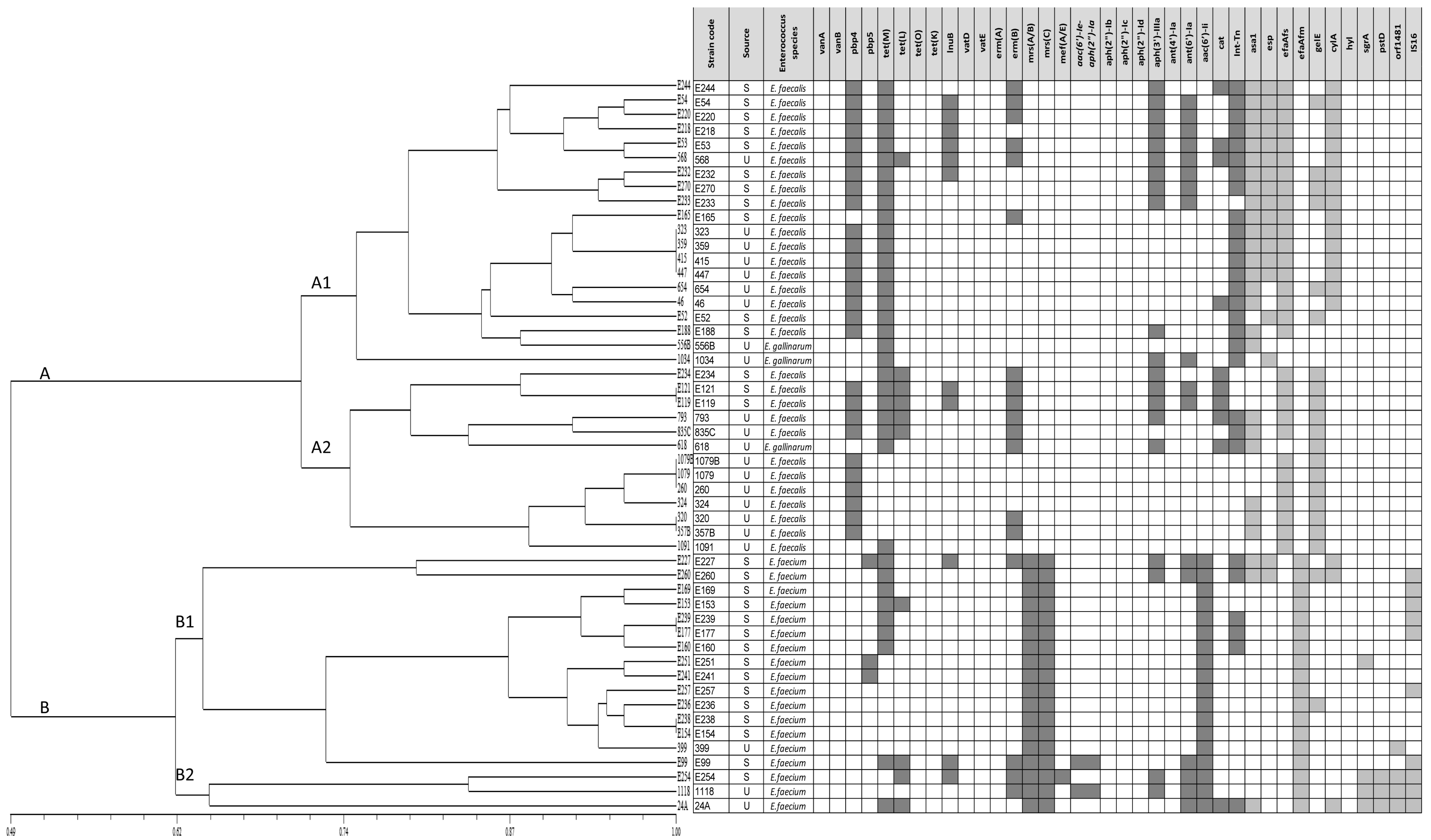
2.6. Clustering Analysis

Safety traits (antibiotic and virulence genes) of tested isolates were examined by cluster analysis with the NTSys ver. 2.02 program (Exeter Software Ltd.). Safety traits were recorded as ‘1′ or ‘0′ if positive or negative, respectively. The similarity distance between gene patterns was calculated using the Dice coefficient and the dendrogram was based on the UPMGA.

2.7. Statistical Analysis

The obtained data were analyzed using Chi-square (X2) test. Statistical tests were used to evaluate the difference between isolates from stools and urine in relation to phenotypic and genotypic antimicrobial resistance, biofilm production and in relation to presence of virulence genes. The statistical significance threshold was set at p-value ≤ 0.05.

3. Results

3.1. Bacteria Strains, Provenience and Identification

Based on identification with the use of the MALDI-TOF MS technique, 30 isolates were classified as E. faecalis (14 from healthy dog stools and 16 from sick dog urine), 18 isolates as E. faecium (15 from feces and 3 from urines) and 3 isolates as E. gallinarum (all from urines). MALDI-TOF MS analysis correctly identified 23 isolates with log (score) values of ≥ 2.3, which indicated ‘highly probable species identification’. In the case of 28 isolates, MALDI-TOF MS gave secure genus identification and probable species identification with a log (score) value of 2.025–2.290. MALDI-TOF MS confirmed biochemical identification of most isolates, with the exception of three E. faecium that were reclassified as E. faecalis, three E. faecium that were reclassified as E. gallinarum and two E. durans that were reclassified as E. faecium. Molecular identification confirmed MALDI-TOF MS results. Identification results obtained with MALDI-TOF MS were considered in the rest of this manuscript.

3.2. Phenotypic Resistance

Considering the results of disk diffusion test, high levels of resistance were detected for aminoglycosides (streptomycin 94.1%, neomycin 90.2%, gentamicin 68.6%), fluoroquinolones (enrofloxacin 74.5%, ciprofloxacin 66.7%), oxacillin (98%), clindamycin (84.3%), tetracycline (78.4%) and quinupristin–dalfopristin (78.4%). Meanwhile, against glycopeptides (vancomycin 13.7%, teicoplanin 3.9%) and amoxicillin/clavulanic acid (11.8%) few of tested isolates resulted as resistant. Most effective compounds included teicoplanin, 84.3% susceptible isolates, amoxicillin–clavulanic acid, 76.5% susceptible isolates and nitrofurantoin, 64.7% susceptible isolates. Table 1 reported detailed data on phenotypic resistance evaluation of all tested strains, in relation to kind of sample, sick dog urines or healthy dog feces. Some differences emerged; in particular, higher resistance was recorded for chloramphenicol, linezolid, nitrofurantoin, trimethoprim and tigecycline in isolates from stools, while isolates from urine were more resistant to quinupristin–dalfopristin, clindamycin and amoxicillin/clavulanic acid (p < 0.05).
A Minimum Inhibitory Concentration test for vancomycin did not confirm resistance in assessed isolates (MIC ≥ 32 µg/mL) and, consequently, all tested enterococci were considered phenotypically susceptible.
Minimum Inhibitory Concentration test for ampicillin confirmed disk diffusion results in all resistant isolates; furthermore, all but three intermediate isolates resulted resistant, presenting a MIC value over the breakpoint (≥ 16 µg/mL). Overall, considering MIC results, 33/51 (64.7%) isolates were ampicillin resistant. However, 24/33 (72.7%) resistant isolates showed a MIC ≥ 64 µg/mL. No statistical differences emerged between isolates from stools and urines (p > 0.05).
High Level Aminoglycoside Resistance was detected in 24/51 (47.1%) isolates. In detail, 11 isolates shoved only high-level streptomycin resistance (HLSR), 3 isolates only high-level gentamicin resistance (HLGR) and 10 isolates showed a high level of resistance to both streptomycin and gentamicin. No statistical differences were observed between isolates from stools and urine (p > 0.05).
Nine isolates showed HLAR in combination with ampicillin MIC ≥ 64 µg/mL: 1 E. faecalis and two E. faecium from stools (all HLSR), two E. faecalis (all HLSR), two E. faecium (one HLGR and one resistant to both streptomycin and gentamicin) and two E. gallinarum (one HLSR and one resistant to both streptomycin and gentamicin) from urine.
Regarding multidrug-resistance, 44 isolates resulted MDR and 6 XDR; 1 E. faecium isolate did not result as multidrug-resistant. XDR isolates were detected only from healthy dog stools; this condition resulted as significantly associated with isolates from this kind of sample (p > 0.05).
Table 2 summarizes data on HLAR, ampicillin MIC and multidrug-resistance classification.

3.3. Genotypic Resistance

The genes vanA, vanB, blaZ, tetO, tetK, ermA, aph(2”)-Ib, aph(2”)-Ic, aph(2”)-Id and ant(4’)-Ia were not detected. The other searched resistance genes were found in at least one isolate (Table 3). Most detected ones were tet(M) (36/51; 70.5%), pbp4 (27/51; 52.9%) and aph(3′)-IIIa (20/51; 39.2%). Furthermore, Int-Tn, part of transposons Tn916/Tn1545, was identified in 28/51 (54.9%).
Some genes responsible for resistance to aminoglycosides (aph(3′)-IIIa, aac(6′)-Ii, ant(6’)-Ia), macrolides (mrsA/B, mrsC) and lincosamides (lnuB) were more often associated with isolates from stools; while, the gene pbp4 related to β lactams resistance was detected more frequently in isolates from sick dog urine (p > 0.05). No statistical differences emerged about the distribution of other genes.
mef(A/E) gene was detected in E. faecium isolate from healthy dog feces and the nucleotide sequence of the product obtained for this gene has been deposited in GenBank under accession number: MN548247.
Sequencing of the C-terminal region of pbp5 gene revealed mutation responsible for amino acid changes at positions 633 E→R, 634 N→T in all studied isolates positive for pbp5. In addition, amino acid changes were found in the positions 471 V→I and 487 Q→L in E227 E. faecium isolate (MIC value of 256 µg/mL), but any changes in the same positions were observed for E251 and E241 E. faecium isolates (MIC values of 128 µg/mL) with respect to reference sequence of E. faecium (GenBank Accession No. CAA59287.1) (Figure 1).

3.4. Resistance Profiles: Comparison of Phenotypic and Genotypic Results

Regarding HLAR, among the 21 isolates showing high level streptomycin resistance, only 14 harbored the related resistance gene (ant(6’)-Ia); furthermore, three isolates were ant(6’)-Ia positive, but did not result HLSR. On the other hand, only two isolates had the gene aac(6’)-Ie-aph(2”)-Ia conferring resistance to high level of gentamycin and only one resulted as phenotypically resistant. Twelve isolates resulted HLGR, but they scored negative for all searched genes.
Sixteen phenotypically ampicillin resistant isolates scored positive for gene pbp4, pbp5 or both. While 17 phenotypically resistant isolates presented anyone of the genes associated with β-lactams resistance. Fourteen isolates were positive for the gene pbp4 but resulted as susceptible to ampicillin and amoxicillin/clavulanic acid.
All but one cat positive isolates were resistant to chloramphenicol; however, 11 out of 20 phenotypically resistant isolates scored negative for resistance gene.
Considering resistance to tetracycline, 36 phenotypically resistant isolates were positive for resistant genes. In particular, 26 harbored tet(M) only, 9 tet(M) and tet(L) in association and 1 isolate tet(L) only. Furthermore, four isolates resulted resistant in a disk diffusion test, but scored negative for all searched genes and one isolate positive for tet(M) was susceptible to tetracycline. All 28 isolates positive for Int-Tn were positive for tet(M) and 4 isolates were also positive for tet(L).
Regarding erythromycin, 19 resistant isolates were positive for one or more resistance genes (erm(B), mrs(A/B), mrs(C), mef(A/E)), whereas 10 resulted negative for all investigated genes. Twelve isolates harboring one or more resistance genes were susceptible to erythromycin; among them, 10 were positive for the mrs(A/B) and mrs(C) genes, 1 for gene mrs(C) and 2 for erm(B) gene.
All isolates harboring lnuB gene resulted resistant to clindamycin. However, 32 resistant isolates scored negative to resistance gene.
Finally, all 40 quinupristin–dalfopristin resistant isolates were negative for both vatE and vatD.
Table S3 reports the phenotypic and genotypic profile of each analyzed isolates as well as data on kind of sample and bacterial species.

3.5. Biofilm Production

All the analyzed enterococci resulted as biofilm producers. In detail, 21/51 (41.2%) were classified as weak biofilm producers, 11/51 (21.5%) as moderate biofilm producers and 19/51 (37.3%) as strong biofilm producers. Table 4 reports the distribution of biofilm production in relation to origin of samples and bacterial species. A statistical difference (p < 0.05) was observed between isolates from stools and UTI: higher level of biofilm production was observed in enterococci from urines. Statistical differences (p < 0.05) emerged also in relation to species: E. faecalis resulted as a higher biofilm producer than other species.

3.6. Virulence Genes and Virulence Profiles

Most detected genes were the ones coding for species-specific surface adhesins, efaAfs and efaAfm, that were present in all investigated E. faecalis and E. faecium isolates, respectively. Other genes encoding for enterococcal surface protein, esp, and aggregation substance, asa1, were largely present in the investigated bacteria: 18/51 (35.3%) and 27/51 (52.9%) positive isolates, respectively.
Other frequently detected genes were gelE, 21/51 (41.2%), and cylA, 19/51 (37.3%), related to gelatinase and cytolysin production, respectively. With the exception of 3 E. faecium isolates positive for gelE and/or cylA and 1 E. gallinarum positive for gelE, these genes were detected mainly in E. faecalis.
The genes sgrA, pstD and orf1481 were detected in less than 10% of isolates; meanwhile, hyl gene was not found in investigated enterococci.
Finally, the IS16 insertion element, often detected in human hospital strains, was detected in 10/51 (19.6%) isolates.
Table 5 reports the detailed distribution of virulence genes and data about phenotypic expression of gelE and cylA.
No statistical differences emerged about the distribution of virulence genes between isolates from healthy dog stools and urine of dogs with UTIs.
Considering the association of virulence genes, 18 different profiles were detected. All isolates were positive for at least one virulence gene. The number of genes found together in an isolate ranged between 1 and 7 (Table S3).
Finally, the genes asa1, cylA and esp more frequently resulted in strong biofilm producer strains (p < 0.05).

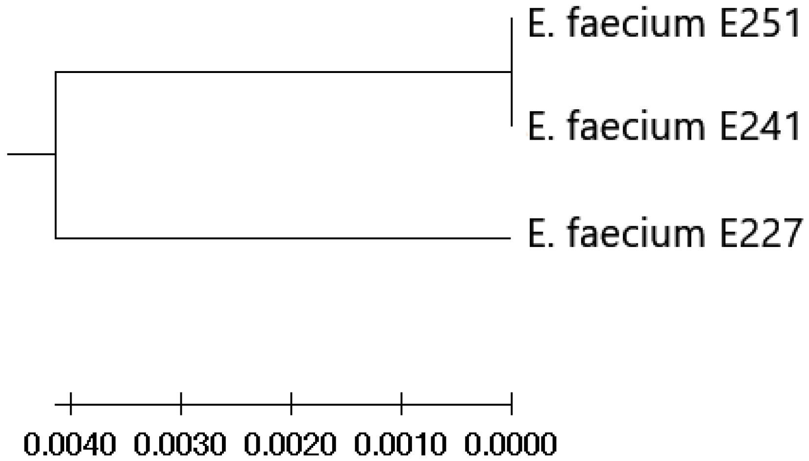
3.7. Clustering Analysis of Relevant Safety Traits

The isolates were clustered in two main groups (A and B). Group A contained E. faecalis and E. gallinarum isolates. Subsequently, two subgroups (A1 and A2) were identified within group A, which accumulated isolates mainly based on the presence of the tet(M), Int-Tn (Tn916/Tn1585), asa1, esp and cylA (A1) and gelE (A2) genes, respectively. Group B contained only E. faecium isolates. This group also had two main subgroups (B1 and B2), which had combined isolates mainly based on the simultaneous presence (B2) or absence (B1) of the sgrA, pstD, orf1481 and IS16 genes. Furthermore, bifunctional (aac(6′)-Ie-aph(2″)-Ia) gene was found within one isolate belonging to subgroup B2 and in the E99 isolate adjacent to this subgroup in contrast to the pbp5 gene, which was present in three isolates pooled within subgroup B1. Figure 2 shows details of the resistance and virulence gene map from which the similarity of the tested isolates was derived.

4. Discussion

In the last few years the opportunistic pathogens belonging to Enterococcus genus obtained a great deal of attention due to their frequent involvement in nosocomial infections linked to high level of antimicrobial resistance. Pets may act as reservoirs of multidrug-resistant enterococci, but may also be infected and develop disease. UTIs are the most frequent infection associated with Enterococcus infections in dogs [13].
In this study, isolates from two distinct kind of samples were analyzed under different points of view and compared. All isolates were collected in the same period of time from animals subjected to routine microbiological analysis. Urine samples were from dogs suffering UTI and Enterococcus spp. was diagnosed as the main responsible pathogen (generally the only one detected in pure culture). Stools from healthy dogs were subjected for routine feces control (presence/absence of Salmonella spp.). Enterococcus faecalis was the species most frequently detected, especially in UTI, with no statistical differences between the two kinds of sample. E. faecium resulted as more frequently present in feces (p < 0.05), and rarely involved in UTI. This is in line with other surveys, suggesting that E. faecalis is the primary species involved in enterococcal bacteriuria [31,32,33]. The two species were equally distributed in stools samples, as reported by other authors [3,7,9]. E. gallinarum is rarely reported as agent of canine UTIs [34], and is probably not a primary pathogen of dogs, as emerged from our investigation.
Antimicrobial resistance is probably the main threat in Enterococcus infections; indeed, these bacteria are frequently resistant to many antimicrobial agents [4]. In the present study, a very high percentage of isolates resulted resistant to aminoglycosides, fluoroquinolones, oxacillin, clindamycin, tetracycline and quinupristin–dalfopristin.
Aminoglycoside resistance in enterococci is well documented; indeed, in humans the therapeutic options are limited to streptomycin and gentamicin, only in combination with a cell-wall–active agent, commonly a β-lactam [6]. Generally, aminoglycoside resistance in enterococci from dogs is low [9,12,33,35,36], but some authors reported data similar to those obtained in the present study [11,37]. About half of resistant strains resulted HLAR and this is an atypical result; indeed, this condition is rarely reported in isolates from animals [12,38], with rare exceptions [39]. Furthermore, streptomycin and gentamicin are rarely employed in pet systemic therapy, due to their toxic effects, to justify a possible selection pressure.
No statistical differences were observed between stools and urine isolates, suggesting that this trait is probably fixed in Enterococcus population from this “ecosystem”. To strengthen this finding, the genes ant(6’)-Ia and aac(6’)-Ie-aph(2”)-Ia were largely detected in tested enterococci. Nevertheless, not all phenotypical resistant isolates scored positive to the investigated resistance genes; this result suggests other mechanisms of resistance, probably linked to other mobile elements or to genome mutations [4,5,6]. Furthermore, all analyzed E. faecium isolates had the aac(6’)-Ii gene, coding for a chromosomal aminoglycoside acetyltransferase specific for E. faecium, associated with the intrinsic low-level resistance to aminoglycosides, which was in agreement with other data [40,41].
In human therapy, ampicillin was generally used in combination with streptomycin or gentamicin to treat Enterococcus infections; resistance to high concentration of ampicillin (MIC ≥ 64 µg/mL) limits this possibility [2,5,6]. In our survey 51.0 and 19.6% of isolates resulted resistant and intermediate, respectively, to ampicillin in a disk diffusion method; all but three of them (64.7%) were confirmed as resistant in the broth microdilution assay. This percentage is higher than those observed in many other surveys on antimicrobial resistance in healthy and sick dogs [10,32,33,37,38,42]; however, some authors obtained similar results to those that emerged from our investigation [3,9,11]. A MIC value equal to or greater than 64 µg/mL was found in 72.7% of resistant isolates (47.1% of the total); this result stresses the hazard that these strains could represent for human health. The gene pbp4 was the second most detected among resistance genes. However, its presence only partially explains the phenotypic results. The gene pbp4 was found in both resistant and susceptible isolates and, among resistant strains, its presence was not associated to higher MIC values (p > 0.05). β-lactams resistance in Enterococcus is generally linked to the production of β-lactamase, to the overexpression of penicillin-binding proteins (PBPs) or to the alterations of PBPs, resulting in the production of PBPs with lower affinity with β-lactams. PBP4 and PBP5, expressed, respectively, by genes pbp4 and pbp5, are generally involved in these two last types of resistance [43,44]. However, some studies showed that these genes could be present in both resistant and susceptible strains; indeed, ampicillin resistance is generally associated to point mutations in some specific position of these genes [45], which is consistent with our results for mutations in the pbp5 gene.
Although the expression of pbp4 gene, alone or in combination with other genes, could mediate cephalosporin resistance too [46], in our study, no relations were found between the resistance to cephalothin and the presence of pbp4 and/or pbp5.
Even though the detected abundant ampicillin resistance, most of strains resulted as susceptible to amoxicillin/clavulanic acid, showing the potential effectiveness of this association; our data are in agreement to the results found in other studies [35,37,42], even if many authors reported a higher percentage of resistant enterococci in dogs, between 20 and 30% [11,32,33,39].
Tetracycline resistance was detected in 78.4% of isolates. This is a common finding, in agreement with other works on dogs reporting similar percentages in Enterococcus from feces [9,12,37,38,39] and other clinical specimens [11,33,42]. High level of tetracycline resistance are probably linked to the abundant use, in past years, of these antimicrobials, not only in pets, but more in general in animals and also in humans [47]. Molecular analyses support phenotypic results: tetracycline resistance genes were the most frequently detected genes and 90% of resistant isolates had tet(M), tet(L) or both genes, similarly to other studies [4,47]. The frequent detection (54.9%) of transposons Tn916/Tn1545, always in association with tet genes, underlines the possibility for these genes to jump from a strain to another and, consequently, the possibility for wide diffusion.
Resistance to oxacillin is well documented in enterococci and it could be considered as intrinsic [32,48,49,50]. Our data seem to confirm this statement, despite not all strains resulting resistant.
Resistance to clindamycin is considered intrinsic in Enterococcus, too; furthermore, this antimicrobial is considered ineffective in vivo also in case of in vitro susceptible isolates [4]. Our results are in line with this statement; however, it is interesting to note that clinical isolates more often resulted resistant. This could be related to the frequent use of this compound in pets therapy. The gene lnuB, conferring resistance to lincosamides and frequently carried by plasmids [51,52], was detected in relatively few isolates (21.6%), mainly E. faecalis, and it alone did not justify the detected level of resistance.
Resistance to quinupristin–dalfopristin is considered intrinsic only in some enterococcal species, in particular E. faecalis and E. gallinarum [26]. All analyzed E. faecalis and E. gallinarum isolates resulted resistant. A percentage of 38.8% of E. faecium were resistant, suggesting a high level of resistance in this species too, among tested isolates. These data are in line with other investigations [3,37]. None of the genes searched for antimicrobial resistance to streptogramins were detected; this finding suggests that other resistance mechanisms are involved in quinupristin–dalfopristin resistance among investigated isolates. In particular, efflux pumps encoded by macrolides resistance genes msrA/B, mefA, lsa, and vga, can confer resistance to streptogramins, too [53]. All E. faecium isolates analyzed in our study were positive for mrs(A/B) and mrs(C); one isolate was also positive for mef(A/E). Similarly, Aun et al. [40] noted that all E. faecium isolates had a chromosomal msr(C) gene associated with macrolide–streptogramin B resistance. In addition, Portillo et al. [41] found that PCR analysis with the msr(A)-specific primers designed for Staphylococcus aureus gives a DNA fragment of the expected size for all E. faecium strains, because it shows 62% identity at the nucleotide level with the msr(A) gene. This probably explains why the E. faecium isolates we tested as positive for the msr(C) gene were also positive for the msr(A/B) gene.
Erythromycin is not used for treatment of human enterococcal infections, but it is widely used in veterinary medicine, especially in production animals [54]. Percentage of resistance to this antimicrobial in Enterococcus strains from sick or healthy dogs is very variable, ranging between 10 and 80%, but it is generally high [3,9,32,33,37,38,39]. Results obtained in our study are in line with this research. Resistance to macrolide could be mediated by production of an enzyme that modifies ribosomal subunit, reducing the affinity of antimicrobial to ribosomes, or by efflux pumps; both of these events could be mediated by mobile genes such as erm or mef and msr, respectively [6,43]. The genes erm(B), msr(A/B) and msr(C) were largely detected in tested strains; however there was not a strong association with phenotypic resistance and this is in agreement with some other studies [52,55,56,57].
A high level of resistance was detected for fluoroquinolones, both in sick and healthy dog samples. This is in line with results of other investigations [3,32,33,37,55], even if some authors reported lower percentages of resistant strains [9,38]. Quinolone resistance mainly occurs via point mutation of bacterial genes for DNA gyrase and topoisomerase IV [43]. Although enterococci have an intrinsic reduced susceptibility to fluoroquinolones, the extensive use of enrofloxacin in veterinary, mainly pet, therapy could have contributed to the spread of resistance [4].
A middle level of resistance was detected for the remaining antimicrobials, rifampicin chloramphenicol, linezolid, nitrofurantoin and trimethoprim, ranging between 25 and 60%. These compounds are not frequently used in veterinary medicine, but are sometimes employed in humans for treatment of different infections, including enterococcal ones [5,6,43,58]. Resistance to these antimicrobials in Enterococcus strains from pet dogs is very variable, as emerged from different studies [9,11,32,33,38,39].
Vancomycin resistant enterococci were not detected in this survey, with both phenotypic and genotypic methods; teicoplanin resulted effective with most tested isolates. These findings are in agreement with most other studies; indeed glycopeptides resistance in pet dogs, and more in general in animals, is generally low [4,9,38,39,55], with some exceptions [3,33].
The last aspect evaluated in relation to antimicrobial resistance was the concurrent resistance to more compounds. All but one isolate resulted as multidrug-resistant; in particular, 44 isolates were MDR and 6 were XDR, following Magiorakos et al.’s classification [28]. This output is in line with different studies on enterococci from dogs [11,33,37,39], confirming that these bacteria are frequently and significantly involved in this hazardous problem for pets and for public health. Antimicrobial resistance in Enterococcus is well documented in the literature and different authors explored the role of animals as reservoirs and sources of multidrug-resistant enterococci for humans [4,59]. However, a great variability has been observed in relation to geographic areas and context, animal species, type of samples and panel of tested antimicrobials. Considering that antimicrobial resistance is a phenomenon in evolution, its monitoring is necessary, mainly in pet dogs that often live in close contact with their owners.
Finally, virulence of isolates was evaluated by biofilm production assay and virulence gene detection. All isolates resulted as biofilm producers. However, isolates from UTI and belonging to E. faecalis showed a higher level of biofilm production. Biofilm production is considered a very important factor in the establishment of urinary tract infections. Indeed, biofilm helps bacteria to adhere to biotic, as well as abiotic, surfaces; furthermore, it protects them from being flushed by urine and from local body defense and antimicrobial substances [14]. Our data confirm the importance of biofilm production in the pathogenesis of UTI; furthermore, the higher level of biofilm production found in E. faecalis could explain why this species is more often isolated from urine samples. Our results are in agreement with data derived from human medicine: generally, Enterococcus isolates from UTI are biofilm producers and E. faecalis, the primary enterococcal species isolated from urinary tract infections, are stronger producer than other species [29,60]. In our investigation, the genes asa1, cylA and esp were found more frequently in strong biofilm producer strains, suggesting their involvement in biofilm production. However, contrasting data emerge from literature about the set of genes leading to biofilm formation in Enterococcus [29,60,61,62,63]; probably, the biofilm formation is a multifactorial process far from being fully understood.
All isolates presented the gene encoding surface adhesins specific for their own species. This is an expected result and data are in accordance with the available literature [12,64,65,66]. Excluding surface adhesins specific genes, the most detected genes were asa1 (52.9%), gelE (41.2%), cylA (37.3%) and esp (35.3%). All these genes improve the ability of enterococci to enter and spread through the body. In particular, asa1 encodes an aggregation substance that facilitates adhesion and sharing of plasmids, gelE codes for a gelatinase capable of hydrolyzing different peptides, cylA is part of an operon elaborating a hemolytic cytolysin and esp is associated with increased virulence linked to enhanced colonization and persistence in body districts and biofilm formation. The percentage of detection in our study was generally higher than in other surveys on pet dogs [12,35,67,68]. All these genes were more associated with E. faecalis than other investigated species (p < 0.05), and this is in line with the literature data [69]. The higher virulence of E. faecalis isolates could explain the higher detection in clinical samples.

5. Conclusions

Data obtained in the present study confirm the double role of pet dogs as reservoirs and target hosts of enterococci. E. faecalis was confirmed as the primary species involved in canine UTIs. No relevant differences emerged between fecal and urinary isolates, with the exception of biofilm production. Although E. faecalis and E. faecium were equally present in stools, E. faecium resulted as less virulent and able to produce biofilm, and these features could explain its less frequent involvement in infections. Moreover, our results confirm the spread of antimicrobial resistance among enterococci, also in clinical isolates from pets. Antibiotic resistance is a severe concern for human and veterinary medicine. Canine enterococcal infections may be very difficult to treat; furthermore, infected dogs may be a source of Enterococcus spp. for their owners who, mainly in case of immunocompromised persons, can develop severe pathologies.

Supplementary Materials

The following are available online at https://www.mdpi.com/article/10.3390/ani11102845/s1, Table S1: Primers of antibiotic resistance used for PCR in the study; Table S2: Target genes and primers used in this study; Table S3: Origin, species, phenotypic and genotypic profile of each Enterococcus spp. isolate tested.

Author Contributions

Conceptualization, D.S.-P. and V.V.E.; methodology, D.S.-P., F.B., M.D. and G.C.; software, D.S.-P., M.D.; validation, D.S.-P. and V.V.E.; formal analysis, D.S.-P.; investigation, D.S.-P., F.B., M.D., G.C. and D.P.-O.; resources, D.S.-P., R.U.-C. and V.V.E.; data curation, D.S.-P. and F.B.; writing—original draft preparation, F.B.; writing—review and editing, D.S.-P., F.B. and V.V.E.; visualization, D.S.-P.; supervision, V.V.E.; funding acquisition, D.S.-P., R.U.-C. and V.V.E. All authors have read and agreed to the published version of the manuscript.

Funding

This research was funded by University of Pisa (Fondi di Ateneo) and University of Life Sciences in Lublin (Financial resources of the University—subvention).

Institutional Review Board Statement

Ethical review and approval were waived for this study, due to no animals being specifically recruited, but analyses were carried out on clinical specimens.

Data Availability Statement

The data presented in this study are contained within the article or supplementary material.

Conflicts of Interest

The authors declare no conflict of interest. The funders had no role in the design of the study; in the collection, analyses, or interpretation of data; in the writing of the manuscript, or in the decision to publish the results.

References

  1. Ramos, S.; Silva, V.; de Lurdes Enes Dapkevicius, M.; Igrejas, G.; Poeta, P. Enterococci, from harmless bacteria to a pathogen. Microorganisms 2020, 8, 1118. [Google Scholar] [CrossRef]
  2. Selleck, E.M.; Van Tyne, D.; Gilmore, M.S. Pathogenicity of Enterococci. Microbiol. Spectr. 2019, 7. [Google Scholar] [CrossRef]
  3. Kubašová, I.; Strompfová, V.; Lauková, A. Safety assessment of commensal enterococci from dogs. Folia Microbiol. 2017, 62, 491–498. [Google Scholar] [CrossRef]
  4. Torres, C.; Alonso, C.A.; Ruiz-Ripa, L.; León-Sampedro, R.; del Campo, R.; Coque, T.M. Antimicrobial Resistance in Enterococcus spp. of animal origin. Antimicrob. Resist. Bact. Livest. Companion Anim. 2018, 6, 185–227. [Google Scholar] [CrossRef]
  5. Miller, W.R.; Murray, B.E.; Rice, L.B.; Arias, C.A. Resistance in Vancomycin-Resistant Enterococci. Infect. Dis. Clin. N. Am. 2020, 34, 751–771. [Google Scholar] [CrossRef] [PubMed]
  6. Kristich, C.J.; Rice, L.B.; Arias, C.A. Enterococcal Infection—Treatment and Antibiotic Resistance. In Enterococci: From Commensals to Leading Causes of Drug Resistant Infection; Gilmore, M., Clewell, D., Ike, Y., Shankar, N., Eds.; Massachusetts Eye and Ear Infirmary: Boston, MA, USA, 2014. [Google Scholar]
  7. Pillay, S.; Zishiri, O.T.; Adeleke, M.A. Prevalence of virulence genes in enterococcus species isolated from companion animals and livestock. Onderstepoort J. Vet. Res. 2018, 85, 1–8. [Google Scholar] [CrossRef] [PubMed] [Green Version]
  8. Bertelloni, F.; Salvadori, C.; Lotti, G.; Cerri, D.; Ebani, V.V. Antimicrobial resistance in enterococcus strains isolated from healthy domestic dogs. Acta Microbiol. Immunol. Hung. 2017, 64, 203–321. [Google Scholar] [CrossRef] [Green Version]
  9. Bondi, M.; Iseppi, R.; Messi, P.; Anacarso, I.; Sabia, C.; Condò, C.; De Niederhausern, S. Antimicrobial resistance and virulence traits in Enterococcus strains isolated from dogs and cats. New Microbiol. 2015, 38, 369–378. [Google Scholar]
  10. Van den Bunt, G.; Top, J.; Hordijk, J.; de Greeff, S.C.; Mughini-Gras, L.; Corander, J.; van Pelt, W.; Bonten, M.J.M.; Fluit, A.C.; Willems, R.J.L. Intestinal carriage of ampicillin- and vancomycin-resistant Enterococcus faecium in humans, dogs and cats in the Netherlands. J. Antimicrob. Chemother. 2018, 73, 607–614. [Google Scholar] [CrossRef]
  11. Oguttu, J.W.; Qekwana, D.N.; Odoi, A. Prevalence and Predictors of Antimicrobial Resistance among Enterococcus spp. from Dogs Presented at a Veterinary Teaching Hospital, South Africa. Front. Vet. Sci. 2021, 7, 589439. [Google Scholar] [CrossRef]
  12. Trościańczyk, A.; Nowakiewicz, A.; Gnat, S.; Łagowski, D.; Osińska, M. Are dogs and cats a reservoir of resistant and virulent Enterococcus faecalis strains and a potential threat to public health? J. Appl. Microbiol. 2021, 131, 2061–2071. [Google Scholar] [CrossRef]
  13. Sykes, J.E. Streptococcal and enterococcal infections. In Canine and Feline Infectious Diseases; Sykes, J.E., Ed.; Elsevier Inc.: St. Louis, MO, USA, 2014; pp. 334–346. ISBN 9781437707953. [Google Scholar]
  14. Byron, J.K. Urinary Tract Infection. Vet. Clin. N. Am.—Small Anim. Pract. 2019, 49, 211–221. [Google Scholar] [CrossRef]
  15. Thompson, M.F.; Litster, A.L.; Platell, J.L.; Trott, D.J. Canine bacterial urinary tract infections: New developments in old pathogens. Vet. J. 2011, 190, 22–27. [Google Scholar] [CrossRef]
  16. Olin, S.J.; Bartges, J.W. Urinary Tract Infections. Treatment/Comparative Therapeutics. Vet. Clin. N. Am.—Small Anim. Pract. 2015, 45, 721–746. [Google Scholar] [CrossRef] [PubMed]
  17. Scarborough, R.; Bailey, K.; Galgut, B.; Williamson, A.; Hardefeldt, L.; Gilkerson, J.; Browning, G. Use of local antibiogram data and antimicrobial importance ratings to select optimal empirical therapies for urinary tract infections in dogs and cats. Antibiotics 2020, 9, 924. [Google Scholar] [CrossRef] [PubMed]
  18. Yamanaka, A.R.; Hayakawa, A.T.; Rocha, Í.S.M.; Dutra, V.; Souza, V.R.F.; Cruz, J.N.; Camargo, L.M.; Nakazato, L. The occurrence of multidrug resistant bacteria in the urine of healthy dogs and dogs with cystitis. Animals 2019, 9, 1087. [Google Scholar] [CrossRef] [Green Version]
  19. Yu, Z.; Wang, Y.; Chen, Y.; Huang, M.; Wang, Y.; Shen, Z.; Xia, Z.; Li, G. Antimicrobial resistance of bacterial pathogens isolated from canine urinary tract infections. Vet. Microbiol. 2020, 241, 108540. [Google Scholar] [CrossRef]
  20. Hernando, E.; Vila, A.; D’Ippolito, P.; Rico, A.J.; Rodon, J.; Roura, X. Prevalence and Characterization of Urinary Tract Infection in Owned Dogs and Cats From Spain. Top. Companion Anim. Med. 2021, 43, 100512. [Google Scholar] [CrossRef] [PubMed]
  21. Stępień-Pyśniak, D.; Hauschild, T.; Różański, P.; Marek, A. MALDI-TOF mass spectrometry as a useful tool for identification of Enterococcus spp. From wild birds and differentiation of closely related species. J. Microbiol. Biotechnol. 2017, 27, 1128–1137. [Google Scholar] [CrossRef]
  22. Kariyama, R.; Mitsuhata, R.; Chow, J.W.; Clewell, D.B.; Kumon, H. Simple and reliable multiplex PCR assay for surveillance isolates of vancomycin-resistant enterococci. J. Clin. Microbiol. 2000, 38, 3092–3095. [Google Scholar] [CrossRef] [Green Version]
  23. Jackson, C.R.; Fedorka-Cray, P.J.; Barrett, J.B. Use of a genus- and species-specific multiplex PCR for identification of enterococci. J. Clin. Microbiol. 2004, 42, 3558–3565. [Google Scholar] [CrossRef] [PubMed] [Green Version]
  24. Clinical and Laboratory Standards Institute. M02-A12—Performance Standards for Antimicrobial Disk Susceptibility Tests. In Approved Standard, 12th ed.; Clinical & Laboratory Standards Institute: Wayne, PA, USA, 2015; pp. 1–96. [Google Scholar]
  25. EUCAST (The European Committee on Antimicrobial Susceptibility Testing). Breakpoint Tables for Interpretation of MICs and Zone Diameters. Version 10.0, 2020; The European Committee on Antimicrobial Susceptibility Testing: Basel, Switzerland, 2020. [Google Scholar]
  26. CLSI (Clinical and Laboratory Standards Institute). M100 Performance Standards for Antimicrobial Susceptibility Testing A CLSI Supplement for Global Application, 28th ed.; CLSI Supplement M100: Clinical and Laboratory Standards Institute, Wayne, PA, USA, 2018. [Google Scholar]
  27. CLSI (Clinical and Laboratory; Standards Institute). M07-A10 Methods for Dilution Antimicrobial Susceptibility Tests for Bacteria That Grow Aerobically. In Approved Standard, 10th ed.; CLSI Document M07-A10: Clinical and Laboratory Standards Institute, Wayne, PA, USA, 2015. [Google Scholar]
  28. Magiorakos, A.P.; Srinivasan, A.; Carey, R.B.; Carmeli, Y.; Falagas, M.E.; Giske, C.G.; Harbarth, S.; Hindler, J.F.; Kahlmeter, G.; Olsson-Liljequist, B.; et al. Multidrug-resistant, extensively drug-resistant and pandrug-resistant bacteria: An international expert proposal for interim standard definitions for acquired resistance. Clin. Microbiol. Infect. 2012, 18, 268–281. [Google Scholar] [CrossRef] [PubMed] [Green Version]
  29. Hashem, Y.A.; Abdelrahman, K.A.; Aziz, R.K. Phenotype–Genotype Correlations and Distribution of Key Virulence Factors in Enterococcus faecalis Isolated from Patients with Urinary Tract Infections. Infect. Drug Resist. 2021, 14, 1713–1723. [Google Scholar] [CrossRef] [PubMed]
  30. Bertelloni, F.; Cagnoli, G.; Ebani, V.V. Virulence and antimicrobial resistance in canine staphylococcus spp. Isolates. Microorganisms 2021, 9, 515. [Google Scholar] [CrossRef] [PubMed]
  31. Wood, M.W.; Lepold, A.; Tesfamichael, D.; Lasarev, M.R. Risk factors for enterococcal bacteriuria in dogs: A retrospective study. J. Vet. Intern. Med. 2020, 34, 2447–2453. [Google Scholar] [CrossRef]
  32. KuKanich, K.S.; Lubbers, B.V. Review of enterococci isolated from canine and feline urine specimens from 2006 to 2011. J. Am. Anim. Hosp. Assoc. 2015, 51, 148–154. [Google Scholar] [CrossRef]
  33. Darwich, L.; Seminati, C.; Burballa, A.; Nieto, A.; Durán, I.; Tarradas, N.; Molina-López, R.A. Antimicrobial susceptibility of bacterial isolates from urinary tract infections in companion animals in Spain. Vet. Rec. 2020, 8, 1118. [Google Scholar] [CrossRef]
  34. Simjee, S.; White, D.G.; McDermott, P.F.; Wagner, D.D.; Zervos, M.J.; Donabedian, S.M.; English, L.L.; Hayes, J.R.; Walker, R.D. Characterization of Tn1546 in vancomycin-resistant Enterococcus faecium isolated from canine urinary tract infections: Evidence of gene exchange between human and animal enterococci. J. Clin. Microbiol. 2002, 40, 4659–4665. [Google Scholar] [CrossRef] [Green Version]
  35. Iseppi, R.; Di Cerbo, A.; Messi, P.; Sabia, C. Antibiotic resistance and virulence traits in vancomycin-resistant enterococci (Vre) and extended-spectrum β-lactamase/ampc-producing (ESBL/ampc) enterobacteriaceae from humans and pets. Antibiotics 2020, 9, 152. [Google Scholar] [CrossRef] [Green Version]
  36. Gómez-Beltrán, D.A.; Villar, D.; López-Osorio, S.; Ferguson, D.; Monsalve, L.K.; Chaparro-Gutiérrez, J.J. Prevalence of antimicrobial resistance in bacterial isolates from dogs and cats in a veterinary diagnostic laboratory in Colombia from 2016–2019. Vet. Sci. 2020, 7, 173. [Google Scholar] [CrossRef]
  37. Bang, K.; An, J.U.; Kim, W.; Dong, H.J.; Kim, J.; Cho, S. Antibiotic resistance patterns and genetic relatedness of enterococcus faecalis and enterococcus faecium isolated from military working dogs in korea. J. Vet. Sci. 2017, 18, 229–236. [Google Scholar] [CrossRef]
  38. Poeta, P.; Costa, D.; Rodrigues, J.; Torres, C. Antimicrobial resistance and the mechanisms implicated in faecal enterococci from healthy humans, poultry and pets in Portugal. Int. J. Antimicrob. Agents 2006, 27, 131–137. [Google Scholar] [CrossRef] [PubMed]
  39. Cinquepalmi, V.; Monno, R.; Fumarola, L.; Ventrella, G.; Calia, C.; Greco, M.F.; De Vito, D.; Soleo, L. Environmental contamination by dog’s faeces: A public health problem? Int. J. Environ. Res. Public Health 2013, 10, 72. [Google Scholar] [CrossRef] [PubMed] [Green Version]
  40. Aun, E.; Kisand, V.; Laht, M.; Telling, K.; Kalmus, P.; Väli, Ü.; Brauer, A.; Remm, M.; Tenson, T. Molecular Characterization of Enterococcus Isolates From Different Sources in Estonia Reveals Potential Transmission of Resistance Genes Among Different Reservoirs. Front. Microbiol. 2021, 12, 585. [Google Scholar] [CrossRef] [PubMed]
  41. Portillo, A.; Ruiz-Larrea, F.; Zarazaga, M.; Alonso, A.; Martinez, J.L.; Torres, C. Macrolide Resistance Genes in Enterococcus spp. Antimicrob. Agents Chemother. 2000, 44, 967. [Google Scholar] [CrossRef] [Green Version]
  42. Rumi, M.V.; Nuske, E.; Mas, J.; Argüello, A.; Gutkind, G.; Di Conza, J. Antimicrobial resistance in bacterial isolates from companion animals in Buenos Aires, Argentina: 2011–2017 retrospective study. Zoonoses Public Health 2021, 00, 1–11. [Google Scholar] [CrossRef]
  43. Miller, W.R.; Munita, J.M.; Arias, C.A. Mechanisms of antibiotic resistance in enterococci. Expert Rev. Anti. Infect. Ther. 2014, 12, 1221–1236. [Google Scholar] [CrossRef]
  44. Ono, S.; Muratani, T.; Matsumoto, T. Mechanisms of resistance to imipenem and ampicillin in Enterococcus faecalis. Antimicrob. Agents Chemother. 2005, 49, 2954–2958. [Google Scholar] [CrossRef] [Green Version]
  45. Conceição, N.; da Silva, L.E.P.; da Costa Darini, A.L.; Pitondo-Silva, A.; de Oliveira, A.G. Penicillin-resistant, ampicillin-susceptible Enterococcus faecalis of hospital origin: Pbp4 gene polymorphism and genetic diversity. Infect. Genet. Evol. 2014, 28, 289–295. [Google Scholar] [CrossRef]
  46. Djorić, D.; Little, J.L.; Kristich, C.J. Multiple low-reactivity class B penicillin-binding proteins are required for cephalosporin resistance in enterococci. Antimicrob. Agents Chemother. 2020, 64, e02273-19. [Google Scholar] [CrossRef]
  47. Choi, J.M.; Woo, G.J. Transfer of Tetracycline Resistance Genes with Aggregation Substance in Food-Borne Enterococcus faecalis. Curr. Microbiol. 2015, 70, 476–484. [Google Scholar] [CrossRef] [Green Version]
  48. Kubašová, I.; Lauková, A.; Hamarová, Ľ.; Pristaš, P.; Strompfová, V. Evaluation of enterococci for potential probiotic utilization in dogs. Folia Microbiol. 2019, 64, 177–187. [Google Scholar] [CrossRef]
  49. Balaei Gajan, E.; Shirmohammadi, A.; Aghazadeh, M.; Alizadeh, M.; Sighari Deljavan, A.; Ahmadpour, F. Antibiotic Resistance in Enterococcus faecalis Isolated from Hospitalized Patients. J. Dent. Res. Dent. Clin. Dent. Prospects 2013, 7, 102–104. [Google Scholar] [CrossRef] [PubMed]
  50. Bertelloni, F.; Salvadori, C.; Moni, A.; Cerri, D.; Mani, P.; Ebani, V.V. Antimicrobial resistance in Enterococcus spp. Isolated from laying hens of backyard poultry flocks. Ann. Agric. Environ. Med. 2015, 22, 665–669. [Google Scholar] [CrossRef] [Green Version]
  51. Si, H.; Zhang, W.J.; Chu, S.; Wang, X.M.; Dai, L.; Hua, X.; Dong, Z.; Schwarz, S.; Liu, S. Novel plasmid-borne multidrug resistance gene cluster including lsa(E) from a Linezolid-Resistant Enterococcus faecium Isolate of Swine Origin. Antimicrob. Agents Chemother. 2015, 59, 7113–7116. [Google Scholar] [CrossRef] [Green Version]
  52. Cho, S.; Barrett, J.B.; Frye, J.G.; Jackson, C.R. Antimicrobial resistance gene detection and plasmid typing among multidrug resistant enterococci isolated from freshwater environment. Microorganisms 2020, 8, 1338. [Google Scholar] [CrossRef] [PubMed]
  53. Mast, Y.; Wohlleben, W. Streptogramins—Two are better than one! Int. J. Med. Microbiol. 2014, 304, 44–50. [Google Scholar] [CrossRef] [PubMed]
  54. Bortolaia, V.; Guardabassi, L. Zoonotic transmission of antimicrobial resistant enterococci: A threat to public health or an overemphasised risk? In Zoonoses-Infections Affecting Humans and Animals: Focus on Public Health Aspects; Springer Netherlands: Dordrecht, The Netherlands, 2015; pp. 407–431. ISBN 9789401794572. [Google Scholar]
  55. Ben Said, L.; Dziri, R.; Sassi, N.; Lozano, C.; Ben Slama, K.; Ouzari, I.; Torres, C.; Klibi, N. Species distribution, antibiotic resistance and virulence traits in canine and feline enterococci in Tunisia. Acta Vet. Hung. 2017, 65, 173–184. [Google Scholar] [CrossRef] [PubMed]
  56. Stȩpień-Pyśniak, D.; Hauschild, T.; Dec, M.; Marek, A.; Urban-Chmiel, R. Clonal structure and antibiotic resistance of enterococcus spp. from wild birds in Poland. Microb. Drug Resist. 2019, 25, 1227–1237. [Google Scholar] [CrossRef] [PubMed]
  57. Grassotti, T.T.; De Angelis Zvoboda, D.; Da Fontoura Xavier Costa, L.; De Araújo, A.J.G.; Pereira, R.I.; Soares, R.O.; Wagner, P.G.C.; Frazzon, J.; Frazzon, A.P.G. Antimicrobial resistance profiles in enterococcus spp. Isolates from fecal samples of wild and captive black capuchin Monkeys (Sapajus nigritus) in South Brazil. Front. Microbiol. 2018, 9, 2366. [Google Scholar] [CrossRef] [PubMed]
  58. Bhat, A.H. Bacterial zoonoses transmitted by household pets and as reservoirs of antimicrobial resistant bacteria. Microb. Pathog. 2021, 155, 104891. [Google Scholar] [CrossRef]
  59. Hammerum, A.M. Enterococci of animal origin and their significance for public health. Clin. Microbiol. Infect. 2012, 18, 619–625. [Google Scholar] [CrossRef]
  60. Zheng, J.X.; Bai, B.; Lin, Z.W.; Pu, Z.Y.; Yao, W.M.; Chen, Z.; Li, D.Y.; Deng, X.B.; Deng, Q.W.; Yu, Z.J. Characterization of biofilm formation by Enterococcus faecalis isolates derived from urinary tract infections in China. J. Med. Microbiol. 2018, 67, 60–67. [Google Scholar] [CrossRef]
  61. Ch’ng, J.H.; Chong, K.K.L.; Lam, L.N.; Wong, J.J.; Kline, K.A. Biofilm-associated infection by enterococci. Nat. Rev. Microbiol. 2019, 17, 82–94. [Google Scholar] [CrossRef]
  62. Fallah, F.; Yousefi, M.; Pourmand, M.R.; Hashemi, A.; Nazari Alam, A.; Afshar, D. Phenotypic and genotypic study of biofilm formation in Enterococci isolated from urinary tract infections. Microb. Pathog. 2017, 108, 85–90. [Google Scholar] [CrossRef]
  63. Mohamed, J.A.; Huang, D.B. Biofilm formation by enterococci. J. Med. Microbiol. 2007, 56, 1581–1588. [Google Scholar] [CrossRef] [PubMed] [Green Version]
  64. Eaton, T.J.; Gasson, M.J. Molecular Screening of Enterococcus Virulence Determinants and Potential for Genetic Exchange between Food and Medical Isolates. Appl. Environ. Microbiol. 2001, 67, 1628–1635. [Google Scholar] [CrossRef] [PubMed] [Green Version]
  65. Kayaoglu, G.; Ørstavik, D. Virulence factors of Enterococcus faecalis: Relationship to endodontic disease. Crit. Rev. Oral Biol. Med. 2004, 15, 308–320. [Google Scholar] [CrossRef] [PubMed] [Green Version]
  66. Dec, M.; Stȩpień-Pyśniak, D.; Gnat, S.; Fratini, F.; Urban-Chmiel, R.; Cerri, D.; Winiarczyk, S.; Turchi, B. Antibiotic Susceptibility and Virulence Genes in Enterococcus Isolates from Wild Mammals Living in Tuscany, Italy. Microb. Drug Resist. 2020, 26, 505–519. [Google Scholar] [CrossRef] [PubMed]
  67. Ferguson, D.M.; Talavera, G.N.; Hernández, L.A.R.; Weisberg, S.B.; Ambrose, R.F.; Jay, J.A. Virulence Genes among Enterococcus faecalis and Enterococcus faecium Isolated from Coastal Beaches and Human and Nonhuman Sources in Southern California and Puerto Rico. J. Pathog. 2016, 2016, 1–7. [Google Scholar] [CrossRef] [Green Version]
  68. Gulhan, T.; Boynukara, B.; Ciftci, A.; Sogut, M.U.; Findik, A. Characterization of Enterococcus faecalis isolates originating from different sources for their virulence factors and genes, antibiotic resistance patterns, genotypes and biofilm production. Iran. J. Vet. Res. 2015, 16, 261–266. [Google Scholar] [CrossRef] [PubMed]
  69. Kiruthiga, A.; Padmavathy, K.; Shabana, P.; Naveenkumar, V.; Gnanadesikan, S.; Malaiyan, J. Improved detection of esp, hyl, asa1, gelE, cylA virulence genes among clinical isolates of Enterococci. BMC Res. Notes 2020, 13, 1–7. [Google Scholar] [CrossRef] [PubMed] [Green Version]
Figure 1. Pbp5 phylogeny coincides with MIC value. An UPGMA tree of pbp5 was constructed using the amino acid sequences from three studied isolates. Pbp5 divides isolates into two groups based on amino acid sequence, and these two groups coincide with the MIC value. E. faecium isolate with an ampicillin MIC of 128 µg/mL was clustered separately from two E. faecium isolates with an MIC of 256 µg/mL in the tree.
Animals 11 02845 g001
Figure 2. Dendrogram generated from safety traits data based on antibiotic and virulence patterns found in analyzed enterococcal isolates. The scale indicates the similarity level. The main groups clustering individual enterococcal species are designated A and B, while their subgroups linking isolates in response to certain genes are described A1, A2, B1 and B2, respectively. S—Stool, U—Urine.
Animals 11 02845 g002
Table 1. Results of disk diffusion antimicrobial test.
AntimicrobialResistant N (%)Intermediate N (%)Susceptible N (%)
ClassAgentTotalStoolUrineTotalStoolUrineTotalStoolUrine
AnsamycinsRD22 (43.1)15 (51.7)7 (31.8)11 (21.6)5 (17.2)6 (27.3)18 (35.3)9 (31.0)9 (40.9)
PhenicolsC21 (41.2)16 (55.2)5 (22.7)8 (15.7)5 (17.2)3 (13.6)22 (43.1)8 (27.6)14 (63.6)
OxazolidinonesLZD21 (41.2)21 (72.4)0 (0.0)5 (9.8)3 (10.3)2 (9.1)25 (49.0)5 (17.2)20 (90.9)
NitrofurantoinsF13 (25.5)13 (44.8)0 (0.0)5 (9.8)3 (10.3)2 (9.1)33 (64.7)13 (44.8)20 (90.9)
Folate pathway antagonistsW31 (60.8)23 (79.3)8 (36.4)19 (37.3)6 (20.7)13 (59.1)1 (2.0)0 (0.0)1 (4.5)
AminoglycosideN46 (90.2)28 (96.6)18 (81.8)4 (7.8)1 (3.4)3 (13.6)1 (2.0)0 (0.0)1 (4.5)
CN35 (68.6)20 (69.0)15 (68.2)4 (7.8)2 (6.9)2 (9.1)12 (23.5)7 (24.1)5 (22.7)
S48 (94.1)28 (96.6)20 (90.9)2 (3.9)1 (3.4)1 (4.5)1 (2.0)0 (0.0)1 (4.5)
CephemsKF32 (62.7)17 (58.6)15 (68.2)14 (27.5)10 (34.5)4 (18.2)5 (9.8)2 (6.9)3 (13.6)
FluoroquinolonesCIP34 (66.7)18 (62.1)16 (72.7)7 (13.7)7 (24.1)0 (0.0)10 (19.6)4 (13.8)6 (27.3)
ENR38 (74.5)22 (75.9)16 (72.7)9 (17.6)5 (17.2)4 (18.2)4 (7.8)2 (6.9)2 (9.1)
GlycopeptidesTEC2 (3.9)2 (6.9)0 (0.0)6 (11.8)3 (10.3)3 (13.6)43 (84.3)24 (82.8)19 (86.4)
VA7 (13.7)2 (6.9)5 (22.7)15 (29.4)7 (24.1)8 (36.4)29 (56.9)20 (69.0)9 (40.9)
Macrolides,
Streptogramins,
Lincosamides
E29 (56.9)16 (55.2)13 (59.1)17 (33.3)11 (37.9)6 (27.3)5 (9.8)2 (6.9)3 (13.6)
QD40 (78.4)19 (65.5)21 (95.5)7 (13.7)6 (20.7)1 (4.5)4 (7.8)4 (13.8)0 (0.0)
DA43 (84.3)21 (72.4)22 (100.0)6 (11.8)6 (20.7)0 (0.0)2 (3.9)2 (6.9)0 (0.0)
PenicillinsOX50 (98.0)28 (96.6)22 (100.0)1 (2.0)1 (3.4)0 (0.0)0 (0.0)0 (0.0)0 (0.0)
AMC6 (11.8)1 (3.4)5 (22.7)6 (11.8)6 (20.7)0 (0.0)39 (76.5)22 (75.9)17 (77.3)
AMP26 (51.0)17 (58.6)9 (40.9)10 (19.6)0 (0.0)10 (45.5)15 (29.4)12 (41.4)3 (13.6)
TetracyclinesTE40 (78.4)24 (82.8)16 (72.7)5 (9.8)2 (6.9)3 (13.6)6 (11.8)3 (10.3)3 (13.6)
TIG15 (29.4)15 (51.7)0 (0.0)8 (15.7)6 (20.7)2 (9.1)28 (54.9)8 (27.6)20 (90.9)
RD = rifampicin; C = chloramphenicol; LZD = linezolid; F = nitrofurantoin; W = trimethoprim; N = neomycin; CN = gentamicin; S = streptomycin; KF = cephalothin; CIP = ciprofloxacin; ENR = enrofloxacin; TEC = teicoplanin; VA = vancomycin; E = erythromycin; QD = quinupristin–dalfopristin; DA = clindamycin; OX = oxacillin; AMC = amoxicillin–clavulanic acid; AMP = ampicillin; TE = tetracycline; TIG = tigecycline.
Table 2. Multidrug-resistance, ampicillin MIC and high level aminoglycosides resistance detected, in relation to species and origin of samples.
Origin
of
Samples
Bacteria
Species
Multidrug-Resistant
Category
Ampicillin MIC ≥
64 µg/mL
HLARHLAR in Isolates with
Ampicillin MIC ≥ 64 µg/mL
MDRXDRHLSRHLGRBothHLSRHLGRBoth
Feces of healthy dogsE. faecalis113112108100
E. faecium11313200200
Urine of dogs with UTIsE. faecalis1606400200
E. faecium302121121
E. gallinarum302211211
Total44624211310832
Table 3. Distribution of resistance genes among Enterococcus spp. isolated from healthy and sick dogs.
AntibioticResistance GeneHealthy Dog FecesSick Dog UrineTotal
n = 51
E. faecalis
n = 14
E. faecium
n = 15
E. faecalis
n = 16
E. faecium
n = 3
E. gallinarum
n = 3
Gentamicin (HL)aac(6’)-Ie-aph(2”)-Ia010102
Kanamycin (HL)aph(3′)-IIIa12321220
Gentamicin
and other aminoglycosides (ML)
aac(6′)-Ii01503018
Streptomycin (HL)ant(6’)-Ia9412117
Chloramphenicolcat5031110
β lactamspbp4120150027
pbp5030003
Tetracyclinestet(M)148101336
tet(L)3331010
LincosamideslnuB7310011
Macrolideserm(B)8351118
mrs(A/B)01503018
mrs(C)11302016
mef(A/B)010001
-Int-Tn10591328
HL—High level; ML—Moderate level.
Table 4. Biofilm production by examined Enterococcus spp. isolates.
SampleSpeciesWBPMBPSBP
Feces of healthy dogsE. faecalis1310
E. faecium1500
Urine of dogs with UTIsE. faecalis358
E. faecium210
E. gallinarum021
Total211119
Legend: WBP = Weak biofilm producers; MBP = Moderate biofilm producers; SB = Strong biofilm producers.
Table 5. Prevalence of virulence genes detected in Enterococcus spp. isolates.
Virulence
Genes
Feces of Healthy DogsUrine of Dogs with UTIsTotal
n= 51 (%)
E. faecalis
n = 14
E. faecium
n = 15
E. faecalis
n = 16
E. faecium
n = 3
E. gallinarum
n = 3
cylA9 (8 a)2 (2 a)7 (2 a)1 (1 a)019 (37.3)
gelE8 (8 b)2 (1 a)10 (5 b)0121 (41.2)
asa110294227 (52.9)
esp10250118 (35.3)
efaAfs140160030 (58.8)
efaAfm01503018 (35.3)
sgrA020204 (7.8)
pstD010203 (5.9)
orf1481010304 (7.8)
IS160802010 (19.6)
a = number of isolates positive for β-hemotytic activity; b = number of isolates showing gelatinase activity.
Publisher’s Note: MDPI stays neutral with regard to jurisdictional claims in published maps and institutional affiliations.

