Article

Arginine Supply Impacts the Expression of Candidate microRNA Controlling Milk Casein Yield in Bovine Mammary Tissue

1
State Key Laboratory of Animal Nutrition, Institute of Animal Science, Chinese Academy of Agricultural Sciences, No. 2 Yuanmingyuan West Road, Beijing 100193, China
2
College of Animal Science and Technology, Yangzhou University, 88 South University Ave., Yangzhou 225009, China
3
School of Clinical Medicine, Southeast University, 87 Dingjiaqiao Road, Nanjing 210009, China
4
Department of Animal Sciences and Division of Nutritional Sciences, University of Illinois, 1207 W. Gregory Drive, Urbana, IL 61801, USA
*
Authors to whom correspondence should be addressed.
Animals 2020, 10(5), 797; https://doi.org/10.3390/ani10050797
Submission received: 15 December 2019 / Revised: 23 April 2020 / Accepted: 28 April 2020 / Published: 5 May 2020
(This article belongs to the Section Animal Nutrition)

Abstract

:

Simple Summary

It has been reported that arginine plays an important role in lactation, including promoting mammary gland development, increasing yields of milk and casein. Recent studies revealed that microRNA could be involved in regulating expression of functional genes related to mammary development. Thus, exploring the role of arginine on the regulation of miRNA related to bovine mammary development and milk production in dairy cows is of importance. The present work revealed that arginine injection increased casein yield by altering the expression of selected microRNA associated with mammary development.

Abstract

Arginine, a semi-essential functional amino acid, has been found to promote the synthesis of casein in mammary epithelial cells to some extent. Data from mouse indicated that microRNA (miRNA) are important in regulating the development of mammary gland and milk protein synthesis. Whether there are potential links among arginine, miRNA and casein synthesis in bovine mammary gland is uncertain. The objective of the present work was to detect the effects of arginine supplementation on the expression of miRNA associated with casein synthesis in mammary tissue and mammary epithelial cells (BMEC). The first study with bovine mammary epithelial cells (BMEC) focused on screening for miRNA candidates associated with the regulation of casein production by arginine. The BMEC were cultured with three different media, containing 0, 1.6 and 3.2 mM arginine, for 24 h. The expression of candidate miRNA was evaluated. Subsequently, in an in vivo study, 6 Chinese Holstein dairy cows with similar BW (mean ± SE) (512.0 ± 19.6 kg), parity (3), BCS (4.0) and DIM (190 ± 10.3 d) were randomly assigned to three experimental groups. The experimental cows received an infusion of casein, arginine (casein plus double the concentration of arginine in casein), and alanine (casein plus alanine, i.e., iso-nitrogenous to the arginine group) in a replicated 3 × 3 Latin square design with 22 d for each period (7 d for infusion and 15 d for washout). Mammary gland biopsies were obtained from each cow at the end of each infusion period. Results of the in vitro study showed differences between experimental groups and the control group for the expression of nine miRNA: miR-743a, miR-543, miR-101a, miR-760-3p, miR-1954, miR-712, miR-574-5p, miR-468 and miR-875-3p. The in vivo study showed that arginine infusion promoted milk protein content, casein yield and the expression of CSN1S1 and CSN1S2. Furthermore, the expression of miR-743a, miR-543, miR-101a, miR-760-3p, miR-1954, and miR-712 was also greater in response to arginine injection compared with the control or alanine group. Overall, results both in vivo and in vitro revealed that arginine might partly influence casein yield by altering the expression of 6 miRNAs (miR-743a, miR-543, miR-101a, miR-760-3p, miR-1954, and miR-712).

1. Introduction

Arginine, a semi-essential functional amino acid, plays an important role in animal nutrition. It has been shown that the addition of arginine to diets effectively increases average daily weight gain and improves the growth performance of animals [1]. Furthermore, some studies also indicated that arginine plays an important role in milk protein synthesis in mammals. For example, Chew et al. [2] found that arginine infusion in late-pregnant cows dramatically increased concentrations of prolactin, growth hormone, and insulin in blood and subsequently increased milk yield. Furthermore, the study by Ma et al. [3] indicated that the supplementation of arginine stimulated protein synthesis and reduced protein degradation in porcine mammary epithelial cells, leading to increases in cell proliferation and the production of major milk proteins. A recent study in lactating cows also found that a deficiency of arginine had a negative effect on milk protein production [4]. In other studies, the effects of arginine on protein synthesis have been attributed to metabolites of arginine, such as nitric oxide and polyamines, which regulate the growth of mammary gland and blood vessels [5,6]. A study in bovine mammary epithelial cells (BMEC) also found that the supplementation of arginine promoted the synthesis of casein and expression of CSN1S1, CSN1S2, CSN2, and CSN3 by regulating the signaling pathway of tyrosine kinase 2-signal transduction and transcriptional activator 5 (JAK2-STAT5) and rapamycin target protein (mTOR) [7].
Studies in dairy cows and rats have revealed that the expression profiles of selected miRNA differ across stages of lactation, underscoring the potential importance of these molecules in milk production [8,9]. A previous study in mice revealed that the expression of miR-431, which is responsible for the sparse spiral ganglion neurons by suppressing EYA4 (Eyes absent homolog 4) translation, was down-regulated as development progressed [10]. However, the expression of miR133, which enhances myoblast proliferation by repressing the serum response factor [11], was up-regulated during pregnancy and lactation. These results suggested that those miRNAs are functionally involved in murine mammary gland development. Murine mammary cell proliferation and β-casein gene expression are partly regulated by miR-101a [12]. In the context of milk components, data indicate that the 3’UTR of the casein genes (CSN1S1, CSN1S2, CSN2, and CSN3) in goats is located in the complementary regions of various predicted miRNA seed sequences [13].
Taking into consideration the results of previous studies, it is possible that the changes in the expression of casein genes and related functional genes that respond to increased arginine concentration are modulated by miRNA. Thus, the objective of this work was to uncover the miRNAs involved in the regulation of milk protein production induced by increased arginine supply.

2. Materials and Methods

2.1. In Vitro Experiment

2.1.1. Experimental Design and Culture Medium

The methods for the in vitro experiment were similar to those described by Wang et al. [14] Briefly, bovine mammary epithelial cells (BMEC) were cultured for 24 h in three different media containing 0 (Control group), 1.60 (Casein group), and 3.20 mM (Arginine group) arginine. The arginine-containing medium (Table 1) had a profile of amino acids similar to casein [15]. In brief, the basic medium devoid of arginine was prepared by combining the individual reagents including vitamins, minerals, and amino acids in ultrapure water (according to the composition of DMEM/F12, Gibco, Invitrogen, Catalog #11320082, Life Technologies Corporation, Burlingont, ONT, Canada). The medium pH was adjusted to 7.2 with NaHCO3. All reagents used in cell culture were purchased from Sigma Chemical Co. (St. Louis, MO, USA). Before the arginine study, the basic medium was used in the cell resuscitation process.

2.1.2. Cell Resuscitation and Treatment

The cell resuscitation process was described in previous papers [7,14]. In brief, second-generation mammary epithelial cells were taken from liquid nitrogen and placed in a 37 °C water bath. Cells were centrifuged at 150× g for 5 min at 4 °C to isolate BMEC, and the precipitate rinsed with fresh medium prior to transferring to a new culture bottle. Once 90% confluence was reached, cultured cells were harvested with 0.25% trypsin EDTA and seeded at a density of 5 × 10⁴ cells/mL into 6-well plates (3335, Corning Life Science, New York, NY, USA), containing growth medium (DMEM/F12 with 10% Fetal bovine serum (FBS (16000-044, Gibco, Carlsbad, CA, USA)), 500 ng/mL hydrocortisone (S0135, Sigma, Saint Louis, MI, USA), 1 µg/L prolactin (L6520, Sigma, Saint Louis, MI, USA), 10 ng/mL epidermal growth factor (AF-100-15, Peprotech, Rocky Hill, CT, USa), and 100 IU/mL penicillin/streptomycin)). Each treatment was replicated 6 times and cultured in an incubator at 37 °C, 95% O2, and 5% CO2. Cells were incubated with DMEM/F12 medium without fetal bovine serum for 16 h, to eliminate the effects from serum, then the cells were incubated in medium with different treatments for another 24 h.

2.1.3. Database Prediction and Result Acquisition

The prediction of miRNA was carried out with PicTar (http://pictar.mdc-berlin.de/) and the TargetScan (http://www.targetscan.org/) database. The first step was to identify highly-conserved miRNA candidates with high context scores, by searching target genes in both PicTar and TargetScan databases. Secondly, the miRNA candidates from both PicTar and TargetScan were listed in the same Excel file, and the miRNA overlapping both databases were selected. Lastly, we selected the miRNA which regulate multiple target genes (at least 2) for further analysis (Table 2).

2.1.4. Detection of miRNA Expression by RT-qPCR

The miRNA were isolated from cells with a miRNA Isolation Kit according to the manufacturer’s protocols (Tian Gen, Catalog # DP501, Beijing, China). The miRNA were polyadenylated using Poly (A) polymerase, according to the manufacturer’s instructions (miRcute miRNA First-Strand Synthesis Kit, Tian Gen, Catalog # KR201, Beijing, China). cDNA was synthesized with Oligo (dT)—universal tag and reverse transcription primer. RT-PCR analysis was done using the SYBR Green method with the miRcute miRNA qPCR Detection Kit (Tian Gen, Catalog # FP401, Beijing, China) in the ABI Prism 7500 Detection Instrument (Applied Biosystems, Foster, CA, USA). Each sample was run in triplicate. The internal reference gene was U6 [16], and the primer sequence from 5′ to 3′ was F—CGC TTC GGC AGC ACA TAT AC & R—TTC ACG AAT TTG CGT GTC AT. The expression of U6 was not altered by treatment. The other primer sequences were the complete miRNA sequences.

2.2. In Vivo Experiment

2.2.1. Experimental Animals and Management

Six lactating Holstein cows with similar BW (mean ± SE) (512.0 ± 19.6 kg), parity (3), BCS (4.0) and DIM (190 ± 10.3 d) were selected for this experiment from the Experimental Farm of Yangzhou University. All animal procedures were approved by the Yangzhou University Animal Care and Use Committee of Jiangsu Province (China).
The basal diet was formulated according to NRC [17]. The diet composition is reported in Table 3. Cows were fed twice a day at 0600 and 2000 h, and feed refusals collected to calculate daily DM intake. The experimental cows were milked 3 times a day at 0700, 1500 and 2300 h. Cows were housed separately in a free-stall barn and had ad libitum access to the TMR and freshwater. Indwelling catheters (137 mm: 1.2 mm i.d.: 2.0 mm o.d., L13712, Jiangxi Huali Medical Instrument Company, Ganzhou, China) were placed in a jugular vein and flushed with heparin and physiological saline (750 IU/mL) twice daily during the week before infusion treatments.

2.2.2. Experimental Design

Six experimental animals were randomly divided into 3 treatments (2 cows per group) in a replicated 3 × 3 Latin square design with 22 days for each period (day 1 to day 7 for infusion and day 8 to day 22 for washout). The treatments were casein group (casein model), arginine group (casein plus double the concentration of arginine in casein), and alanine group (added alanine to make the it iso-nitrogenous to arginine group). The perfusates (Table 4) were manufactured by Cambridge Biological Company (Nanjing, China), and infused continuously through a peristaltic pump (Longer, Hebei, China) for 8 h/d (from 0600 to 1400 h). The total perfusion for each cow was 4 L in 8 h.

2.2.3. Samples and Data Collection

Milk yield during infusion was recorded every day, and milk samples taken thrice daily at 0700, 1500 and 2300 h and mixed to create a pool in proportion to milk yield at each milking. A subsample of the milk was used for measuring protein, fat, and non-fat milk solids (Bentley FTS/FCM 400 Combi; Bentley Instrument Inc., Chaska, MN, USA). Another subsample was centrifuged at 2810× g at 4 °C for 10 min to separate milk fat, for the determination of casein concentrations in milk. The concentrations of α-casein, β-casein and κ-casein were detected using the bovine ELISA kits from Cloud-Clone Corp. (SEJ333Bo, SEJ332Bo, and SEJ331Bo, respectively, Cloud-Clone Corp., Houston, TX, USA).
At the end of each infusion period, around 200 mg mammary gland tissue were taken from each cow using a published biopsy method [18] and placed in liquid N prior to storage at −80 °C until RNA extraction. Frozen mammary tissue was quickly minced and immediately subjected to RNA extraction with ice-cold TRIzol (15596018, ThermoFisher, Carlsbad, CA, USA) as described previously, including a DNAse digestion step [19]. The RNA integrity was assessed via electrophoresis analysis and the RNA concentration was measured with a Nanodrop spectrophotometer (ThermoFisher). The RNA was diluted to 100 ng/µL before reverse-transcription and cDNA synthesis with the High-Capacity cDNA Reverse Transcription Kit (4368813, Applied Biosystems, Waltham, MA, USA).
Subsequently, the qRT-PCR analysis of target genes screened out in the in vitro experiment (Table 5) was performed using the Power SYBR Green PCR Master Mix (4367659, Applied Biosystems). Each sample was run in triplicate and the reactions were performed in the ABI Prism 7500 Detection Instrument (Applied Biosystems) using the protocol below: 30 s at 95 °C, 10 s at 95 °C, 20 s annealing temperature, and 30 s at 72 °C for 40 cycles. The same conditions were used on an equal amount of RNAse-free water as a negative control. Primers were synthesized by Biotech Bioengineering Co. Ltd. (Shanghai, China). Glyceraldehyde-3-phosphate dehydrogenase (GAPDH) and β-actin (ACTB) were selected as internal control genes. The expression of GAPDH and ACTB was not altered by treatments.

2.3. Statistical Analysis

The mRNA expression was assessed with the 2-ΔΔCt method [20], where ΔΔCt = (Ct target gene of the experimental group − Ct internal control of experimental group) − (Ct target gene of the control group − Ct internal control of the control group). The statistical analysis was carried out with SPSS (v16.0). Variance homogeneity and normality tests were performed, and the 4 variables (miRNA-181d, miRNA-329, miR-3062, α-Casein) were transformed using the square root transformation to normalize distributions prior to analysis. A one-way ANOVA was used to test differences among groups, while Tukey’s multiple comparison test was used to determine differences between treatments. The fixed effect considered in the model was the treatment, and cow represented the random effect. Significance was declared at p < 0.05. The same statistical approach was used for both in vitro and in vivo data.

3. Results

3.1. miRNA Expression in Mammary Cells In Vivo

The expression of detectable miRNA was reported in Table 6. Only 15 of the 53 miRNA screened from the casein synthesis pathway by database prediction were detectable. Among those detectable miRNA in vitro, the expression of miR-101b, miR-181b, miR-181d, miR-329, miR-3062 and miR-3065 was similar among different groups. However, the expression of miR-743a, miR-543, miR-101a, miR-1954, miR-712, miR-574-5p and miR-468 was higher in the arginine group compared with the control (p < 0.05). Furthermore, expression of miR-543, miR-101a, miR-1954, miR-712, miR-574-5p and miR-468 in the arginine group was higher compared with the control or casein groups (p < 0.05). The expression of miR-760-3p and miR-1954 was lowest in the casein group, compared with both control and arginine groups (p < 0.05). However, the expression of miR-875-3p was the lowest in the arginine group compared with both the casein and control groups. In addition, miR-875-3p in response to the casein group was lower compared to the control group.

3.2. Effects of Arginine Infusion on Milk Production in Lactating Cows

The milk protein concentration in the arginine group was greater compared to the casein or alanine groups (p < 0.05) (Table 7). Compared with the control (p < 0.05), milk and milk protein yield were higher in the arginine and alanine group. Compared with the casein group, the concentration of α-casein was higher in the arginine (p < 0.05) and alanine (p < 0.05) groups. No difference was observed in β-casein concentrations among treatments (p > 0.05). Compared with the alanine and the casein group, the concentration of κ-casein was the highest with arginine (p < 0.05). The total concentration of casein in milk (α-, β-, and κ-casein) and its daily yield were greater with the arginine compared with the alanine and casein groups (p < 0.05). The proportion of casein protein to milk protein was in the range of 73.18% to 92.63%, and greater in the arginine group, with 92.63% compared with the casein group. The mRNA expression of CSN1S1 and CSN1S2 was lower in the casein and alanine groups compared with the arginine group (p < 0.05). However, no difference was observed in the expression of CSN2 and CSN3 among treatments.

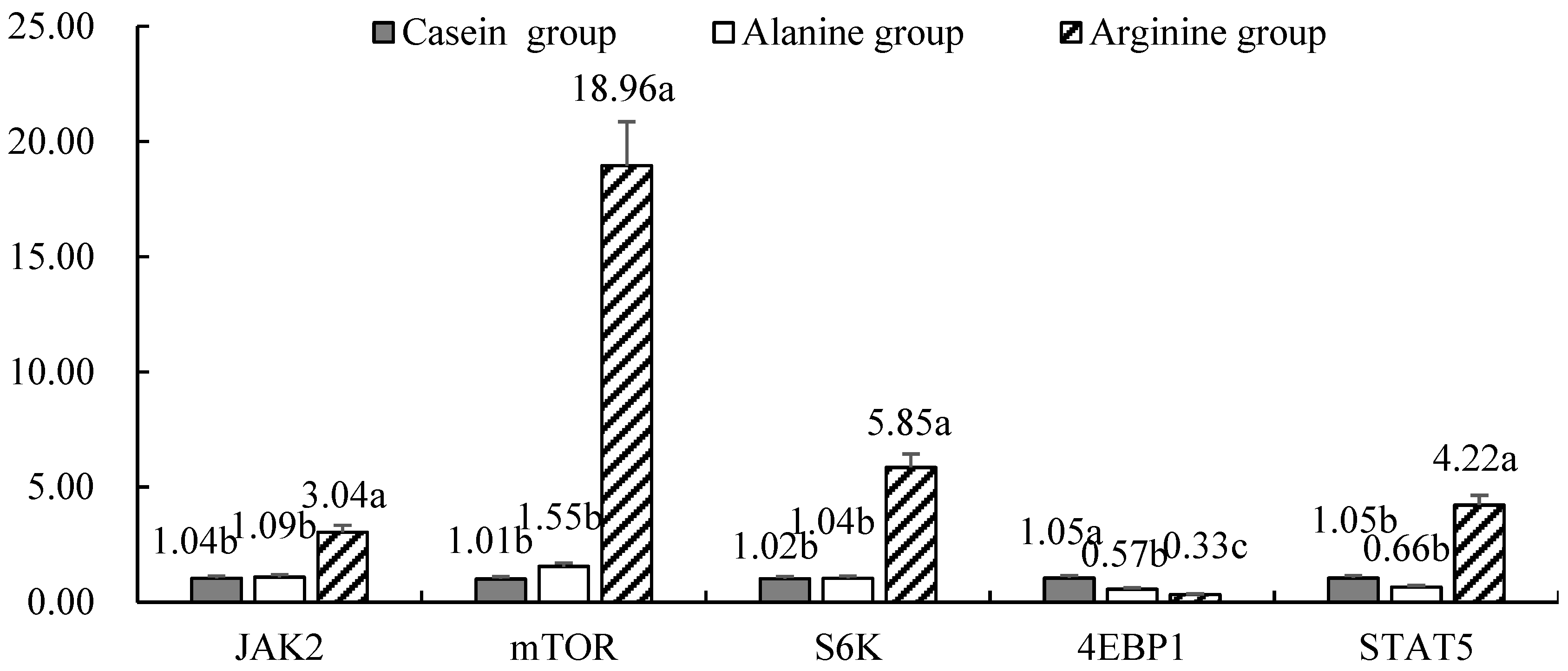
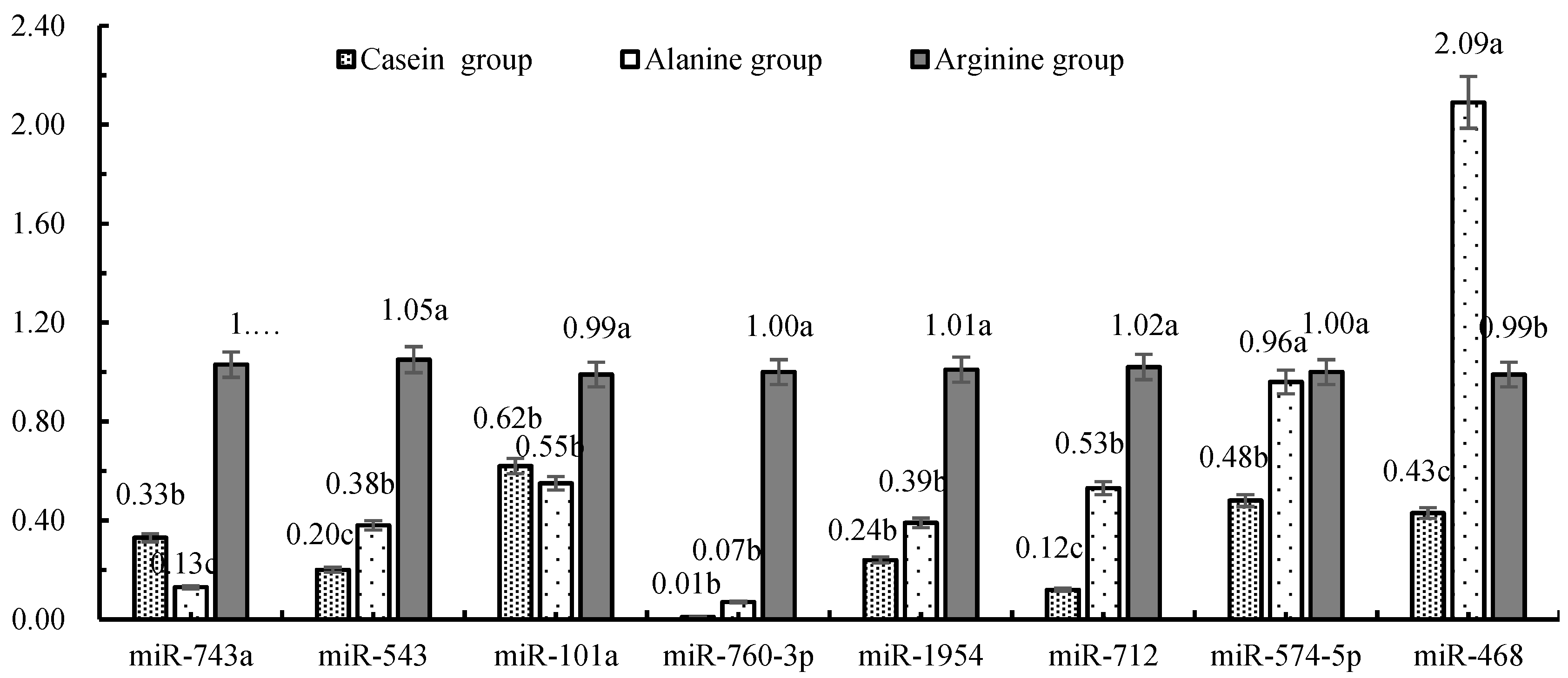
3.3. Effects of Arginine Infusion on Expression of mRNA and miRNA Involved in Milk Production

The mRNA expression of JAK2, STAT5, mTOR and S6K was greater in cows infused with arginine (p < 0.05) (Figure 1), but the mRNA expression of 4EBP1 was lower (p < 0.05) compared with the casein group. As described in Figure 2, the expression of miR-743a, miR-543, miR-101a and miR-760-3p in response to arginine infusion was greater compared with alanine or casein groups (p < 0.05). The expression of miR-1954 and miR-712 was greatest in the arginine group (p < 0.05). The expression of miR-574-5p in response to arginine and alanine infusion was similar, but greater than that in the casein group (p < 0.05). The expression of miR-468 was greatest in response to alanine (p < 0.05), and higher in the arginine compared with the casein group.

4. Discussion

4.1. Effects of Arginine Infusion on Milk Protein production

The casein gene is a cluster composed of CSN1S1, CSN1S2, CSN2, and CSN3, that encode the αs1-, αs2-, β-, and κ-casein proteins, respectively. The αs1-casein and β-casein are the main casein types in milk. Although the content of κ-casein is low, it is an important component of milk casein. The proportion of α- casein, β- casein and κ-casein in the present study varied from 52.70% to 62.96%, 27.96% to 38.56% and 9.01% to 10.24%, respectively, which was similar to previous data [21]. Arginine plays an important role in casein synthesis in BMEC, by regulating the transcription of casein genes [7,22]. A study in lactating cows also found that arginine supplementation increased both milk yield and milk protein yield [23]. Although in the present study arginine supplementation did not alter the content of β-casein, which differed from our previous in vitro results in mammary epithelial cells [7], the observed responses for α-casein and the mRNA expression of CSN1S1 and CSN1S2 agreed with previous in vitro work with bovine mammary cells [7] and rat mammary tissue [24]. The increase in κ-casein content, as well as CSN3 expression in response to arginine, was consistent with the previous result [22]. The discrepancies among studies may be due to the inherent differences in metabolism between in vivo and in vitro systems [25], along with potential species-specific response [21,26].
Unlike data from the present study, enhanced EAA supply increased milk protein yield, but had no effects on the expression of csn2 in the mammary tissue of lactating cows [27]. Additionally, in the review by Cant et al. [28] evidence indicated that shifts in milk protein yield were not always accompanied by changes in the mammary expression of csn1, csn2, or csn3. Authors suggested that NEL source and supply do not generally affect the expression of milk protein genes, but impact milk protein synthesis primarily through other mechanisms. The discrepancies between previous studies described and the present work may be attributed to specific mechanisms induced by EAA or energy availability, compared with functional amino acids such as arginine. Clearly, the regulation of casein and milk synthesis likely occurs through multifaceted mechanisms that response to the availability of EAA, cellular energy, and functional amino acids.

4.2. miRNA Regulation of Casein Gene Expression in Response to Arginine Supplementation

Shi and Gibson [29] reported that the up-regulation of mitochondrial malate dehydrogenase (MDH) by oxidative stress was mediated by miR-743a in mouse hippocampal cells. Therefore, we speculate that the up-regulation of miR-743a in response to arginine supply in the present study might have increased casein yield by inhibiting MDH activity. Because arginine has a protective effect on oxidative stress in rodents [30], it could also encompass the maintenance of a normal non-stress metabolic state in bovine mammary cells.
A previous study in BMEC showed that proliferation and casein protein concentration were increased by arginine [7,22]. A study in Wistar rats demonstrated an increase of both mammary acinar area and β-casein content in response to the dietary supply of arginine [24]. Similarly, Tan et al. [31] reported that 0.3 mM arginine in the medium was more suitable for intestinal epithelial cell proliferation and protein turnover. Together, the above data indicate that protein synthesis might be influenced by cell proliferation. miR-543 targets and inhibits SIRT1, a class III histone deacetylase, and promotes gastric cancer cell proliferation and cell cycle progression [32], suggesting that this miRNA plays a role in cell proliferation. In the context of arginine supply, the up-regulation of miR-543 expression and casein yield might have been associated with the stimulation of cell proliferation. It is generally believed that the dysregulation of miR-543 plays crucial roles in various human cancers, not only to promote cell proliferation and carcinoma [33], but also to inhibit cancer [34]. Based on data from the present study, however, miR-543 appears to be associated with the former.
Both proline dehydrogenase (PRODH) and ornithine aminotransferase (OAT) are the predicted target genes for miR-760-3p and miR-1954 (Table 2), which agrees with the greater expression of miR-760-3p and miR-1954 in response to arginine, both in vivo and in vitro. Thus, we speculate that the negative effect of miR-760-3p and miR-1954 on the proline pathway could help channel arginine through the polyamine pathway. As discussed above, it is possible that arginine enhances mammary cell proliferation and casein yield via polyamines [35] produced through the arginine-polyamine pathway.
miR-574-5p is a candidate oncogene in various types of cancers, such as colorectal cancer [36] and thyroid cancer [37,38]. miR-712 is generally believed to induce vascular endothelial inflammation and atherosclerosis [39], and has been shown to cause aortic aneurysms by stimulating matrix metalloproteinase activity [40]. In this study, the greater expression of miR-574-5p and miR-712 in response to arginine supply suggested that its effect on mammary cells could be related to the promotion of mammary cell proliferation, thereby further inducing casein synthesis in this group. However, the mechanisms by which they are linked to the mammary gland and lactation require further study.
The overexpression of miR-101a inhibited β-casein synthesis in mouse mammary cells (HC11) [12] suggesting this miRNA would be expected to suppress casein synthesis at least in part through targeting mTOR and ARGⅡ (Table 2). A previous study with Wistar rats showed that arginine supplementation in the diet increased mammary acinar area and β-casein content [24]. However, the abundance of α-casein and κ-casein in mammary gland tissue was not affected. Because in mouse mammary cells the overexpression of miR-101a also inhibits the cyclooxygenase 2 (COX-2)—prostaglandin pathway, it could be possible that more than one regulatory mechanism for casein synthesis encompassing the action of miR-101a exists. The fact that in a previous study [7], mTOR expression was also increased in response to arginine, seems to suggest that miR-101a up-regulation can play the same role in casein synthesis in the bovine as in the mouse mammary gland. This idea is supported by the fact that the increase of total casein protein yield in response to arginine was mainly derived from increases of α-casein and κ-casein, but not β-casein. We speculate that one reason for the impact of arginine on casein protein might be that this amino acid can regulate α-casein and κ-casein in bovine through a different mechanism than the one for β-casein reported in the mouse. Further research will have to be conducted to clarify the underlying mechanisms.
The present in vivo work showed that arginine infusion increased the content of α-casein and κ-casein in milk and the expression of CSN1S1 and CSN1S2, both of which are beneficial to the improvement of milk protein content. In addition, both in vitro and in vivo studies indicated that the differential expression of miR-743a, miR-543, miR-101a, miR-760-3p, miR-1954, and miR-712 in response to arginine supply might be a coordinated response that culminates in the regulation of casein synthesis. Clearly, physiological regulation in vivo is complex and in vitro studies cannot fully mimic the in vivo events. Thus, the precise in vivo regulatory mechanisms involving miRNA in the context of amino acid supply to the mammary gland merit further research.

Author Contributions

Conceptualization, M.W. and X.Z.; methodology, D.B.; software, Y.W.; validation, X.Z., G.Z., L.C. and L.D.; formal analysis, L.C. and L.D.; investigation, X.Z., G.Z., L.C. and L.D.; resources, D.B.; data curation, L.D.; writing—original draft preparation, X.Z.; writing—review and editing, X.Z., M.W. and J.L.; supervision, D.B. All authors have read and agreed to the published version of the manuscript.

Funding

This research was funded by the Open fund of the State Key Laboratory of Animal Nutrition (2004DA125184F1715), the National Key Research and Development Program of China (2018YFD0501600, 2018YFD0502100), and Beijing Dairy Industry Innovation Team (BAIC06-2020), as well as the Agriculture Science and Technology Innovation Program (ASTIP-IAS07).

Acknowledgments

The authors gratefully acknowledge grants from the experimental farm at Yangzhou University for providing the cows for this study.

Conflicts of Interest

The authors declare no conflict of interest.

References

  1. Kim, S.W.; McPherson, R.L.; Wu, G. Dietary arginine supplementation enhances the growth of milk-fed young pigs. J. Nutr. 2004, 134, 625–630. [Google Scholar] [CrossRef] [PubMed]
  2. Chew, B.P.; Eisenman, J.R.; Tanaka, T.S. Arginine infusion stimulates prolactin, growth hormone, insulin, and subsequent lactation in pregnant dairy cows. J. Dairy Sci. 1984, 67, 2507–2518. [Google Scholar] [CrossRef]
  3. Ma, Q.; Hu, S.; Bannai, M.; Wu, G. L-Arginine regulates protein turnover in porcine mammary epithelial cells to enhance milk protein synthesis. Amino Acids 2018, 50, 621–628. [Google Scholar] [CrossRef] [PubMed]
  4. Tian, W.; Wang, H.; Wu, T.; Ding, L.; Zhao, R.; Khas, E.; Wang, C.; Zhang, F.; Mi, F.; Wang, L.; et al. Milk protein responses to balanced amino acid and removal of Leucine and Arginine supplied from jugular-infused amino acid mixture in lactating dairy cows. J. Anim. Physiol. Anim. Nutr. (Berl.) 2017, 101, e278–e287. [Google Scholar] [CrossRef] [PubMed]
  5. Kim, S.W.; Wu, G. Regulatory role for amino acids in mammary gland growth and milk synthesis. Amino Acids 2009, 37, 89–95. [Google Scholar] [CrossRef]
  6. Wu, G.; Bazer, F.W.; Davis, T.A.; Kim, S.W.; Li, P.; Rhoads, J.; Satterfield, M.; Smith, S.B.; Spencer, T.E.; Yin, Y. Arginine metabolism and nutrition in growth, health and disease. Amino Acids 2009, 37, 153–168. [Google Scholar] [CrossRef] [Green Version]
  7. Wang, M.; Xu, B.; Wang, H.; Bu, D.; Wang, J.; Loor, J.J. Effects of Arginine concentration on the in vitro expression of Casein and mTOR pathway related genes in mammary epithelial cells from dairy cattle. PLoS ONE 2014, 9, e95985. [Google Scholar] [CrossRef]
  8. Wang, C.; Li, Q. Identification of differentially expressed microRNAs during the development of Chinese murine mammary gland. J. Genet. Genom. 2007, 34, 966–973. [Google Scholar] [CrossRef]
  9. Wang, M.; Moisá, S.; Khan, M.J.; Wang, J.; Bu, D.; Loor, J.J. MicroRNA expression patterns in the bovine mammary gland are affected by stage of lactation 1. J. Dairy Sci. 2012, 95, 6529–6535. [Google Scholar] [CrossRef] [Green Version]
  10. Fan, Y.; Zhang, Y.; Wu, R.; Chen, X.; Zhang, Y.; Chen, X.; Zhu, D. miR-431 is involved in regulating cochlear function by targeting Eya4. Biochim. Biophys. Acta 2016, 1862, 2119. [Google Scholar] [CrossRef]
  11. Chen, J.F.; Mandel, E.M.; Thomson, J.M.; Wu, Q.; Callis, T.E.; Hammond, S.M.; Conlon, F.L.; Wang, D.Z. The role of microRNA-1 and microRNA-133 in skeletal muscle proliferation and differentiation. Nat. Genet. 2005, 38, 228–233. [Google Scholar] [CrossRef] [PubMed]
  12. Tanaka, T.; Haneda, S.; Imakawa, K.; Sakai, S.; Nagaoka, K. A microRNA, miR-101a, controls mammary gland development by regulating cyclooxygenase-2 expression. Differentiation 2009, 77, 181–187. [Google Scholar] [CrossRef] [PubMed]
  13. Zidi, A.; Amills, M.; Tomás, A.; Vidal, O.; Ramírez, O.; Carrizosa, J.; Urrutia, B.; Serradilla, J.M.; Clop, A. Short communication: Genetic variability in the predicted microRNA target sites of caprine casein genes. J. Dairy Sci. 2010, 93, 1749–1753. [Google Scholar] [CrossRef] [PubMed] [Green Version]
  14. Wang, M.Z.; Ding, L.Y.; Wang, C.; Chen, L.M.; Loor, J.J.; Wang, H.R. Short communication: Arginase inhibition reduces the synthesis of casein in bovine mammary epithelial cells. J. Dairy Sci. 2017, 100, 4128–4133. [Google Scholar] [CrossRef] [PubMed]
  15. Vickery, H.B.; White, A. The basic amino acids of casein. J. Biol. Chem. 1933, 103, 413–415. [Google Scholar]
  16. Thomson, J.M.; Newman, M.; Parker, J.S.; Morinkensicki, E.M.; Wright, T.; Hammond, S.M. Extensive post-transcriptional regulation of microRNAs and its implications for cancer. Genes Dev. 2006, 20, 2202–2207. [Google Scholar] [CrossRef] [Green Version]
  17. NRC. Nutrient Requirements of Dairy Cattle, 7th ed.; National Research Council, National Academy Press: Washington, DC, USA, 2001. [Google Scholar]
  18. Bionaz, M.; Loor, J.J. Identification of reference genes for quantitative real-time PCR in the bovine mammary gland during the lactation cycle. Physiol. Genom. 2007, 29, 312–319. [Google Scholar] [CrossRef]
  19. Loor, J.J.; Dann, H.M.; Guretzky, N.A.; Everts, R.E.; Oliveira, R.; Green, C.A.; Litherland, N.B.; Rodriguez-Zas, S.L.; Lewin, H.A.; Drackley, J.K. Plane of nutrition prepartum alters hepatic gene expression and function in dairy cows as assessed by longitudinal transcript and metabolic profiling. Physiol. Genom. 2006, 27, 29–41. [Google Scholar] [CrossRef]
  20. Livak, K.J.; Schmittgen, T.D. Analysis of relative gene expression data using real-time quantitative PCR and the 2-△△Ct method. Methods 2000, 25, 4. [Google Scholar]
  21. Hallén, E.; Wedholm, A.; Andrén, A.; Lundén, A. Effect of beta-casein, kappa-casein and beta-lactoglobulin genotypes on concentration of milk protein variants. J. Anim. Breed Genet. 2008, 125, 119–129. [Google Scholar] [CrossRef]
  22. Chen, L.M.; Li, Z.T.; Wang, M.Z.; Wang, H.R. Preliminary report of arginine on synthesis and gene expression of casein in bovine mammary epithelial cell. Int. Res. J. Agric. Sci. Soil Sci. 2013, 3, 17–23. [Google Scholar]
  23. Ding, L.Y.; Chen, L.M.; Wang, M.Z.; Zhang, J.; Loor, J.J.; Zhou, G.; Zhang, X.; Wang, H.R. Inhibition of arginase via jugular infusion of Nω-hydroxy-nor-l-arginine inhibits casein synthesis in lactating dairy cows. J. Dairy Sci. 2018, 101, 3514–3523. [Google Scholar] [CrossRef]
  24. Hu, L.Y.; Wu, J.C.; Wang, M.Z.; Li, P.F.; Feng, D.; Zhang, J. Effects of dietary arginine on mammary gland development and casein synthesis in Wistar rats after delivery. Chin. J. Anim. Nutr. 2015, 27, 1326–1332. [Google Scholar]
  25. Lorian, V. Differences between in vitro and in vivo studies. Antimicrob. Agents Chemother. 1988, 32, 1600–1601. [Google Scholar] [CrossRef] [PubMed] [Green Version]
  26. Bionaz, M.; Hurley, W.; Loor, J. Milk protein synthesis in the lactating mammary gland: Insights from transcriptomics analyses. In Milk Proteinn; Hurley, W., Ed.; InTech North America: New York, NY, USA, 2012; pp. 285–324. [Google Scholar]
  27. Nichols, K.; Doelman, J.; Kim, J.J.M.; Carson, M.; Metcalf, J.A.; Cant, J.P. Exogenous essential amino acids stimulate an adaptive unfolded protein response in the mammary glands of lactating cows. J. Dairy Sci. 2017, 100, 5909–5921. [Google Scholar] [CrossRef] [PubMed] [Green Version]
  28. Cant, J.P.; Kim, J.J.M.; Cieslar, S.R.L.; Doelman, J. Symposium review: Amino acid uptake by the mammary glands: Where does the control lie? J. Dairy Sci. 2018, 101, 5655–5666. [Google Scholar] [CrossRef] [PubMed] [Green Version]
  29. Shi, Q.; Gibson, G.E. Up-regulation of the mitochondrial malate dehydrogenase by oxidative stress is mediated by miR-743a. J. Neurochem. 2011, 118, 440–448. [Google Scholar] [CrossRef] [PubMed] [Green Version]
  30. Dasgupta, T.; Hebbel, R.P.; Kaul, D.K. Protective effect of arginine on oxidative stress in transgenic sickle mouse models. Free Radic. Biol. Med. 2006, 41, 1771–1780. [Google Scholar] [CrossRef] [Green Version]
  31. Tan, B.; Yin, Y.; Kong, X.; Li, P.; Li, X.; Huang, R.; Wu, G. L-Arginine stimulates proliferation and prevents endotoxin-induced death of intestinal cells. Amino Acids 2010, 38, 1227–1235. [Google Scholar] [CrossRef] [Green Version]
  32. Li, J.; Dong, G.; Wang, B.; Gao, W.; Yang, Q. miR-543 promotes gastric cancer cell proliferation by targeting SIRT1. Biochem. Biophys. Res. Commun. 2016, 469, 15–21. [Google Scholar] [CrossRef]
  33. Chen, Z.Y.; Du, Y.; Wang, L.; Liu, X.H.; Guo, J.; Weng, X.D. MiR-543 promotes cell proliferation and metastasis of renal cell carcinoma by targeting Dickkopf 1 through the Wnt/β-catenin signaling pathway. J. Cancer 2018, 9, 3660–3668. [Google Scholar] [CrossRef] [PubMed] [Green Version]
  34. Liu, X.; Gan, L.; Zhang, J. miR-543 inhibites cervical cancer growth and metastasis by targeting TRPM7. Chem. Biol. Interact. 2019, 302, 83–92. [Google Scholar] [CrossRef]
  35. Wu, G.; Morris, S.M., Jr. Arginine metabolism: Nitric oxide and beyond. Biochem. J. 1998, 336, 1–17. [Google Scholar] [CrossRef] [PubMed]
  36. Ji, S.; Ye, G.; Zhang, J.; Wang, L.; Wang, T.; Wang, Z.; Zhang, T.; Wang, G.; Guo, Z.; Luo, Y. Original article: miR-574-5p negatively regulates Qki6/7 to impact β-catenin/Wnt signalling and the development of colorectal cancer. Gut 2013, 62, 716–726. [Google Scholar] [CrossRef] [PubMed] [Green Version]
  37. Zhang, Z.; Li, X.; Xiao, Q. MiR-574-5p mediates the cell cycle and apoptosis in thyroid cancer cells via Wnt/β-catenin signaling by repressing the expression of Quaking proteins. Oncol. Lett. 2018, 15, 5841–5848. [Google Scholar] [CrossRef]
  38. Wang, X.; Lu, X.; Geng, Z.; Yang, G.; Shi, Y. LncRNA PTCSC3/miR-574-5p Governs Cell Proliferation and Migration of Papillary Thyroid Carcinoma via Wnt/β-Catenin Signaling. J. Cell. Biochem. 2017, 118, 4745–4752. [Google Scholar] [CrossRef]
  39. Dong, J.S.; Kumar, S.; Takabe, W.; Chan, W.K.; Ni, C.W.; Albertsgrill, N.; Jang, I.H.; Kim, S.; Kim, W.; Kang, S.W. The atypical mechanosensitive microRNA-712 derived from pre-ribosomal RNA induces endothelial inflammation and atherosclerosis. Nat. Commun. 2013, 4, 3000. [Google Scholar]
  40. Kim, C.W.; Kumar, S.; Son, D.J.; Jang, I.H.; Griendling, K.K.; Jo, H. Prevention of abdominal aortic aneurysm by anti–microrna-712 or anti–microrna-205 in angiotensin ii–infused mice. Arterioscler. Thromb. Vasc. Biol. 2014, 34, 1412. [Google Scholar] [CrossRef] [Green Version]
Figure 1. Expression of functional genes regulating casein yield in cow mammary tissue in the in vivo trial. Values with lower case letters were not different (p > 0.05). Values with different lower case letters differed (p < 0.05). JAK2 = Janus kinase 2; mTOR = Mechanistic target of rapamycin; S6K = Ribosomal protein S6 kinase; 4EBP1 = eIF4E-binding protein 1; STAT5 = Signal transducer and activator of transcription 5.
Animals 10 00797 g001
Figure 2. Expression of target miRNA in mammary tissue from lactating Holstein cows in response to jugular infusions. Values with different lower case letters differed (p < 0.05).
Animals 10 00797 g002
Table 1. Composition of amino acids in culture media of bovine mammary epithelial cells (mM).
Amino AcidsControl GroupCasein GroupArginine Group
Tyr3.253.253.25
Ala2.762.762.76
Gly0.570.570.57
Glu15.9415.9415.94
Ser5.095.095.09
Cys0.350.350.35
Phe3.573.573.57
Leu11.7511.7511.75
Ile4.244.244.24
His1.381.381.38
Lys4.394.394.39
Thr4.144.144.14
Met2.222.222.22
Trp1.051.051.05
Val4.844.844.84
Pro7.537.537.53
Asp3.383.383.38
Arg01.603.20
Table 2. Candidate miRNA in various genes related to casein protein yield in dairy cows.
miRNAsTarget Gene Symbol
miR-325CSN1S1CSN3PRODHASL
miR-3062CSN1S1CSN3
miR-471-3pCSN1S1ARGⅡ
miR-186CSN1S1CSN3
miR-465c-5pCSN1S1OAT
miR-465b-5pCSN1S1OAT
miR-465a-5pCSN1S1OAT
miR-1942CSN1S1ARGⅡ
miR-743b-3pCSN1S1OAT
miR-743aCSN1S1OAT
miR-543CSN1S1ARGⅡOAT
miR-362-3pCSN1S1OAT
miR-329CSN1S1OAT
miR-694CSN1S1ARGⅠOAT
miR-145CSN2
miR-335-3pCSN3ARG1
miR-208a-5pCSN3OAT
miR-3095-5pCSN3OAT
miR-875-3pCSN3OAT
miR-669bCSN3ARG1
miR-3071CSN3ARGⅡ
miR-574-5pCSN3PRODH
miR-181dCSN3OAT
miR-181aCSN3OAT
miR-181bCSN3OAT
miR-181cCSN3OAT
miR-434-3pCSN3ARGⅠ
miR-3067CSN3ARGⅡ
miR-374CSN3OAT
miR-384-3pCSN3ARGⅠOATPRODH
miR-410CSN3OAT
miR-344dCSN3OAT
miR-344eCSN3OAT
miR-686CSN3ARGⅠ
miR-141CSN3GLS
miR-200aCSN3GLS
miR-340-5pCSN3ARGⅠ
miR-712CSN3ARGⅡ
miR-205CSN3ARGⅡ
miR-467fCSN3ARGⅠ
miR-669m-3pCSN3ARGⅠ
miR-409-3pCSN3ARGⅡ
miR-101bmTORARGⅡ
miR-101amTORARGⅡ
miR-466c-3pARGⅠOAT
miR-466p-3pARGⅠOAT
miR-466b-3pARGⅠOAT
miR-532-5pARGⅡOAT
miR-3065ARGⅡOAT
miR-5098ARGⅡOAT
miR-468ARGⅡOAT
miR-760-3pOATPRODH
miR-1954OATPRODH
CSN1S1 = αs1 - casein; CSN2 = β - casein; CSN3 = κ -casein; PRODH = proline dehydrogenase; OAT = Ornithine aminotransferase; ASL = argininosuccinate lyase; ARGⅠ = Arginase Ⅰ; ARGⅡ = Arginase Ⅱ; mTOR = Mechanistic target of rapamycin; GLS = Glutaminase.
Table 3. Composition and nutrient levels of basal diets (DM basis).
IngredientsPercentage/%NutrientsLevel/%
Alfalfa15.30NEL/(MJ/kg) 2)4.66
Chinese wildrye10.47CP14.08
Silage28.80NFC40.27
Corn21.50NDF34.99
Cottonseed meal6.10ADF21.09
Soybean meal6.80EE3.96
Distillers Dried Grains with Soluble9.40Ca0.91
CaHPO40.60Total P0.59
NaCl0.50
Premix1)0.53
Total100.00
1 The premix provided following per kg of diet: CuSO4 25 mg, FeSO4·H2O 75 mg, ZnSO4·H2O 105 mg, Co 0.0024 mg, Na2SeO3 0.016 mg, Vitamin A 12,000 IU, Vitamin D3 10,000 IU, Vitamin E 25 mg, Nicotinic acid 36 mg, Choline 1000 mg. 2 NEL in the diet was calculated according to the NEL of ingredients and their percentages; concentrations of the other nutrients were measured values.
Table 4. Composition of the treatment perfusates (g/L).
Amino AcidsCasein GroupArginine GroupAlanine Group
Lys21.5021.5086.00
Met13.9213.9213.92
Phe12.8812.8812.88
Ile4.754.754.75
Arg0.009.420.00
Ala0.000.0019.31
Table 5. Primers for real-time PCR analysis of target genes and casein in bovine mammary epithelial cells.
GeneAccession No.Gene DescriptionSequence (5′ → 3′)Sources
CSN1S1BC109618αs1-caseinFTCA CAG TAT GAA AGA GGG AABos taurus
RAGC CAA TAG GAT TAG GGA
CSN1S2NM_174528.2αs2-caseinFAGG AAC GCA AAT GAA GAGBos taurus
RGGA GTA ATG GGA ACA GCA
CSN2NM_181008β-caseinFTGA GGA ACA GCA GCA AACBos taurus
RCAG AGG CAG AGG AAG GTG
CSN3NM_174294κ-caseinFCGC TGT GAG AAA GAT GAABos taurus
RAGA CCG CAG TTG AAG TAA
mTORXM_001788228.1Mechanistic Target
of Rapamycin
FCAT GTG CGA ACA CAG CAA CABos taurus
RTGC CTT TCA CGT TCC TCT CC
JAK2XM_002689603.1Janus kinase 2FACA GGG ATC TGG CAA CAA GGBos taurus
RCGC ATA AAT TCC GCT GGT GG
STAT5NM_001012673.1Signal transducer and
activator of transcription 5
FCAA TGG ACA GTC TGG AGC CCBos taurus
RCCT GCA CAC TGG GGA TTG TT
S6KNM_205816.1Ribosomal protein S6 kinaseFCGG AAC AGT CAC ACA CAC CTBos taurus
RTGG CTT CTT GCG TGA GGT AG
4EBP1BC120290.1EIF4E binding protein1FCGG AAC TCA CCT GTG ACC AABos taurus
RAGG TGA TTC TGC CTG GCT TC
GAPDHXM_001252479.1Glyceraldehyde-3-phosphate
dehydrogenase
FCCC CGC GCT CTA ATG TTC ABos taurus
RGAA GGG GTC ATT GAT GGC GA
ACTBNM_173979.3β-actinFACT GTT AGC TGC GTT ACA CCC TTBos taurus
RTGC TGT CAC CTT CAC CGT TCC
Note: All the primers used in this experiment were synthesized in Invitrogen (Nanjing, China).
Table 6. Expression of miRNA screened from PicTar and TargetScan databases in bovine mammary epithelial cells in vitro.
miRNAsTreatmentsSEMp-Value
ControlCaseinArginine
miR-101b1.321.280.980.110.342
miR-181b0.820.930.840.0630.749
miR-181d1.231.031.220.0830.792
miR-3291.792.141.960.1890.993
miR-30622.271.742.470.170.463
miR-30650.901.161.020.0830.212
miR-743a1.20b2.10ab2.73a0.213<0.001
miR-5430.68b0.61b1.60a0.1030.012
miR-101a0.22b0.37b3.99a0.1570.003
miR-760-3p1.76a0.82b2.35a0.1270.034
miR-19541.99b0.7c5.63a0.183<0.001
miR-7120.42b0.52b2.44a0.1470.01
miR-574-5p0.95b0.35b5.63a0.107<0.001
miR-4680.23b0.30b1.48a0.0470.041
miR-875-3p48.26a17.17b2.97c1.6530.001
a,b Values with different letters in the same row differed significantly (p < 0.05).
Table 7. Effect of jugular vein arginine infusion on the yield of milk protein and milk casein protein in Holstein cows.
ItemsTreatmentsSEMp-Value
CaseinAlanineArginine
Average daily intake/(kg)22.8221.6422.31.6250.461
Milk protein/%3.04b3.11b3.17a0.0770.046
Milk yield/(kg/d)21.45b23.65a24.16a0.8230.039
Milk protein yield/(kg/d)0.65b0.74a0.77a0.0270.04
α-Casein/(g/L)11.70b16.37a17.24a1.4170.008
β-Casein/(g/L)8.567.279.090.5230.985
κ-Casein/(g/L)2.00b2.37b3.00a0.1940.024
Casein content/%2.23c2.60b2.93a0.1670.031
Casein yield/(kg/d)0.48c0.62b0.71a0.0530.044
Casein/milk protein/%73.18b83.85ab92.63a6.6130.003
αs1-casein0.96b1.00b1.54a0.1130.028
αs2-casein0.98b1.03b1.39a0.1370.037
β-casein1.031.431.220.1170.832
κ-casein1.001.031.360.1030.327
a,b Values with different letters in the same row differed significantly (p < 0.05).

