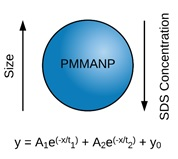
Formula-Driven, Size-Tunable Synthesis of PMMA Nanoparticles by Varying Surfactant Concentration

Abstract
:
1. Introduction
2. Materials and Methods
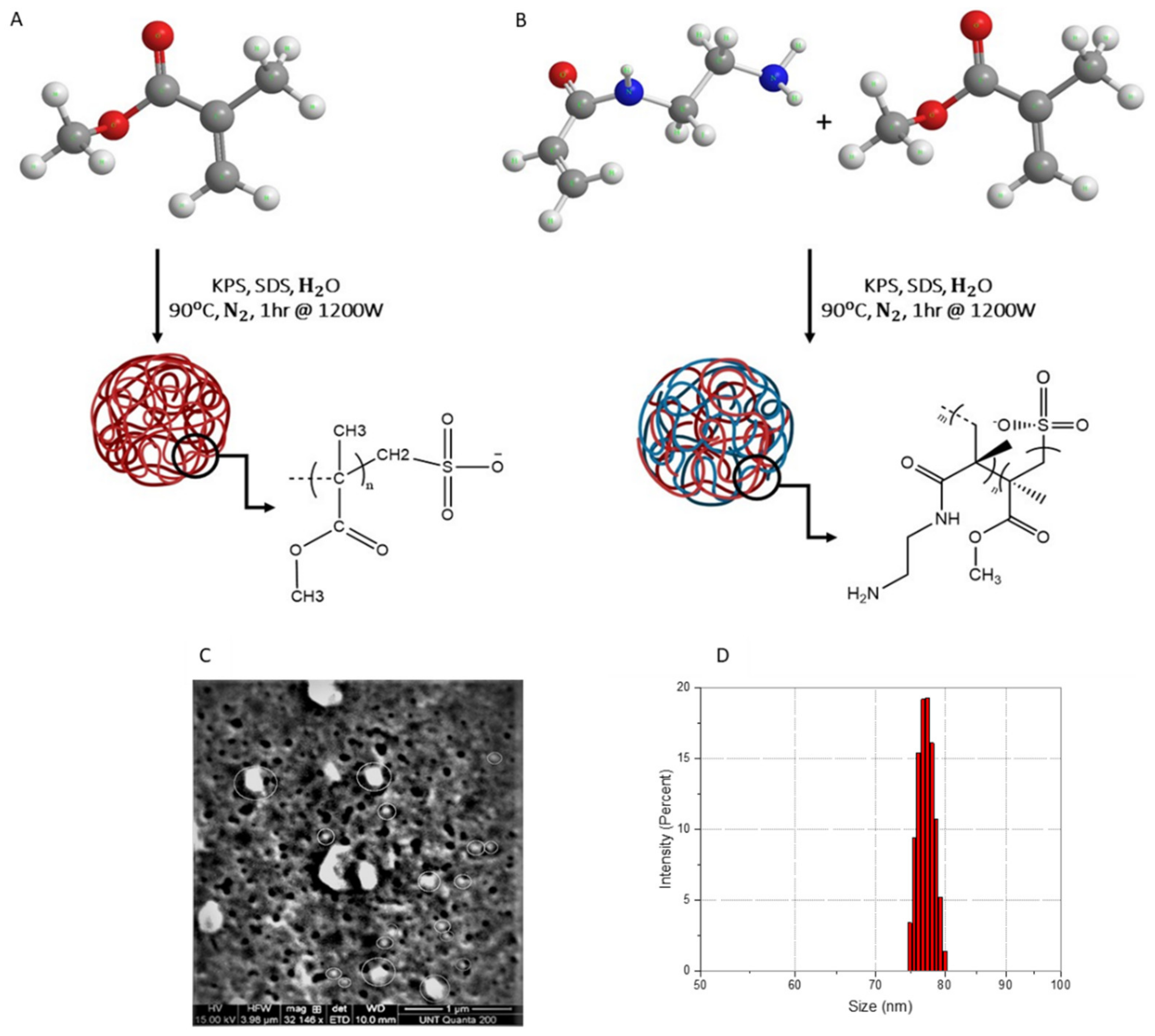
3. Results and Discussion
Author Contributions
Funding
Acknowledgments
Conflicts of Interest
References
- Albanese, A.; Tang, P.S.; Chan, W.C. The Effect of Nanoparticle Size, Shape, and Surface Chemistry on Biological Systems. Annu. Rev. Biomed. Eng. 2012, 14, 1–16. [Google Scholar] [CrossRef] [PubMed] [Green Version]
- Zhang, S.; Li, J.; Lykotrafitis, G.; Bao, G.; Suresh, S. Size-Dependent endocytosis of nanoparticles. Adv. Mater. 2009, 21, 419–424. [Google Scholar] [CrossRef] [PubMed] [Green Version]
- Xie, X.; Liao, J.; Shao, X.; Li, Q.; Lin, Y. The Effect of shape on Cellular Uptake of Gold Nanoparticles in the forms of Stars, Rods, and Triangles. Sci. Rep. 2017, 7, 1–9. [Google Scholar] [CrossRef] [PubMed]
- Chithrani, B.D.; Ghazani, A.A.; Chan, W.C. Determining the Size and Shape Dependence of Gold Nanoparticle Uptake Into Mammalian Cells. Nano Lett. 2006, 6, 662–668. [Google Scholar] [CrossRef]
- Jindal, A.B. The Effect of Particle Shape on Cellular Interaction and Drug Delivery Applications of Micro- and Nanoparticles. Int. J. Pharm. 2017, 532, 450–465. [Google Scholar] [CrossRef]
- Hollick, E.J.; Spalton, D.J.; Ursell, P.G.; Pande, M.V. Biocompatibility of poly (methylmethacrylate), silicone, and AcrySof intraocular lenses: Randomized comparison of the cellular reaction on the anterior lens surface. J. Cataract Refract. Surg. 1998, 24, 361–366. [Google Scholar] [CrossRef]
- Kreuter, J. Nanoparticles-A historical perspective. Int. J. Pharm. 2007, 331, 1–10. [Google Scholar] [CrossRef]
- Kreuter, J. Nanoparticulate systems for brain delivery of drugs. Adv. Drug Deliv. Rev. 2001, 47, 65–81. [Google Scholar] [CrossRef]
- Vollrath, A.; Schallon, A.; Pietsch, C.; Schubert, S.; Nomoto, T.; Matsumoto, Y.; Kataoka, K.; Schubert, U.S. A toolbox of differently sized and labeled PMMA nanoparticles for cellular uptake investigations. Soft Matter 2013, 9, 99–108. [Google Scholar] [CrossRef]
- Yuan, L.; Wang, Y.; Pan, M.; Rempel, G.L.; Pan, Q. Synthesis of poly (methyl methacrylate) nanoparticles via differential microemulsion polymerization. Eur. Polym. J. 2013, 49, 41–48. [Google Scholar] [CrossRef]
- Chen, Y.; Yang, D.; Yoon, Y.J.; Pang, X.; Wang, Z.; Jung, J.; He, Y.; Harn, Y.W.; He, M.; Zhang, S.; et al. Hairy Uniform Permanently Ligated Hollow Nanoparticles with Precise Dimension Control and Tunable Optical Properties. J. Am. Chem. Soc. 2017, 139, 12956–12967. [Google Scholar] [CrossRef] [PubMed]
- Reis, C.P.; Neufeld, R.J.; Veiga, F. Nanomedicine in Cancer; Pan Stanford Publishing: Singapore, 2017; pp. 197–240. [Google Scholar]
- Bao, J.; Zhang, A. Poly (methyl methacrylate) nanoparticles prepared through microwave emulsion polymerization. J. Appl. Polym. Sci. 2004, 93, 2815–2820. [Google Scholar] [CrossRef]
- Song, Y.; Hormes, J.; Kumar, C.S.S.R. Microfluidic Synthesis of Nanomaterials. Small 2008, 4, 698–711. [Google Scholar] [CrossRef] [PubMed]
- Karnik, R.; Gu, F.; Basto, P.; Cannizzaro, C.; Dean, L.; Kyei-Manu, W.; Langer, R.; Farokhzad, O.C. Deterniming the Size and Shape Dependence of Gold Nanoparticles Into Mammalian Cells. Nano Lett. 2008, 8, 2906–2912. [Google Scholar] [CrossRef] [PubMed]
- Timmins, M.R.; Lenz, R.W.; Fuller, R.C. Heterogeneous Kinetics of the Enzymatic Degradation of Poly (β-Hydroxyalkanoates). Polymer 1997, 38, 551–562. [Google Scholar] [CrossRef]
- Goldstein, J.I.; Newbury, D.E.; Echlin, P.; Joy, D.C.; Lifshin, E.; Fiori, C. Scanning Electron Microscopy and X-ray Microanalysis: A Text for Biologists, Materials Scientists and Geologists; Springer Science Business Media: New York, NY, USA, 2014. [Google Scholar]
- Williams, D.B.; Carter, B.C. Transmission Electron Microscopy; Springer: New York, NY, USA, 2009. [Google Scholar]
- Zanetti-Ramos, B.G.; Fritzen-Garcia, M.B.; Creczynski-Pasa, T.B.; Oliveira, C.S.D.; Pasa, A.A.; Soldi, V.; Borsali, R. Characterization of Polymeric Particles with Electron Microscopy, Dynamic Light Scattering, and Atomic Force Microscopy. Part. Sci. Technol. 2010, 28, 472–484. [Google Scholar] [CrossRef]
- Rao, J.P.; Geckeler, K.E. Polymer nanoparticles: Preparation techniques and size control parameters. Prog. Polym. Sci. (Oxf.) 2011, 36, 887–913. [Google Scholar] [CrossRef]
- Marpu, S.; Upadhyay, P.K.; Nguyen, D.T.; Oswald, I.W.H.; Arvapally, R.K.; Petros, R.A.; Hu, Z.; Omary, M.A. Self-Assembly of Linear Polymers into Phosphorescent Nanoparticles: Optimization toward Non-Cytotoxic Bioimaging and Photonic Devices. J. Phys. Chem. C 2015, 119, 12551–12561. [Google Scholar] [CrossRef]
- Bootz, A.; Vogel, V.; Schubert, D.; Kreuter, J. Comparison of Scanning Electron Microscopy, Dynamic Light Scattering and Analytical Ultracentrifugation for the Sizing of Poly(Butyl Cyanoacrylate) Nanoparticles. Eur. J. Pharm. Biopharm. 2004, 57, 369–375. [Google Scholar] [CrossRef]
- Mirkin, M.V.; Amemiya, S. Nanoelectrochemistry; CRC Press, Taylor & Francis Group: Boca Raton, FL, USA, 2015. [Google Scholar]
- Tuoriniemi, J.; Johnsson, A.-C.J.H.; Holmberg, J.P.; Gustafsson, S.; Gallego-Urrea, J.A.; Olsson, E.; Pettersson, J.B.C.; Hassellöv, M. Intermethod Comparison of the Particle Size Distributions of Colloidal Silica Nanoparticles. Sci. Technol. Adv. Mater. 2014, 15, 035009. [Google Scholar] [CrossRef] [Green Version]
- Rodriguez-Lorenzo, L.; Rothen-Rutishauser, B.; Petri-Fink, A.; Balog, S. Nanoparticle polydispersity can strongly affect in vitro dose. Part. Part. Syst. Charact. 2015, 32, 321–333. [Google Scholar] [CrossRef]
- Korir, D.K.; Gwalani, B.; Joseph, A.; Kamras, B.; Arvapally, R.K.; Omary, M.A.; Marpu, S.B. Facile Photochemical Syntheses of Conjoined Nanotwin Gold-Silver Particles within a Biologically-Benign Chitosan Polymer. Nanomaterials 2019, 9, 596. [Google Scholar] [CrossRef] [PubMed] [Green Version]
- Marpu, S.; Kolailat, S.S.; Korir, D.; Kamras, B.L.; Chaturvedi, R.; Joseph, A.; Smith, C.M.; Palma, M.C.; Shah, J.; Omary, M.A. Photochemical formation of chitosan-stabilized near-Infrared-Absorbing silver Nanoworms: A Green synthetic strategy and activity on Gram-negative pathogenic bacteria. J. Colloid Interface Sci. 2017, 507, 437–452. [Google Scholar] [CrossRef] [PubMed]
- Tcholakova, S.; Mitrinova, Z.; Golemanov, K.; Denkov, N.D.; Vethamuthu, M.; Ananthapadmanabhan, K.P. Control of Ostwald Ripening by Using Surfactants with High Surface Modulus. Langmuir 2011, 27, 14807–14819. [Google Scholar] [CrossRef] [PubMed]
- Shang, L.; Nienhaus, K.; Nienhaus, G. Engineered Nanoparticles Intercating with Cells: Size Matters. J. Nanobiotechnol. 2014, 12, 3155–3170. [Google Scholar] [CrossRef] [Green Version]


| SDS (wt./wt.%) | PMMANP Z-Ave (nm) | PDI | |
|---|---|---|---|
| Actual | Calculated | ||
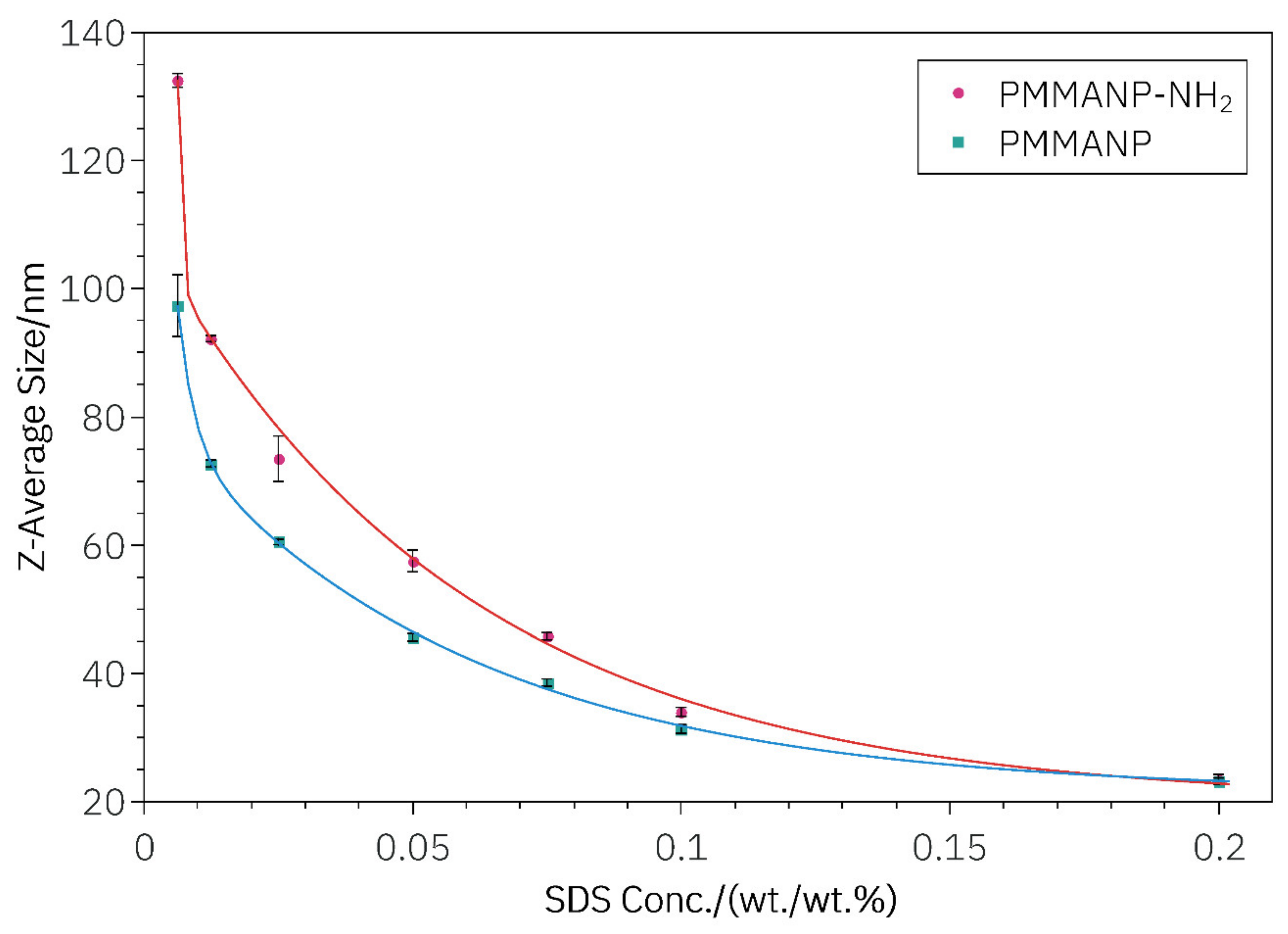
| 0.200 | 23.19 ± 1.57 | 24.97 | 0.09 |
| 0.100 | 31.31 ± 1.64 | 31.88 | 0.06 |
| 0.075 | 38.57 ± 2.74 | 37.44 | 0.03 |
| 0.050 | 45.61 ± 2.73 | 46.29 | 0.05 |
| 0.025 | 60.49 ± 2.93 | 60.37 | 0.04 |
| 0.013 | 77.70 ± 2.95 | 72.47 | 0.04 |
| 0.008 | 97.36 ± 11.77 | 91.74 | 0.03 |
| SDS (wt./wt.%) | H2N-PMMANP Z-Ave (nm) | PDI | |
|---|---|---|---|
| Actual | Calculated | ||
| 0.200 | 23.40 ± 1.53 | 24.68 | 0.08 |
| 0.100 | 34.00 ± 1.60 | 35.06 | 0.06 |
| 0.075 | 45.76 ± 2.81 | 42.46 | 0.03 |
| 0.050 | 57.50 ± 4.67 | 54.53 | 0.05 |
| 0.025 | 73.56 ± 6.35 | 74.28 | 0.04 |
| 0.013 | 92.22 ± 2.97 | 95.03 | 0.04 |
| 0.008 | 132.53 ± 3.23 | 125.22 | 0.03 |
| Constant | PMMANP | H2N-PMMANP |
|---|---|---|
| A1 (nm) | 1.67 × 10² | 8.78 × 10⁶ |
| A2 (nm) | 6.04 × 10 | 8.96 × 10 |
| t1 (wt./wt.%) | 3.07 × 10⁻³ | 6.12 × 10⁻⁴ |
| t2 (wt./wt.%) | 5.68 × 10⁻² | 5.82 × 10⁻² |
| y0 (nm) | 21.43 | 19.93 |
© 2020 by the authors. Licensee MDPI, Basel, Switzerland. This article is an open access article distributed under the terms and conditions of the Creative Commons Attribution (CC BY) license (http://creativecommons.org/licenses/by/4.0/).
Share and Cite
Kamras, B.L.; Mirzanasiri, N.; Korir, D.K.; Mandal, S.; Hariharakumar, S.L.; Petros, R.A.; Marpu, S.B.; Simmons, D.P.; Omary, M.A. Formula-Driven, Size-Tunable Synthesis of PMMA Nanoparticles by Varying Surfactant Concentration. Materials 2020, 13, 1834. https://doi.org/10.3390/ma13081834
Kamras BL, Mirzanasiri N, Korir DK, Mandal S, Hariharakumar SL, Petros RA, Marpu SB, Simmons DP, Omary MA. Formula-Driven, Size-Tunable Synthesis of PMMA Nanoparticles by Varying Surfactant Concentration. Materials. 2020; 13(8):1834. https://doi.org/10.3390/ma13081834
Chicago/Turabian StyleKamras, Brian L., Nooshin Mirzanasiri, Daniel K. Korir, Sujata Mandal, Shreya L. Hariharakumar, Robby A. Petros, Sreekar B. Marpu, Denise P. Simmons, and Mohammad A. Omary. 2020. "Formula-Driven, Size-Tunable Synthesis of PMMA Nanoparticles by Varying Surfactant Concentration" Materials 13, no. 8: 1834. https://doi.org/10.3390/ma13081834