Dynamics of Photogenerated Charge Carriers in TiO2/MoO3, TiO2/WO3 and TiO2/V2O5 Photocatalysts with Mosaic Structure

 ,
,
Abstract
:1. Introduction
2. Results and Discussion
2.1. Characterization of the Samples
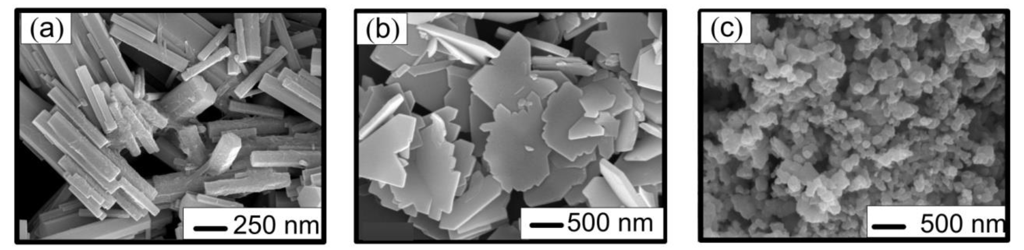
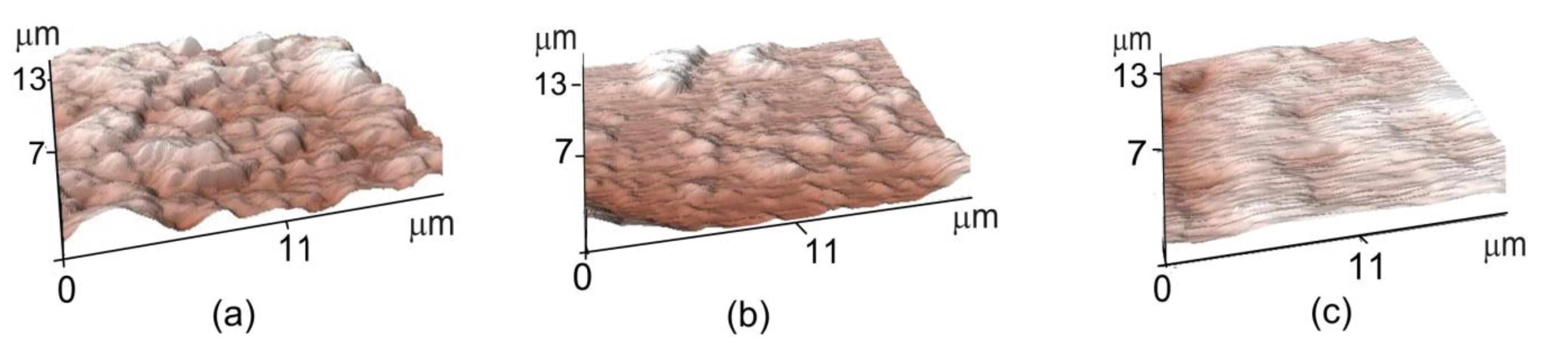
2.1.1. Microscopy
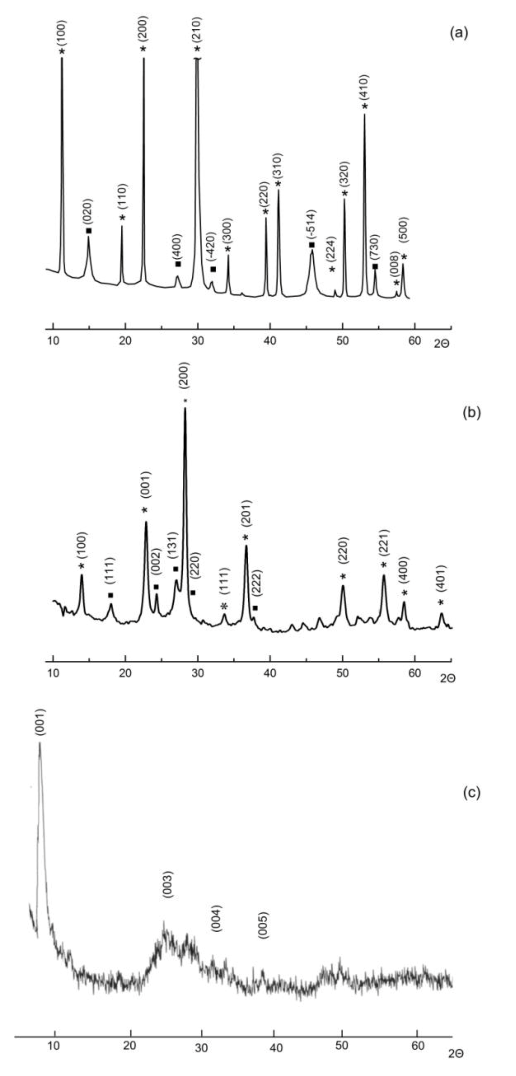
2.1.2. XRD
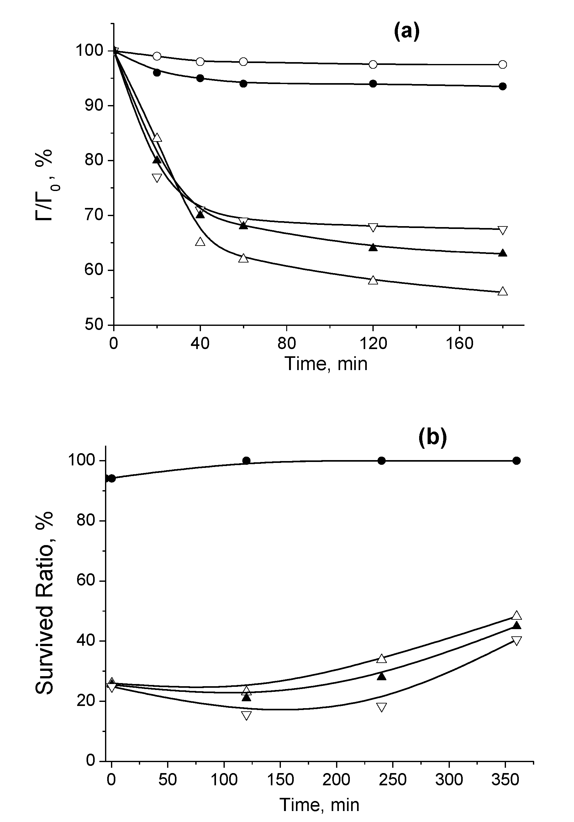
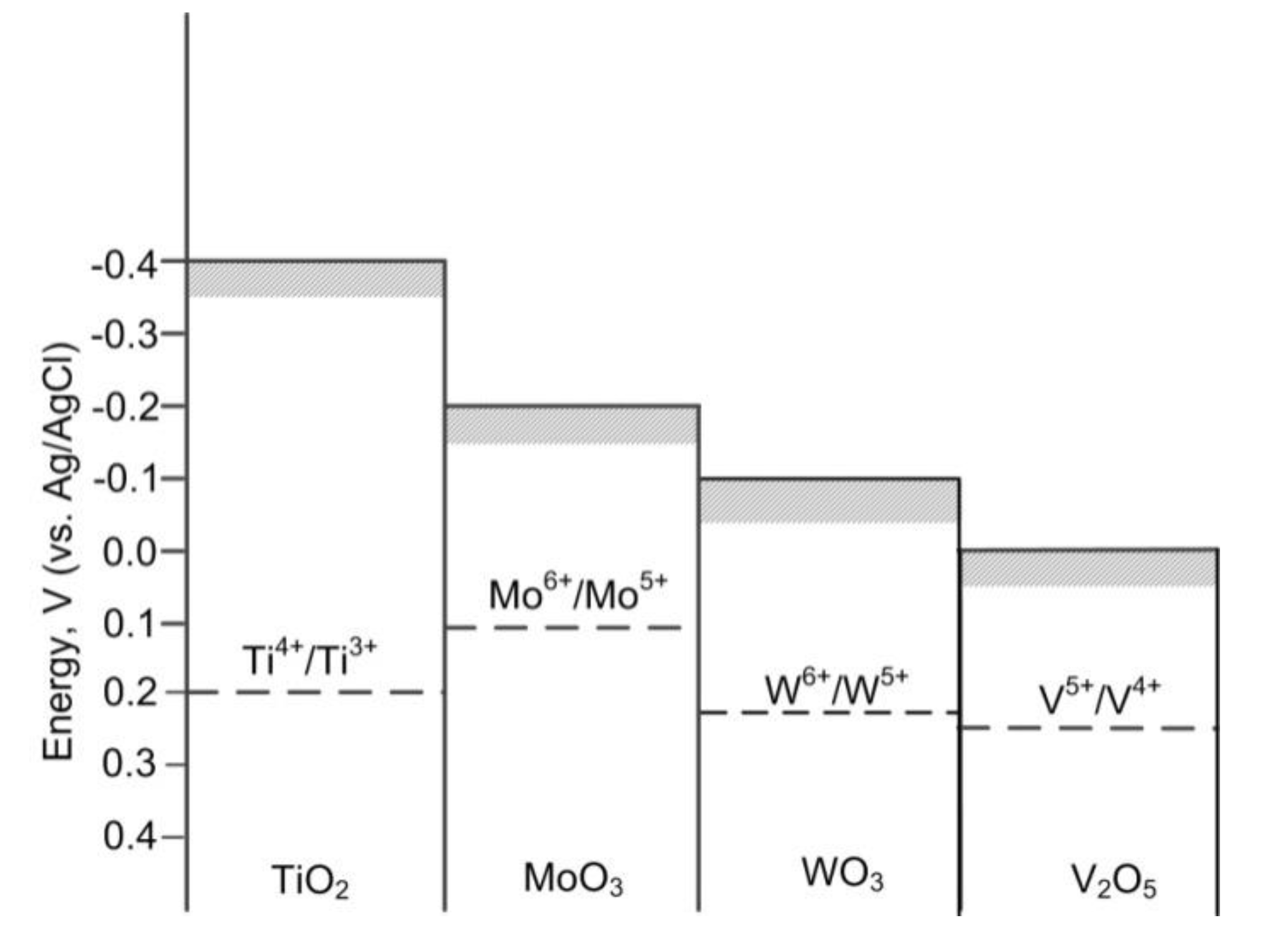
2.2. Photocatalytic and Pathphysiological Activity of TiO2/MoO3, TiO2/WO3, and TiO2/V2O5 Heterostructure Photocatalysts
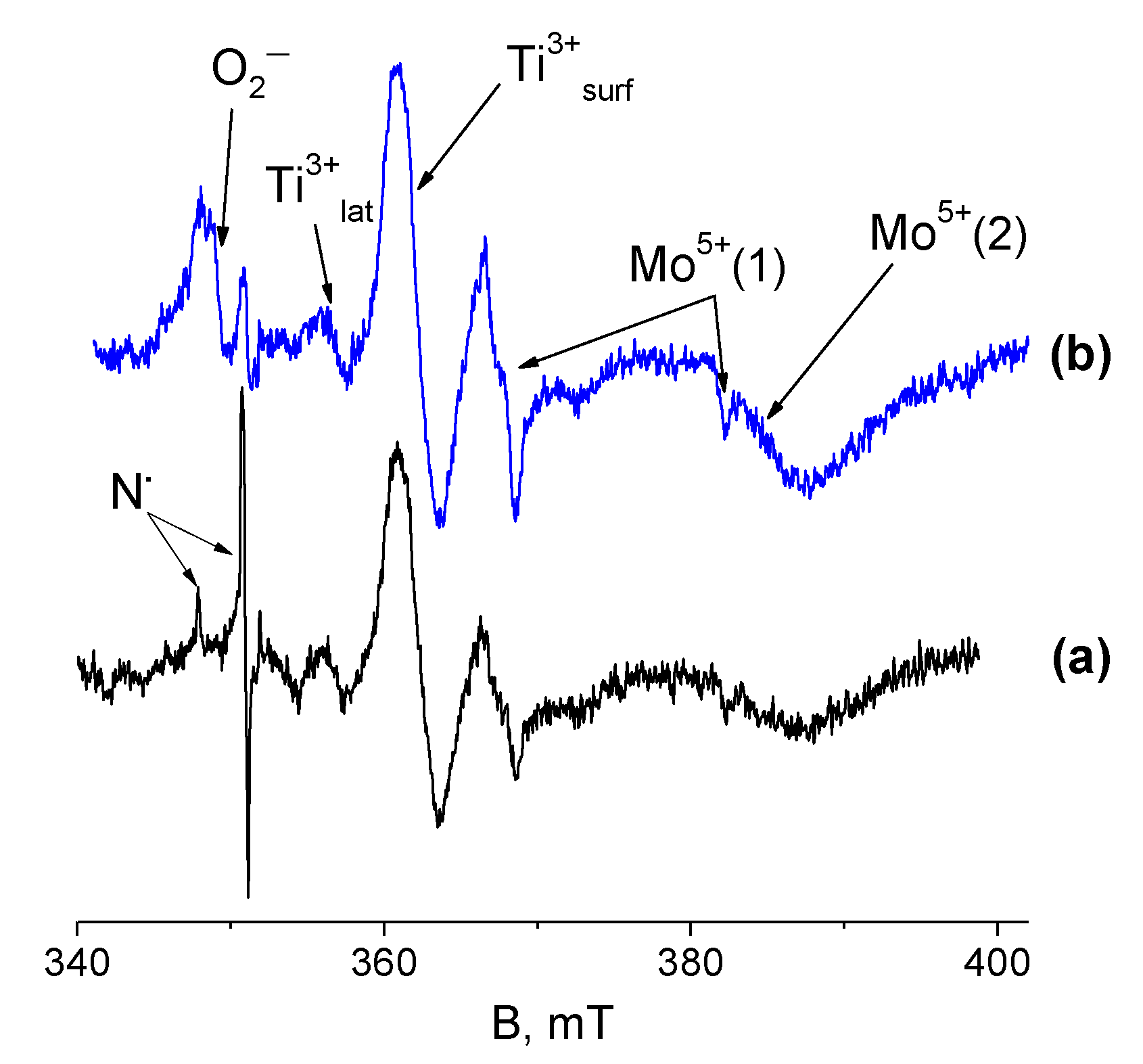
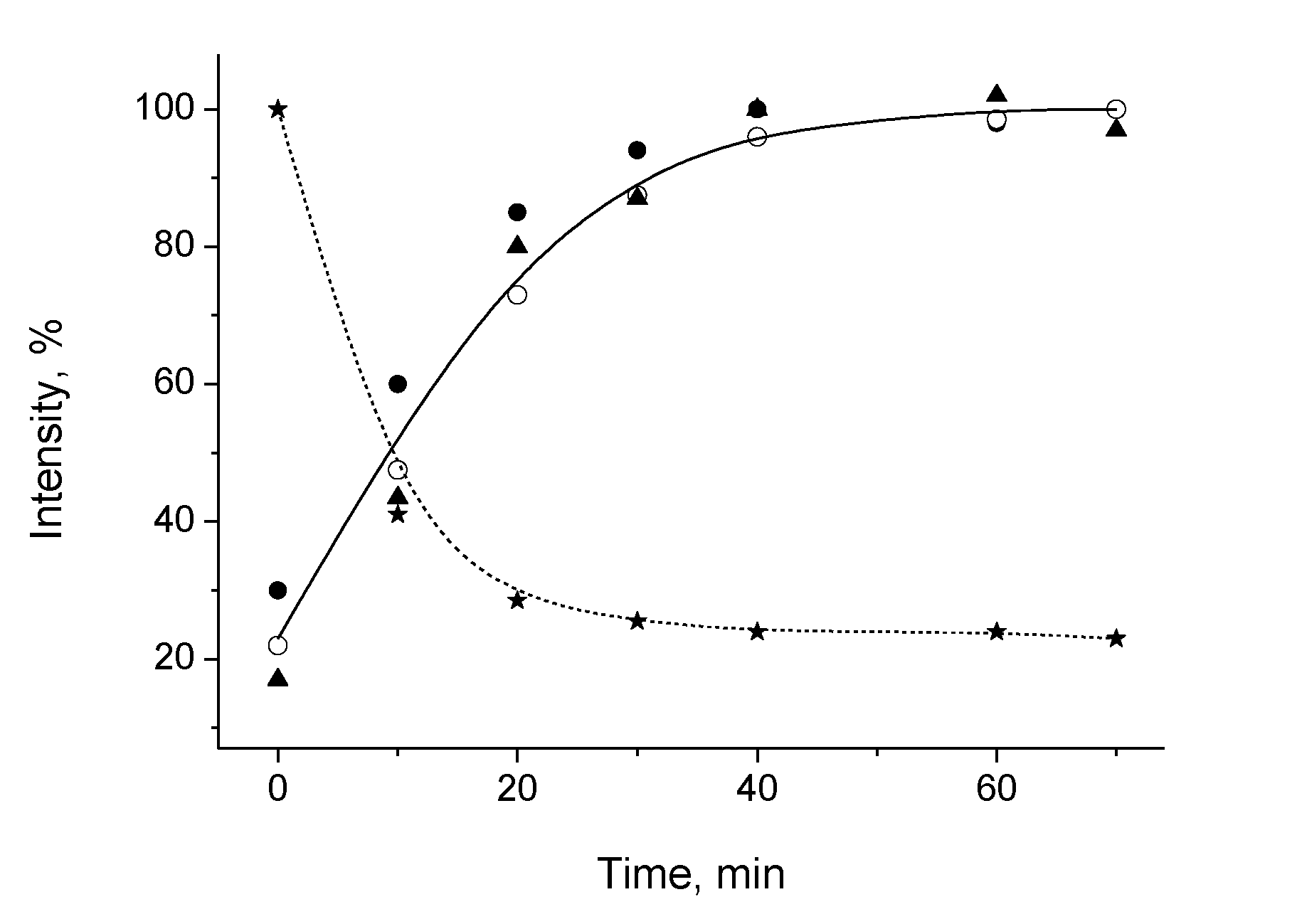
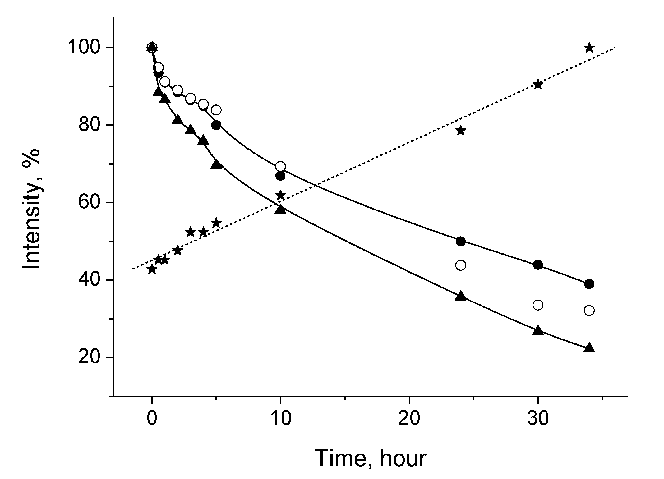
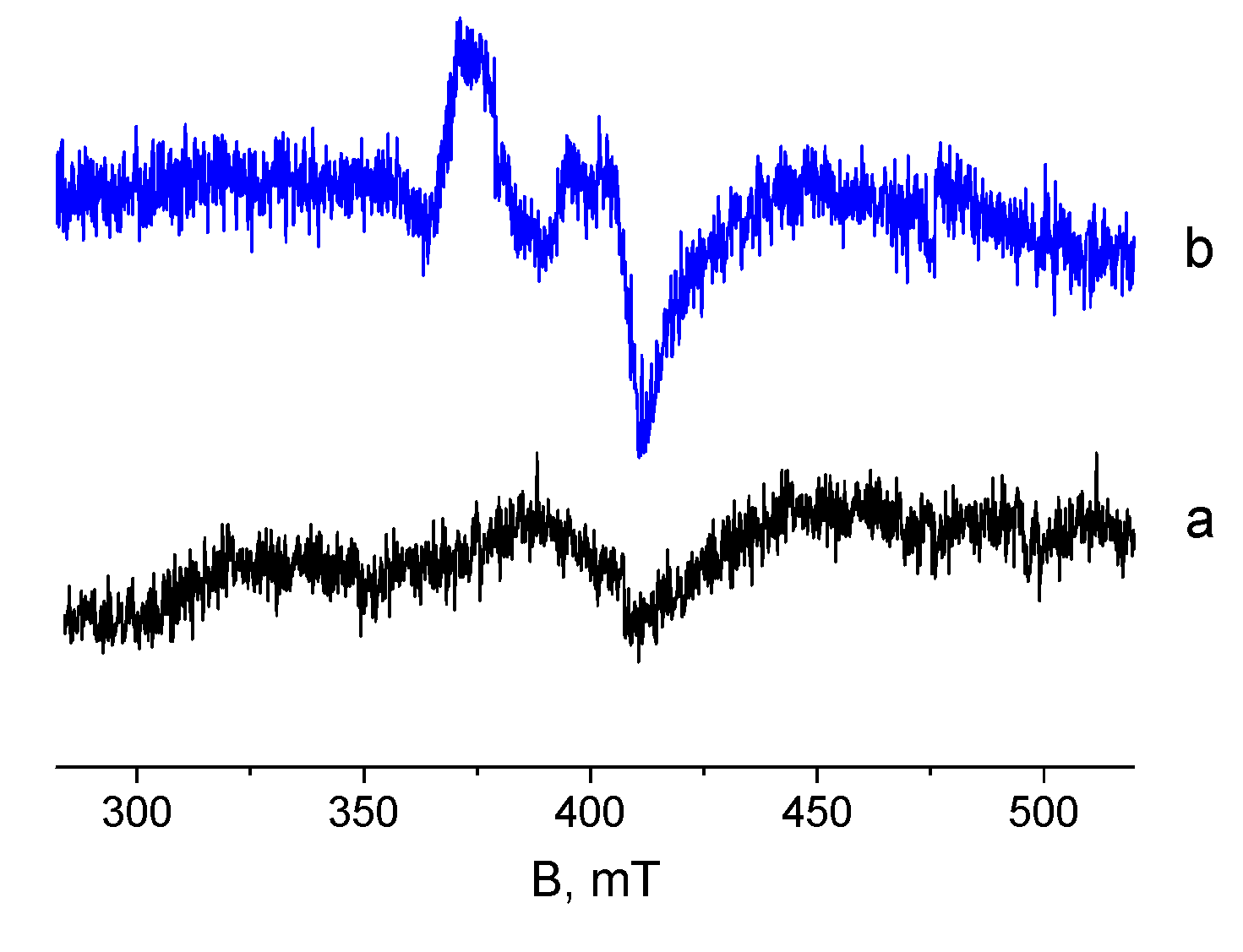
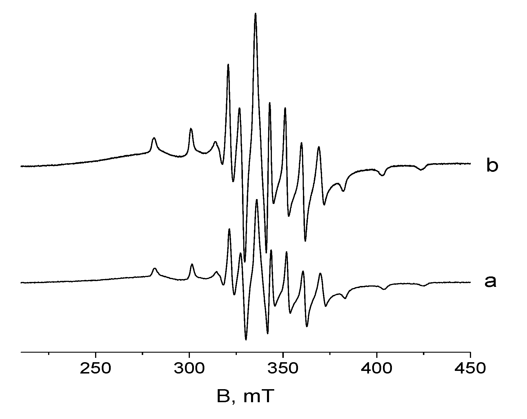
2.3. EPR Measurements of Heterostructure Photocatalysts
3. Materials and Methods
4. Conclusions
Author Contributions
Funding
Acknowledgments
Conflicts of Interest
References
- Ge, M.; Li, Q.; Cao, C.; Huang, J.; Li, S.; Zhang, S.; Chen, Z.; Zhang, K.; Al-Deyab, S.S.; Lai, Y.; et al. One-dimensional TiO2 nanotube photocatalysts for solar water splitting. Adv. Sci. 2017, 4, 1–31. [Google Scholar]
- Hoffmann, M.R.; Martin, S.T.; Choi, W.; Bahnemann, D.W. Environmental applications of semiconductor photocatalysis. Chem. Rev. 1995, 95, 69–96. [Google Scholar] [CrossRef]
- Hagfeldt, A.; Graetzel, M. Light-Induced redox reactions in nanocrystalline systems. Chem. Rev. 1995, 95, 49–68. [Google Scholar] [CrossRef]
- Ibhadon, A.O.; Fitzpatrick, P. Heterogeneous photocatalysis: Recent advances and applications. Catalysts 2013, 3, 189–218. [Google Scholar] [CrossRef] [Green Version]
- Bahnemann, D. Photocatalytic water treatment: Solar energy applications. Solar Energy 2004, 77, 445–459. [Google Scholar] [CrossRef]
- Chen, X.; Mao, S. Titanium dioxide nanomaterials: Synthesis, properties, modifications, and applications. Chem. Rev. 2007, 107, 2891–2959. [Google Scholar] [CrossRef]
- Zhang, H.; Chen, G.; Bahnemann, D. Photoelectrocatalytic materials for environmental applications. J. Mat. Chem. 2009, 19, 5089–5121. [Google Scholar] [CrossRef]
- Chen, W.F.; Koshy, P.; Huang, Y.; Adabifiroozjaei, E.; Yao, Y.; Sorrell, C.C. Effects of Precipitation, Liquid. Formation, and Intervalence Charge Transfer on the Properties and Photocatalytic Performance of Cobalt-. or Vanadium-Doped TiO2 Thin Films. Int. J. Hydrog. Energy 2016, 41, 19025–19056. [Google Scholar] [CrossRef]
- Schneider, J.; Matsuoka, M.; Takeuchi, M.; Zhang, J.; Horiuchi, Y.; Anpo, M.; Bahnemann, D.W. Understanding TiO2 photocatalysis: Mechanisms and materials. Chem. Rev. 2014, 114, 9919–9986. [Google Scholar] [CrossRef]
- Tatsuma, T.; Takeda, S.; Saitoh, S.; Ohko, Y.; Fujishima, A. Bactericidal effect of an energy storage TiO2–WO3 photocatalyst in dark. Electrochem. Comm. 2003, 5, 793–796. [Google Scholar] [CrossRef]
- Takahashi, Y.; Ngaotrakanwiwat, P.; Tatsuma, T. Energy storage TiO2-MoO3 photocatalysts. Electrochim. Acta 2004, 49, 2025–2029. [Google Scholar] [CrossRef]
- Yang, F.; Takahashi, Y.; Sakai, N.; Tatsuma, T. Visible light driven photocatalysts with oxidative energy storage abilities. J. Mat. Chem. 2011, 21, 2288–2293. [Google Scholar] [CrossRef]
- Ngaotrakanwiwat, P.; Saitoh, S.; Ohko, Y.; Tatsuma, T.; Fujishima, A. TiO2-phosphotungstic acid photocatalysis systems with an energy storage ability. J. Electrochem. Soc. 2003, 150, A1405–A1407. [Google Scholar] [CrossRef]
- Sviridova, T.V.; Sadovskaya, L.Y.; Shchukina, E.M.; Logvinovich, A.S.; Shchukin, D.G.; Sviridov, D.V. Nanoengineered Thin-Film TiO2/h-MoO3 Photocatalysts Capable to Accumulate Photoinduced Charge. J. Photochem. Photobiol. A 2016, 327, 44–50. [Google Scholar] [CrossRef] [Green Version]
- Sviridova, T.V.; Antonova, A.A.; Kokorin, A.I.; Degtyarev, E.N.; Sviridov, D.V. Thermally Induced Transformations in Nanostructured Molybdenum-Vanadium Oxides Synthesized by a Solvothermal Method. Russ. J. Phys. Chem. B 2015, 9, 36–42. [Google Scholar] [CrossRef]
- Konstantinova, E.A.; Kokorin, A.I.; Minnekhanov, A.A.; Sviridova, T.V.; Sviridov, D.V. EPR Study of Photoexcited Charge Carrier Behavior in TiO2/MoO3 and TiO2/MoO3:V2O5 Photocatalysts. Catal. Lett. 2019, 149, 2256–2267. [Google Scholar] [CrossRef]
- Sviridova, T.V.; Sadovskaya, L.Y.; Konstantinova, E.A.; Belyasova, N.A.; Kokorin, A.I.; Sviridov, D.V. Photoaccumulating TiO2–MoO3, TiO2–V2O5, and TiO2–WO3 Heterostructures for Self-Sterilizing Systems with the Prolonged Bactericidal Activity. Catal. Lett. 2019, 149, 1147–1153. [Google Scholar] [CrossRef]
- Kokorin, A.I.; Sviridova, T.V.; Kolbanev, I.V.; Sadovskaya, L.Y.; Degtyarev, E.N.; Vorobyeva, G.A.; Streletskii, A.N.; Sviridov, D.V. Structure and Photocatalytic Properties of TiO2/MoO3 and TiO2/V2O5 Nanocomposites Obtained by Mechanochemical Activation. Russ. J. Phys. Chem. B 2018, 12, 330–335. [Google Scholar] [CrossRef]
- Choi, W.; Termin, A.; Hoffmann, M.R. The Role of Metal Ion Dopants in Quantum-Sized TiO2: Correlation between Photoreactivity and Charge Carrier Recombination Dynamics. J. Phys. Chem. 1994, 98, 13669–13679. [Google Scholar] [CrossRef]
- Polliotto, V.; Livraghi, S.; Giamello, E. Electron magnetic resonance as a tool to monitor charge separation and reactivity in photocatalytic materials. Res. Chem. Intermed. 2018, 44, 3905–3921. [Google Scholar] [CrossRef]
- Sviridova, T.V.; Stepanova, L.I.; Sviridov, D.V. Molybdenum: Characteristics, Production and Applications in Nano- and Microcrystals of Molybdenum Trioxide and Metal-Matrix Composites on Their Basis; Ortiz, M., Herrera, T., Eds.; Nova Science Publishers: New York, NY, USA, 2012; p. 147. [Google Scholar]
- Sviridova, T.V.; Stepanova, L.I.; Sviridov, D.V. Electrochemical synthesis of Ni-MoO3 composite films: Redox-mediated mechanism of electrochemical growth of metal-matrix composite. J. Sol. St. Electrochem. 2012, 16, 3799–3803. [Google Scholar] [CrossRef]
- Ishibashi, K.; Fujishima, A.; Watanabe, T.; Hashimoto, K. Quantum yields of active oxidative species formed on TiO2 photocatalyst. J. Photochem. Photobiol. A 2000, 134, 139–142. [Google Scholar] [CrossRef]
- Ishibashi, K.; Fujishima, A.; Watanabe, T.; Hashimoto, K. Generation and deactivation processes of superoxide formed on TiO2 film illuminated byvery weak UV light in air or water. J. Phys. Chem. B 2000, 104, 4934–4938. [Google Scholar] [CrossRef]
- Konstantinova, E.A.; Minnekhanov, A.A.; Kokorin, A.I.; Sviridova, T.V.; Sviridov, D.V. Determination of the Energy Levels of Paramagnetic Centers in the Band Gap of Nanostructured Oxide Semiconductors Using EPR Spectroscopy. J. Phys. Chem. C 2018, 122, 10248–10254. [Google Scholar] [CrossRef]
- Konstantinova, E.A.; Kokorin, A.I.; Lips, K.; Sakthivel, S.; Kisch, H. EPR study of illumination effect on properties of paramagnetic centers in nitrogen–doped TiO2, active in visible light photocatalysis. Appl. Magn. Reson. 2009, 35, 421–427. [Google Scholar] [CrossRef]
- Sviridova, T.V.; Sadovskaya, L.Y.; Kokorin, A.I.; Konstantinova, E.A.; Agabekov, V.E.; Sviridov, D.V. Photoaccumulating Film Systems Based on TiO2/MoO3 and TiO2/MoO3:V2O5 Nanoheterostructures. Russ. J. Phys. Chem. B 2017, 11, 348–353. [Google Scholar] [CrossRef]
- Kokorin, A.I. Electron Spin Resonance of Nanostructured Oxide Semiconductors in Chemical Physics of Nanostructured Semiconductors; Kokorin, A.I., Bahnemann, D.W., Eds.; VSP-Brill Academic Publishers: Utrecht, The Netherlands; Boston, MA, USA, 2003; p. 203. [Google Scholar]
- Occhiuzzi, M.; Cordischi, D.; Gazzoli, D.; Valigi, M.; Heydorn, P.C. WOx/ZrO2 catalysts: Part 4. Redox properties as investigated by redox cycles, XPS and EPR. Appl. Catal. A 2004, 269, 169–177. [Google Scholar] [CrossRef]
- Folli, A.; Blohb, J.Z.; Macphee, D.E. Band structure and charge carrier dynamics in (W,N)-codoped TiO2 resolved by electrochemical impedance spectroscopy combined with UV–vis and EPR spectroscopies. J. Electroanalyt. Chem. 2016, 780, 367–372. [Google Scholar] [CrossRef] [Green Version]
- Gazzinelli, R.; Schirmer, O.F. Light induced W5+ ESR in WO3. J. Phys. C Sol. St. Phys. 1977, 10, L145–L149. [Google Scholar] [CrossRef]
- Hashimoto, S.; Matsuoka, H. Mechanism of electrochromism for amorphous WO3 thin films. J. Appl. Phys. 1991, 69, 933–937. [Google Scholar] [CrossRef]
- Wedland, W.; Hecht, H. Reflectance Spectroscopy; Interscience Publishers: New York, NY, USA, 1966. [Google Scholar]
- Stoll, S.; Schweiger, A. EasySpin, a Comprehensive Software Package for Spectral Simulation and Analysis in EPR. J. Magn. Reson. 2006, 178, 42–55. [Google Scholar] [CrossRef]


| Center | g⊥ | g|| | C, Spin/g |
|---|---|---|---|
| Ti3+lat | 1.971 | 1.967 | 2 × 10¹⁵ |
| Ti3+surf | 1.939 | 1.929 | (4–5) × 1017 |
| Mo5+(1) | 1.918, 1.891 a | 1.819 | (7–9) × 1017 |
| Mo5+(2) | 1.804 b | - | - |
| W5+ | 1.74, 1.625 | 1.568 | <1015 |
| V4+ A, mT | 1.980 7.7 | 1.931 20.3 | >(2–3) × 1019 |
| N• <A>, mT | 2.007, 2.0057 a 0.13, 0.36 c | gzz = 2.0043 Azz = 3.29 | 1.7 × 10¹⁶ |
| O2− | 2.022, 2.011 d | g3 = 1.999 | - |
| Charge Accepting States | Energy, eV |
|---|---|
| Ti4+/Ti3+ * | 2.9 * |
| Mo6+/Mo5+ | 2.7 * |
| W6+/W5+ | 2.4 ** |
| V5+/V4+ | 2.2 * |
| Oxide | Eg *, eV | Eon, V |
|---|---|---|
| TiO2 | 3.50 | –0.4 |
| MoO3 | 3.01 | –0.2 |
| WO3 | 2.75 | –0.1 |
| V2O5 | 2.45 | 0.0 |
© 2020 by the authors. Licensee MDPI, Basel, Switzerland. This article is an open access article distributed under the terms and conditions of the Creative Commons Attribution (CC BY) license (http://creativecommons.org/licenses/by/4.0/).
Share and Cite
Kokorin, A.I.; Sviridova, T.V.; Konstantinova, E.A.; Sviridov, D.V.; Bahnemann, D.W. Dynamics of Photogenerated Charge Carriers in TiO2/MoO3, TiO2/WO3 and TiO2/V2O5 Photocatalysts with Mosaic Structure. Catalysts 2020, 10, 1022. https://doi.org/10.3390/catal10091022
Kokorin AI, Sviridova TV, Konstantinova EA, Sviridov DV, Bahnemann DW. Dynamics of Photogenerated Charge Carriers in TiO2/MoO3, TiO2/WO3 and TiO2/V2O5 Photocatalysts with Mosaic Structure. Catalysts. 2020; 10(9):1022. https://doi.org/10.3390/catal10091022
Chicago/Turabian StyleKokorin, Alexander I., Tatyana V. Sviridova, Elizaveta A. Konstantinova, Dmitry V. Sviridov, and Detlef W. Bahnemann. 2020. "Dynamics of Photogenerated Charge Carriers in TiO2/MoO3, TiO2/WO3 and TiO2/V2O5 Photocatalysts with Mosaic Structure" Catalysts 10, no. 9: 1022. https://doi.org/10.3390/catal10091022