
- 1 Cognitive Neuroscience Sector, International School for Advanced Studies, Trieste, Italy
- 2 Center for Advanced Research in Education, University of Chile, Santiago, Chile
The measurement of newborns’ brain hemodynamic activity has improved our understanding of early cognitive processes, in particular of language acquisition. In this paper, we describe two experimental protocols adapted to study neonates’ speech-processing capacities using functional near-infrared spectroscopy (fNIRS): the block design and the familiarization-recognition design. We review some of their benefits and disadvantages, and refer to research issues that can be explored by means of these protocols. We also illustrate the use of the two experimental designs through representative fNIRS studies that reveal specific patterns of activation of the newborn brain during speech perception, learning of repetition structures, and word recognition.
Introduction
Neonates represent an ideal population for the investigation of the biological dispositions that guide humans to acquire their native language. Neonate studies reveal how we begin processing speech, and inform us about the cognitive faculties that we possess before the accumulation of significant linguistic experience.
However, working with this population is a great challenge. Newborns stay awake only for short periods of time; and when they are awake they eat, interact with caretakers, cry, or are in a quiet state of drowsiness. These changes of state make it difficult to obtain behavioral measures useful for research. In addition, most of the current neuroimaging techniques are impractical for testing healthy young participants because these techniques a) use machines that produce high levels of acoustic noise, b) require the application of liquids or gel in the newborn’s head, or c) have very low tolerance to facial and body movement. Functional near-infrared spectroscopy (fNIRS) is an imaging technique that has been employed for clinical purposes (Wolf et al., 2007) and more recently in cognitive research as well. It is regarded as one of the most appropriate to study language faculties and cognitive capacities in the newborn’s brain (see Aslin and Mehler, 2005; Minagawa-Kawai et al., 2008; Lloyd-Fox et al., 2010; Obrig et al., 2010, for reviews on fNIRS developmental studies, but see also Dehaene-Lambertz and Peña, 2001; Kushnerenko et al., 2001; Cheour et al., 2002, 2004; Imada et al., 2006, for examples of language studies in newborns using magnetoencephalography and electroencephalography). fNIRS is non-invasive, and there is no need to use any substance, not even to keep the device in place on the infant’s head. It is ideal to study how neonates process auditory stimuli because the device makes hardly any noise. Moreover, we have observed that the number of participants providing useful fNIRS data is very high compared to many behavioral methods used to investigate neonatal cognition: in typical behavioral paradigms used to study auditory capacities, rejection rate may reach beyond 50%. In our fNIRS studies, the number of infants that are excluded from the analysis ranges between 20 and 25%. One reason for this is that, in fNIRS studies regarding auditory perception, participants need not be excluded because of behaviors like falling asleep1 or refusing the pacifier, very common in neonates. However, infants must be relatively quiet because head movements can easily displace the fNIRS probes.
In what follows, we describe some studies carried out with newborns in our laboratory using fNIRS, with particular focus on the technical and methodological aspects.
Equipment and Experimental Setup
Functional near-infrared spectroscopy works by measuring changes in cerebral blood flow volume and oxygen saturation using optical means. It is based on the emission of near-infrared laser light on the subject’s scalp. Lloyd-Fox et al. (2010) and Gervain et al. (2011) provide excellent reviews about the workings of fNIRS and its applications to infant research.
Our laboratory is located in the Hospital Azienda Ospedaliera Santa Maria della Misericordia in Udine, in the same building as the Obstetrics and Neonatology Department. We use the ETG-4000 machine (Hitachi Medical Corporation, Tokyo, Japan), which emits continuous near-infrared light at two wavelengths-695 nm and 830 nm-through optical fibers. The sampling rate is 10 Hz, and the total laser power output per fiber we use is 0.75 mW.
A silicon holder usually called “probe” keeps the optical fibers at a fixed distance from one another. In each probe, five fibers act as emitters and four as detectors, allowing simultaneous recording from 12 points per probe. The separation between each emitter–detector couple is 3 cm. Two probes are used, one for each of the cerebral hemispheres (Figure 1), providing a total of 24 recording sites.
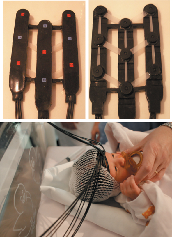
Figure 1. Probes containing the emitters (red marks) and detectors (blue marks) of near-infrared light, and their positioning on the neonate’s head. Bottom picture by F. Giraldi.
Because we are interested in normal development and language acquisition, we focus our studies on healthy hearing newborns. Infants are considered eligible to participate in our studies if their gestational age is between 38 and 42 weeks, their Apgar score is at least eight in the first minute of life, no problems were observed in the hearing test, and do not present hematomes. Moreover, to maximize the likelihood of monitoring the same areas of the brain across different infants, we recruit only those whose head diameter ranges between 33.5 and 36.0 cm. Personnel of the Hospital carry out the whole recruitment process.
Each infant is tested individually inside a dimly lit sound-attenuated booth (Figure 2). The placement of the probes on the infant’s head takes a few minutes. The use of pacifier is usually not necessary in this stage. Newborns come to the test session when they are in a quiet state of rest, either awake or sleeping. They remain in their own nursery crib and are tested lying in it, without receiving any reinforcement. A nurse or a pediatrician assists the neonates.
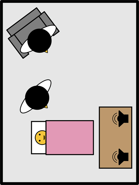
Figure 2. Scheme of the experimental setup. The neonate rests in his o her crib, assisted by a nurse or pediatrician. Sounds are presented by two loudspeakers located about 70 cm in front of the neonate’s head. One of the parents may be present during the experimental session, sitting on an armchair.
Because of our focus on language-related processes, we have mostly worked with auditory stimuli. These are presented at an appropriate intensity via two loudspeakers inside the experimental booth. The fNIRS machine and the control computer are placed outside the booth, to avoid undesired noise and heating in the testing room. An infrared videocamera is used to monitor the infant’s behavior. It is common that one or both parents attend the testing session. They choose whether to be inside the testing booth (provided that they will remain still and silent throughout the session) or outside, observing the infant’s behavior through the online video recording. Parents are informed of how fNIRS functions, and they sign a consent form after they have understood how the experiment works and all their questions have been answered.
Reliability of the Method
A long tradition in developmental research has used behavioral methods such as the high-amplitude sucking paradigm for tackling many infant speech perception questions. Thus, during our first experiences with fNIRS, we aimed to determine whether previous results obtained with these behavioral methods could be reproduced. While replicating previous findings, the fNIRS studies aimed to provide a link between behavioral observations and their underlying brain mechanisms.
Our very first study assessed the specialization of the brain to process speech stimuli at birth. In healthy adult participants, the left-hemisphere dominance for speech is a well-known fact supported by a wealth of clinical, behavioral, and brain imaging studies. The question that arose among developmental cognitive scientists was whether the left and right hemispheres are equal in function at birth and then specialized through experience, or whether at birth both hemispheres are already predisposed to process distinct types of stimuli. Subsequent research with young infants suggested that the second alternative was the most probable (Segalowitz and Chapman, 1980; Best, 1988; Bertoncini et al., 1989; Holowka and Petitto, 2002). Many researchers used indirect behavioral measures to assess a diversity of questions: for instance, Bertoncini et al. (1989) tested 2-weeks-old infants with a dichotic listening technique during a high-amplitude sucking experimental session. Two stimuli, a syllable and a musical stimulus, were presented simultaneously to the infant, one in each ear. When infants listened to repeated syllables with their right ear, they displayed a change in behavior when a different syllable was presented, which was interpreted as a discrimination response. Instead, no discrimination occurred if infants listened to the linguistic material with their left ear. The converse was observed when the musical stimulus was changed.
Peña et al. (2003) used fNIRS to investigate the patterns of brain activation in response to auditory stimulation in full-term neonates. Their study thus provided a direct measure to assess hemispheric specialization while participants listened to normal speech, backward speech, or silence. Peña et al. (2003) found that the newborn left-hemisphere shows greater hemodynamic activity in response to normal speech than to backward speech2 or silence. In addition, no area of the right hemisphere showed differential activation when contrasting forward and backward speech.
Peña et al.’s (2003) findings have yielded important theoretical and methodological implications. First, their study supported the notion of an early left-hemisphere specialization for speech, a fact intrinsically related to the emergence of the language faculty. Second, it validated fNIRS as a technique capable of both replicating previous behavioral results and enriching them by providing direct measurements of the activity in left and right perisylvian areas when processing speech.
Being a relatively young technique (see Wolf et al., 2007, for a history of near-infrared spectroscopy techniques), fNIRS must be evaluated also in terms of the reliability of the provided measures. In this respect, researchers have showed promising results for certain procedures based on fNIRS. For instance, Plichta et al. (2006) reported a high reproducibility at the group level, but not when re-testing single participants. Our studies do not address the issue of re-testing yet. However, the results of Peña et al. (2003) have been successfully reproduced in our laboratory in several occasions after the original work.
Experimental Protocols
Below we present in detail the two main testing protocols that we use in our research with neonates.
Block Design
We started our exploration of cognitive core language acquisition mechanisms by borrowing block designs from the fMRI tradition. We have used them in the aforementioned study by Peña et al. (2003), and also in Gervain et al. (2008).
Newborns are presented with sets of stimuli (blocks) corresponding to the different experimental conditions. The duration of the blocks and their content vary according to the purposes of each study. For example, Peña et al. (2003) presented neonates with continuous stimuli: backward or forward infant-directed utterances in blocks of 15 s of duration. Instead, in the Gervain et al. (2008) studies, blocks were composed of 10 discrete items separated by pauses of varying length (0.5–1.5 s), yielding blocks of about 18 s. There were two kinds of blocks, which were presented in an interleaved fashion, avoiding the presentation of more than two consecutive blocks of the same condition (Figure 3A). From the first published study, our group used and continued using a variable separation of blocks (25–35 s) to avoid the effects of spontaneous oscillations frequently detected in fNIRS recordings (Diehl et al., 1998). For the statistical analyses we usually consider oxyhemoglobin or total hemoglobin concentration changes during the time window spanning from 10 to 20 s after the onset of stimulation, which roughly coincides with the plateau of the hemodynamic response. In each experimental block a given channel is rejected either because of movement artifact–that is, if the hemodynamic signal shows a variation per unit of time above a given threshold level–or saturation of the optical channel due to displacement of the probes. Only infants with a minimum amount of accepted block-channel pairs are further considered.
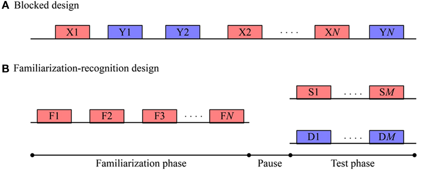
Figure 3. Scheme depicting the experimental protocols used in our laboratory. Each rectangle represents a block, consisting in either continuous auditory stimulation or a series of discrete sounds separated by short pauses of about 1 s. Consecutive blocks are separated by silent intervals of varying duration. (A) The block design consists in the intermixed presentation of N blocks belonging to each of the conditions X and Y. (B) The familiarization-recognition design consists in a Familiarization phase (F) in which all neonates listen to N blocks of stimulation. After a silent retention interval–Pause–that can last for some minutes, a Test phase follows. Here, half of the group of infants listens to M stimuli blocks of the same kind (S) of the familiarization, whereas the other half listens to a different kind of stimuli (D).
Block designs are useful to contrast brain responses to two or more experimental conditions (e.g., backward speech, forward speech, silence). One advantage is that one can compare conditions without the need of additional ad hoc control groups. However, a trade-off between number of contrasts and length of the experimental session should be considered, because the longer the session, the higher is the probability of the infant becoming fussy, and therefore of rejection.
It is important to highlight that experiments using this design have proven sensible not only for the study of prosodic or acoustic properties, but also for assessing learnability of specific structures in the first days of life: Gervain et al. (2008) examined newborns’ ability to learn and detect repetition patterns (e.g., words like “mubaba,” “penana”), showing that these sequences evoke greater activation in specific areas of the newborn brain as compared to non-repeating sequences (e.g., words like “mubage,” “penaku”).
Familiarization-Recognition Design
Behavioral methods have delivered very useful data that increased our understanding of cognitive development and language acquisition. The habituation-dishabituation method was the most frequently used to test neonates, yielding important findings that advanced our knowledge of how infants acquire language3. Among many other colleagues that applied this paradigm, we highlight the work by Eimas et al. (1971), the cardinal study who first discovered that 1- and 4-month-olds distinguish phonemes that differ by one feature, for example [b] from [p] or [b] from [d]. Furthermore, some years later Kuhl (1983) explored how infants categorize syllables despite the variability of the speech stimuli, and Mehler et al. (1978) discovered that newborns recognize their own mother’s voice (see also Jusczyk, 1997, for an extended review on infant’s speech-processing studies based on behavioral methods).
Notwithstanding the important findings obtained with the habituation-dishabituation paradigm, the studies using it suffered from the aforementioned problems of behavioral methods such as high rejection rate. Moreover, using only behavioral paradigms restricted investigations to discrimination capacities mostly, leaving practically unattended other cognitive functions equally important for language acquisition. For instance, very little is known about the memory capacities of neonates, in spite of the great progress that cognitive scientists and neuroscientists have made in identifying brain regions underlying memory processes in adults (e.g., Nyberg and Cabeza, 2000; Baddeley et al., 2009).
One of the most important challenges of fNIRS is to address questions that are difficult to assess behaviorally. Benavides-Varela, Gómez, Bion, Macagno, Peretz and Mehler (in preparation) adapted a habituation-deshabituation paradigm for testing newborn’s memory for speech sounds with fNIRS, which could also provide indications of neural activity associated with encoding and recognition at birth.
Benavides-Varela and Colleagues tested neonates between 2 and 5 days old in a recognition memory task. Newborns listened to a single CVCV word repeated for 10 blocks (each block contained six identical words) for a total of 6 min of exposure4 (see Figure 3B). A 2-min-long silent pause separated the familiarization and test phases, providing a means of tapping infants’ memory. After the pause, the same word of the familiarization phase was presented to half of the infants, whereas the other half listened to a novel word. The test consisted of five blocks (3 min). Differential responses in hemoglobin concentration between the two groups of newborns would indicate that the familiar stimulus is remembered. Results were auspicious: participants hearing a novel word showed higher relative concentrations of oxyhemoglobin in the first block of the test phase, as compared to neonates who heard again the familiar word. fNIRS revealed these differences in a bilaterally distributed network, involving temporo-parietal and anterior areas. These results open a field of possibilities for studying and better understanding the development of early memory capacities, and represent a promising step forward with respect to behavioral approaches.
The use of fNIRS has the advantage that the experimental session can be shortened or lengthened to study important variables such as amount of exposure or long-lasting retention. This represented a problem in behavioral studies, because infants often fall asleep during long silent pauses, or because the behavioral responses of the control groups may become noisy (see Jusczyk et al., 1995). Furthermore, the findings of Benavides-Varela and Colleagues suggest that 6 min of familiarization(see footnote 4) are enough for newborns to form a lasting representation of the presented word. This exposure time is considerably shorter than the ones used in comparable behavioral studies.
The usual implementation of the habituation-dishabituation procedure requires the adoption of a criterion for shifting from habituation phase to test phase, depending on each infant’s behavior. The amount of exposure required to achieve this criterion differs considerably across infants. We point out that the fNIRS version of this method allowed us to equate the amount of exposure each infant received in the familiarization phase. This is particularly important in memory studies, in which the amount of exposure partly determines the robustness of the memory trace, and therefore of the recognition response. Finally, we stress the fact that the fNIRS technique not only provides a yes/no answer to the experimental question at hand, but also informs us of the activation of several cortical areas.
We believe that this type of design will prove useful for studies aiming beyond infants’ discrimination abilities and preferences, representing an opportunity to track the time course of learning, and the encoding and recognition processes in newborns and infants at different stages of development.
Concluding Remarks
Developmental scientists face the challenge of devising reliable methodologies to assess cognitive capacities in neonates. The incorporation of the fNIRS imaging technique promotes our understanding of early language acquisition and memory capacities. We hope that the successful experiences with fNIRS in several laboratories (e.g., Peña et al., 2003; Bortfeld et al., 2007; Gervain et al., 2008; Nakano et al., 2009; Lloyd-Fox et al., 2010; Obrig et al., 2010) will encourage further exploitation of this valuable tool for studying early human development.
An important line of methodological progress is focusing on designs that combine different paradigms (behavioral and neuroimaging) or techniques (e.g., fNIRS and electroencephalography), either across different experimental sessions or in simultaneous recordings. The use of two or more techniques has the potential to provide complementary information about the functioning of the newborn’s brain. A pioneer study by Telkemeyer et al. (2009) has already used NIRS and EEG concurrently to test auditory processing in newborns. We are confident that such adaptations will play a central role in improving our knowledge about human innate cognitive capacities.
Conflict of Interest Statement
The authors declare that the research was conducted in the absence of any commercial or financial relationships that could be construed as a potential conflict of interest.
Acknowledgments
We thank Laurence White for helpful comments on earlier versions of this manuscript; Marijana Sjekloća, Francesca Gandolfo, and Alessio Isaja for their permanent administrative and technical support; Dr. Francesco Macagno and the personnel of Udine’s Hospital Neonatology and Obstetrics Departments for their assistance in the recruitment of neonates. We also thank the parents of the young participants for their collaboration. This work was supported by McDonnell Foundation Grant 21002089, and a grant from Ministerio de Ciencia y Tecnología (MICIT) and Consejo Nacional de Investigaciones Científicas y Tecnológicas (CONICIT) of Costa Rica to the first author.
Footnotes
- ^For studies with auditory stimulation, it is not considered as an issue that infants sleep during the experimental session. Indeed, sleeping newborns can discriminate and learn speech sounds (Cheour et al., 2002), and sleeping 3-month-olds show brain responses associated with memory for previously heard sentences (Dehaene-Lambertz et al., 2002).
- ^Backward speech is widely considered a very good control to compare whether infants specifically react to language stimuli and not to some physical features such as intensity, duration, or pitch. Indeed, a snippet of language played backward has the same energy of the corresponding stimuli played forward, but all the phonetic, phonological, syntactical, and prosodic transitions are lost.
- ^We refer to the vast majority of studies concerning language acquisition and infants’ general auditory capacities. In this field, the most common behavioral procedure was high-amplitude sucking.
- ^Of this total duration, however, the fraction corresponding to effective acoustic stimulation was about 1 min.
References
Aslin, R. N., and Mehler, J. (2005). Near-infrared spectroscopy for functional studies of brain activity in human infants: promise, prospects, and challenges. J. Biomed. Opt. 10, 0001–0003.
Bertoncini, J., Morais, J., Bijeljac-Babic, R., McAdams, S., Peretz, I., and Mehler, J. (1989). Dichotic perception and laterality in neonates. Brain Lang. 37, 591–605.
Best, C. T. (1988). “The emergence of cerebral asymmetries in early human development: a literature review and a neuroembryological model,” in Brain Lateralization in Children, eds D. L. Molfese and S. J. Segalowitz (New York: Guilford), 3–5.
Bortfeld, H., Wruck, E., and Boas, D. A. (2007). Assessing infants’ cortical response to speech using near-infrared spectroscopy. Neuroimage 34, 407–415.
Cheour, M., Imada, T., Taulu, S., Ahonen, A., Salonen, J., and Kuhl, P. (2004). Magnetoencephalography is feasible for infant assessment of auditory discrimination. Exp. Neurol. 190, S44–S51.
Cheour, M., Martynova, O., Näätänen, R., Erkkola, R., Sillanpää, M., Kero, P., Raz, A., Kaipio, M.-L., Hiltunen, J., Aaltonen, O., Savela, J., and Hämäläinen, H. (2002). Speech sounds learned by sleeping newborns. Nature 415, 599–600.
Dehaene-Lambertz, G., Dehaene, S., and Hertz-Pannier, L. (2002). Functional neuroimaging of speech perception in infants. Science 298, 2013–2015.
Dehaene-Lambertz, G., and Peña, M. (2001). Electrophysiological evidence for automatic phonetic processing in neonates. Neuroreport 12, 3155–3158.
Diehl, R. R., Linden, D., Lücke, D., and Berlit, P. (1998). Spontaneous blood pressure oscillations and cerebral autoregulation. Clin. Auton. Res. 8, 7–12.
Eimas, P. D., Siqueland, E. R., Juszcyk, P. W., and Vigorito, J. (1971). Speech perception in infants. Science 171, 303–306.
Gervain, J., Macagno, F., Cogoi, S., Peña, M., and Mehler, J. (2008). The neonate brain detects speech structure. Proc. Natl. Acad. Sci. U.S.A. 105, 14222–14227.
Gervain, J., Mehler, J., Werker, J. F., Nelson, C. A., Csibra, G., Lloyd-Fox, S., Shukla, M., and Aslin, R. N. (2011). Near-infrared spectroscopy: a report from the McDonnell infant methodology consortium. Dev. Cogn. Neurosci. 1, 22–46.
Holowka, S., and Petitto, L. A. (2002). Left hemisphere cerebral specialization for babies while babbling. Science 297, 1515.
Imada, T., Zhang, Y., Cheour, M., Taulu, S., Ahonen, A., and Kuhl, P. (2006). Infant speech perception activates Broca’s area: a developmental magnetoencephalography study. Neuroreport 17, 957–962.
Jusczyk, P. W., Kennedy, L. J., and Jusczyk, A. M. (1995). Young infants’ retention of information about syllables. Infant. Behav. Dev. 18, 27–41.
Kuhl, P. K. (1983). Perception of auditory equivalence classes for speech in early infancy. Infant. Behav. Dev. 6, 263–285.
Kushnerenko, E., Cheour, M., Ceponiene, R., Fellman, V., Renlund, M., Soininen, K., Alku, P., Koskinen, M., Sainio, K., and Näätänen, R. (2001). Central auditory processing of durational changes in complex speech patterns by newborns: an event-related brain potential study. Dev. Neuropsychol. 19, 83–97.
Lloyd-Fox, S., Blasi, A., and Elwell, C. E. (2010). Illuminating the developing brain: the past, present and future of functional near infrared spectroscopy. Neurosci. Biobehav. Rev. 34, 269–284.
Mehler, J., Bertoncini, J., Barriere, M., and Jassik-Gerschenfeld, D. (1978). Infant recognition of mother’s voice. Perception 7, 491–497.
Minagawa-Kawai, Y., Mori, K., Hebden, J. C., and Dupoux, E. (2008). Optical imaging of infants’ neurocognitive development: recent advances and perspectives. Dev. Neurobiol. 68, 712–728.
Nakano, T., Watanabe, H., Homae, F., and Taga, G. (2009). Prefrontal cortical involvement in young infants’ analysis of novelty. Cereb. Cortex 19, 455–463.
Nyberg, L., and Cabeza, R. (2000). “Brain imaging of memory,” in The Oxford Handbook of Memory, eds E. Tulving and F. I. M. Craik (Oxford University Press, New York), 501–519.
Obrig, H., Rossi, S., Telkemeyer, S., and Wartenburger, I. (2010). From acoustic segmentation to language processing: evidence from optical imaging. Front. Neuroenergetics 2:13. doi: 10.3389/fnene.2010.00013
Peña, M., Maki, A., Kovacčicč, D., Dehaene-Lambertz, G., Koizumi, H., Bouquet, F., and Mehler, J. (2003). Sounds and silence: an optical topography study of language recognition at birth. Proc. Natl. Acad. Sci. U.S.A. 100, 11702–11705.
Plichta, M. M., Herrmann, M. J., Baehne, C. G., Ehlis, A.-C., Richter, M. M., Pauli, P., and Fallgater, A. J. (2006). Event-related functional near-infrared spectroscopy (fNIRS): are the measurements reliable? Neuroimage 31, 116–124.
Segalowitz, S. J., and Chapman, J. S. (1980). Cerebral asymmetry for speech in neonates: a behavioral measure. Brain Lang. 9, 281–288.
Telkemeyer, S., Rossi, S., Koch, S. P., Nierhaus, T., Steinbrink, J., Poeppel, D., Obrig, H., and Wartenburger, I. (2009). Sensitivity of newborn auditory cortex to the temporal structure of sounds. J. Neurosci. 29, 14726–14733.
Keywords: cognitive development, language acquisition, newborns’ memory, fNIRS
Citation: Benavides-Varela S, Gómez DM and Mehler J (2011) Studying neonates’ language and memory capacities with functional near-infrared spectroscopy. Front. Psychology 2:64. doi: 10.3389/fpsyg.2011.00064
Received: 19 January 2011; Accepted: 28 March 2011;
Published online: 18 April 2011.
Edited by:
Judit Gervain, Université Paris Descartes, FranceReviewed by:
Isabell Wartenburger, University of Potsdam, GermanyRamon Mariano Guevara, Basque Center for Brain, Language and Cognition, Spain
Copyright: © 2011 Benavides-Varela, Gómez and Mehler. This is an open-access article subject to a non-exclusive license between the authors and Frontiers Media SA, which permits use, distribution and reproduction in other forums, provided the original authors and source are credited and other Frontiers conditions are complied with.
*Correspondence: Jacques Mehler, Cognitive Neuroscience Sector, International School for Advanced Studies, Via Bonomea 265, 34136 Trieste, Italy. e-mail: mehler@sissa.it