Iron deprivation enhances transcriptional responses to in vitro growth arrest of Mycobacterium tuberculosis
 Sogol Alebouyeh1†
 Jorge A. Cárdenas-Pestana2,3†
 Lucia Vazquez1
 Rafael Prados-Rosales1
 Patricia Del Portillo4
 Joaquín Sanz2,3*
 Maria Carmen Menéndez1*
 Maria J. García1*
- 1Department of Preventive Medicine and Public Health and Microbiology, School of Medicine, Autonomous University of Madrid, Madrid, Spain
- 2Department of Theoretical Physics, University of Zaragoza, Zaragoza, Spain
- 3Institute for Biocomputation and Physics of Complex Systems (BIFI), University of Zaragoza, Zaragoza, Spain
- 4Corporación CorpoGen, Bogota, Colombia
A corrigendum on
Iron deprivation enhances transcriptional responses to in vitro growth arrest of Mycobacterium tuberculosis
by Alebouyeh, S., Cárdenas-Pestana, J. A., Vazquez, L., Prados-Rosales, R., Del Portillo, P., Sanz, J., Menéndez, M. C., and García, M. J. (2022). Front. Microbiol. 13:956602. doi: 10.3389/fmicb.2022.956602
In the published article, there was an error in the legend of Figure 7B as published.
Analysis of lipid content by thin layer chromatography. (A) Mycolic acids. FAME, fatty acids methyl esters. (B) Polar lipids: PGL, phenolic glycolipid; GPL, glycopeptidolipids; TMM, trehalose monomycolate; PIMs, phosphatidyl-inositol mannosides; Cl, chloroform; Met, methanol; W, water. (C) Apolar lipids: PDIM, phthiocerol dymycocerosate; TAG, triacylglycerol; MQs, menaquinones; PE, petroleum ether; DE, diethyl ether. Lanes: a, Exp5–Fe; b, Exp5+Fe; c, Stat6–Fe; and d, Stat6+Fe.
The corrected legend appears below.
In the published article, there was an error in Figure 7B as published. Names of lipids are wrongly label.
The corrected Figure 7 and its caption appear below.
FIGURE 7
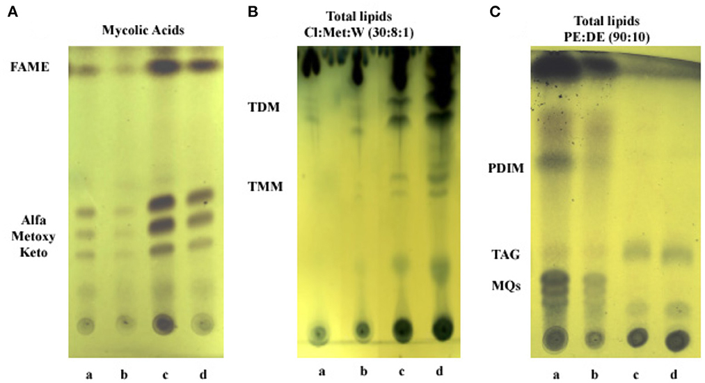
Figure 7. Analysis of lipid content by thin layer chromatography. (A) Mycolic acids. FAME, fatty acids methyl esters. (B) Polar lipids: TDM, trehalose dimycolate; TMM, trehalose monomycolate; Cl, chloroform; Met, methanol; W, water. (C) Apolar lipids: PDIM, phthiocerol dymycocerosate; TAG, triacylglycerol; MQs, menaquinones; PE, petroleum ether; DE, diethyl ether. Lanes: a, Exp5–Fe; b, Exp5+Fe; c, Stat6–Fe; and d, Stat6+Fe.
In the published article, there was an error. Page 10, Results.
A correction has been made to section Results, subsection Lipids characterization. This sentence previously stated:
“To gain insight into lipid changes linked to the effect of iron and growth arrest, TLC analysis was performed on whole Mtb cells submitted to the four different conditions under study: Exp5 and Stat6, any of them; with (+Fe) and without iron (−Fe). No differences were detected in the mycolic acid composition of the bacteria under the different conditions used (Figure 7A). Concerning total lipid analysis, conditions to develop polar and non-polar lipids were applied (Figures 7B,C). The analysis of polar lipids showed a higher abundance of PIMs and glycopeptidolipids (GPL) at Stat6 phase compared to Exp5 phase (Figure 7B). The opposite result was observed when apolar lipids were analyzed (Figure 7C). Further characterization to confirm the detection of PIMs in Exp5 phase, was performed by using two-dimensional TLC (Supplementary Figure 6). Interestingly, by applying conditions aimed at resolving apolar lipids, we observed that the band corresponding to PDIM was visible at Exp5 phase but was not detected at Stat6 phase independently of the iron content (Figure 7C). Similar to previous data (Bacon et al., 2007) increased levels of MQs were detected in iron starvation during exponential phase (Figure 7C). We also detected increased levels of TAG in stationary phase, in agreement with the detected higher proportion of red-nile stained bacilli (Figure 2).”
The corrected sentence appears below:
“To gain insight into lipid changes linked to the effect of iron and growth arrest, TLC analysis was performed on whole Mtb cells submitted to the four different conditions under study: Exp5 and Stat6, any of them; with (+Fe) and without iron (-Fe). No differences were detected in the mycolic acid composition of the bacteria under the different conditions used (Figure 7A). Concerning total lipid analysis, conditions to develop polar and non-polar lipids were applied (Figures 7B,C). The analysis of polar lipids showed a higher abundance of trehalose mono- (TMM) and dimycolates (TDM) at Stat6 phase compared to Exp5 phase (Figure 7B). The opposite result was observed when apolar lipids were analyzed (Figure 7C). Further characterization to confirm the detection of PIMs in Exp5 phase, was performed by using two-dimensional TLC (Supplementary Figure 6). Interestingly, by applying conditions aimed at resolving apolar lipids, we observed that the band corresponding to PDIM was visible at Exp5 phase but was not detected at Stat6 phase independently of the iron content (Figure 7C). Similar to previous data (Bacon et al., 2007) increased levels of MQs were detected in iron starvation during exponential phase (Figure 7C). We also detected increased levels of TAG in stationary phase, in agreement with the detected higher proportion of red-nile stained bacilli (Figure 2).”
The authors apologize for this error and state that this does not change the scientific conclusions of the article in any way. The original article has been updated.
Publisher's note
All claims expressed in this article are solely those of the authors and do not necessarily represent those of their affiliated organizations, or those of the publisher, the editors and the reviewers. Any product that may be evaluated in this article, or claim that may be made by its manufacturer, is not guaranteed or endorsed by the publisher.
References
Bacon, J., Dover, L. G., Hatch, K. A., Zhang, Y., Gomes, J. M., Kendall, S., et al. (2007). Lipid composition and transcriptional response of Mycobacterium tuberculosis grown under iron-limitation in continuous culture: identification of a novel wax ester. Microbiology 153 (Pt. 5), 1435–1444. doi: 10.1099/mic.0.2006/004317-0
Keywords: Mycobacterium tuberculosis, iron availability, transcriptomics, growth arrest, metabolic changes
Citation: Alebouyeh S, Cárdenas-Pestana JA, Vazquez L, Prados-Rosales R, Del Portillo P, Sanz J, Menéndez MC and García MJ (2022) Corrigendum: Iron deprivation enhances transcriptional responses to in vitro growth arrest of Mycobacterium tuberculosis. Front. Microbiol. 13:1081051. doi: 10.3389/fmicb.2022.1081051
Received: 26 October 2022; Accepted: 27 October 2022;
Published: 29 November 2022.
Edited and reviewed by: Nuno S. Osório, ICVS/3B's Associate Laboratory (AL), Portugal
Copyright © 2022 Alebouyeh, Cárdenas-Pestana, Vazquez, Prados-Rosales, Del Portillo, Sanz, Menéndez and García. This is an open-access article distributed under the terms of the Creative Commons Attribution License (CC BY). The use, distribution or reproduction in other forums is permitted, provided the original author(s) and the copyright owner(s) are credited and that the original publication in this journal is cited, in accordance with accepted academic practice. No use, distribution or reproduction is permitted which does not comply with these terms.
*Correspondence: Maria J. García, mariaj.garcia@uam.es; Maria Carmen Menéndez, carmen.menendez@uam.es; Joaquín Sanz, jsanz@bifi.es
†These authors have contributed equally to this work and share first authorship