 Hai Gao1†Yanan Wang1†Qiao Luo1Liyuan Yang1Xingxing He1
 Jun Wu2
 Konthorn Kachanuban3Pongthep Wilaipun3
 Weiming Zhu1*
 Yi Wang1*
- 1School of Medicine and Pharmacy, Ocean University of China, Laboratory for Marine Drugs and Bioproducts of Qingdao National Laboratory for Marine Science and Technology, Qingdao, China
- 2School of Pharmaceutical Sciences, Southern Medical University, Guangzhou, China
- 3Faculty of Fisheries, Kasetsart University, Bangkok, Thailand
Despite being potentially useful extremophile resources, there have been few reports on acid-tolerant fungi and their bioactive metabolites. Acidophilic/aciduric fungi (n = 237) were isolated from Thai mangrove sediments in an acidic medium. Using fungal identification technology (including morphologic observation, chemical screening, and sequence comparisons) all the isolates were identified and 41 representative isolates were selected for analysis of the phylogenetic relationships (ITS rDNA, β-tubulin, calmodulin, and actin gene sequences). There were seven genera identified – Penicillium; Aspergillus; Talaromyces; Cladosporium; Allophoma; Alternaria; and Trichoderma – in four taxonomic orders of the phylum Ascomycota, and Penicillium, Aspergillus, and Talaromyces were the dominant genera. Acidity tolerance was evaluated and 95% of the isolates could grow under extremely acidic conditions (pH 2). Six strains were classed as acidophilic fungi that cannot survive under pH 7, all of which had an extraordinarily close genetic relationship and belonged to the genus Talaromyces. This is the first report on the acidophilic characteristics of this genus. The antimicrobial, anti-tumor, and antiviral activities of the fermentation extracts were evaluated. Nearly three-quarters of the extracts showed cytotoxic activity, while less than a quarter showed antimicrobial or anti-H1N1 activity. The typical aciduric fungus Penicillium oxalicum OUCMDZ-5207 showed similar growth but completely different chemical diversity at pH 3 and 7. The metabolites of OUCMDZ-5207 that were obtained only at pH 3 were identified as tetrahydroauroglaucin (1), flavoglaucin (2), and auroglaucin (3), among which auroglaucin showed strong selective inhibition of A549 cells with an IC50 value of 5.67 μM. These results suggest that acid stress can activate silent gene clusters to expand the diversity of secondary metabolites, and the bioprospecting of aciduric/acidophilic microorganism resources in Thai mangrove sediments may lead to the discovery of compounds with potential medicinal applications.
Introduction
Microbes are important drug producers, however, it is becoming more difficult to obtain new drugs and drug leads from common environmental microorganisms, with less discovery of new microorganisms occurring and duplication of research on their metabolites. Over the last 10 years, we have been investigating microbes and their metabolites from extreme environments. Extreme microorganisms are those that live in a special environment, including places with high temperatures (>50°C), low temperatures (<20°C), high pressure (>35 MPa), high salinity (>3% NaCl), high pH (>pH 9), and low pH (<pH 4) (Wilson and Brimble, 2009), where ordinary microbes cannot survive. The different physiological characteristics of extreme microorganisms can create unique metabolic pathways and their secondary metabolites can have remarkable chemical diversity and interesting bioactivity (Wang et al., 2009, 2011a,b,c, 2013; Zheng et al., 2009, 2010, 2013; Peng et al., 2011a,b, 2018; Fan et al., 2013, 2018; Lin et al., 2015; Qin et al., 2016; Wang M. M. et al., 2017; Jin et al., 2018).
Acid-tolerant microbes are a type of extreme microorganisms, which include acidophilic and aciduric microbes. The optimum pH for the growth of acidophiles is <pH 3, while aciduric microorganisms can tolerate both extremely acidic and neutral conditions (Johnson, 1998). There have been few studies on acidophilic fungi resources. In 2019, Martina et al. summarized the taxonomically identified indigenous acidophilic fungi, but only nine species (Acidea extrema, Acidiella bohemica, Acidiella uranophila, Acidomyces acidophilus, Acidomyces acidothermus, Acidothrix acidophila, Coniochaeta fodinicola, Neohortaea acidophila, and Soosiella minima) were listed. All these fungi were obtained from highly acidic habitats, including highly acidic soil, rivers, hot springs, mines, drainage, and acidophilic algae (Hujslová et al., 2019). Furthermore, Berkeley Pit Lake in the United States and mangrove sediments in China are acid-tolerant fungal habitats (Wang Y. et al., 2017). Although there has been a lack of investigation regarding the diversity of acidophilic fungi, this microbiota has the potential for a variety of applications, including bioremediation technologies (Deshmukh et al., 2016). For instance, acidophilic fungi have been reported to play an important role in the passive remediation of acid mine drainage and the degradation of environmental pollutants, such as phenolic compounds and polymerized rubber. The metabolites of acidophilic fungi also have potential medicinal value. In 2017, the Stierle group published a review of their work on the bioactive metabolites isolated from the abandoned acid metal mine, Berkeley Pit Lake (Stierle et al., 2017). By 2017, new compounds and bioactive compounds occupied 65 and 68% of all the compounds from aciduric microorganisms, respectively, suggesting that acid-tolerant fungi are potential medicinal resources (Wang Y. et al., 2017).
Mangroves are a special marine plant community distributed in the intertidal zone of tropical and subtropical regions. The particular nature of the environment means that mangrove sediments possess strong acidity and have the characteristics of high nutrition, high salinity, and strong reducibility. Although there have been some reports regarding microorganisms from mangroves, there have been few investigations of acidophilic or aciduric microorganisms. In our previous work, some novel bioactive compounds were obtained from acid-tolerant fungi in the soil of the mangrove rhizosphere of the China Mangrove Nature Reserve (Fan et al., 2013; Lin et al., 2015; Qin et al., 2016; Fan et al., 2018; Jin et al., 2018). Examples include new indole-diterpenoids with Anti-influenza virus activity from Penicillium camemberti OUCMDZ-1492 (Fan et al., 2013) and new anthraquinone derivatives with Anti hepatitis B virus activity from Penicillium sp. OUCMDZ-4736 (Jin et al., 2018). These results suggest that further investigation of the aciduric fungi obtained from mangrove sediments and their bioactive secondary metabolites are worthy of attention.
Previously, most studies of microbes from Thai mangroves have focused on bacteria and yeast (Limtong et al., 2007, 2008; Am-In et al., 2008, 2011; Atipan et al., 2012; Songsumanus et al., 2013; Wongwongsee et al., 2013; Hwanhlem et al., 2014; H-Kittikun et al., 2015; Wanapaisan et al., 2018). Fungi from Thai mangroves, especially acid-tolerant fungi and their bioactive metabolites under conditions of acidic stress, have not previously been investigated. In this paper, we report acid-tolerant fungi and their bioactive metabolites obtained from Thailand mangrove sediments. Using morphologic observations, chemical diversity comparisons, and analysis of the phylogenetic relationships of genes including internal transcribed spacer (ITS)-ribosomal DNA (rDNA), β-tubulin, calmodulin, partial actin gene sequences, the acid-tolerant fungi were identified and classified, and the biodiversity and biological characteristics were evaluated. Furthermore, acid tolerance and antibiotic biological activity (antibacterial, cytotoxicity, and antiviral activity) were also studied. A typical aciduric fungus – Penicillium sp. OUCMDZ-5207 – was analyzed under acidic and neutral conditions, and the metabolites at pH 3 and their cytotoxicity were studied.
Materials and Methods
General Experimental Procedures
UV spectra were recorded on a Thermo Fisher Scientific NanoDrop One micro-spectrophotometer (Thermo Fisher Scientific, Waltham, MA, United States). 1H NMR, 13C NMR, and 2D NMR spectra were recorded on a Bruker Avance 500 MHz spectrometer with tetramethylsilane (TMS; Alfa Chemistry, United States) as an internal standard. Mass spectra were recorded on an Agilent 6200 Q-TOF MS system. TLC was performed on plates precoated with silica gel GF254 (10–40 μm, Qingdao Marine Chemical Ltd., Qingdao, China). Column chromatography was performed on silica gel (100–200, 200–300, and 300–400 mesh; Qingdao Marine Chemical Ltd., Qingdao, China), RP-18 gel (20–45 μm), and Sephadex LH-20 (Amersham Biosciences, United Kingdom). Medium-pressure liquid chromatography (MPLC; CXTH-3000, China) was performed using columns packed with RP-18 gel and an LC3000 system equipped with P3000A pump modules. Semipreparative high performance liquid chromatography (HPLC; Waters 1525, United States) was performed using a column (YMC-pakODS-A, 10 × 250 mm, 5 μm, 4 mL/min).
Fungi Isolation
Fungi strains were isolated from the sediment around the roots of Hibiscus tiliaceus. Samples were collected from the mangrove habitat at PakMeng Beach, Thailand in October 2016. The samples were stored at –20°C and processed immediately for isolation and cultivation of the fungus after transport to the laboratory. The samples were then frozen at –20°C.
The gradient dilution method was used for fungi isolation. To remove superficial sediments and microbes, samples (1.0 g) were sterilized with 75% ethanol and rapidly washed three times with sterile water under sterile conditions. The samples were homogenized thoroughly and diluted to three different concentrations (0.1, 0.01, and 0.001) with sterile seawater. Then, 100-μL samples of each dilution were plated onto the corresponding isolation medium (Table 1) in triplicate. The acid medium was adjusted with citric acid and disodium hydrogen phosphate buffer solution to obtain the desired pH. The neutral medium was adjusted with NaOH. Chloramphenicol (100 μg/mL) was added to the medium to prevent the growth of bacteria during the incubation. The fungi were cultured at 28°C for at least 5 days. To ensure strain diversity, timely observations were conducted as much as possible. All the colonies were collected and purified on the plate repeatedly until a single colony was obtained, then inoculated onto slope media and stored at 4°C. Furthermore, these strains were also preserved in 20% glycerin liquid medium at −80°C.
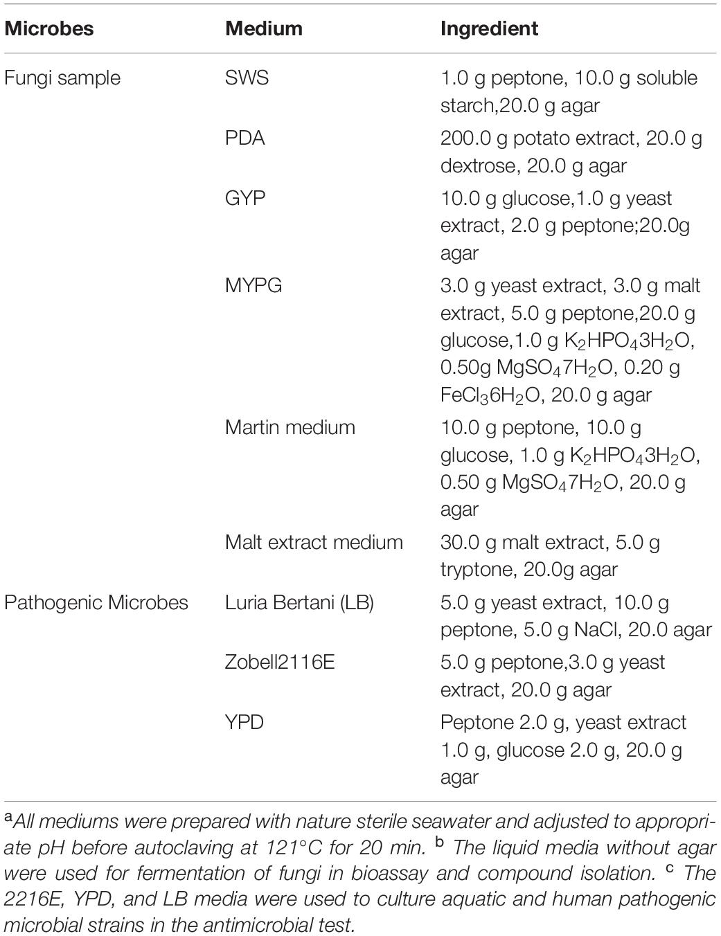
Table 1. Ingredient of media used for isolation and fermentation of microbial strains a,b,c.
DNA Extraction, PCR Amplification, and Sequencing
Single fungal colonies were transferred with a sterilized toothpick to 50 μL of a lysis buffer for PCR with a Direct PCR kit (Takara) in a sterilized microtube. The microtubes were placed at 80°C for pre-degeneration for 15 min and then centrifuged at low speed. A 2-μL sample of the supernatant was used as the template to amplify the fungal gene fragment in a polymerase chain reaction (PCR). internal transcribed spacer (ITS)-ribosomal DNA (rDNA) sequences were amplified with the primers ITS1F (5′-CTTGGTCATTTAGAGGAAGTAA-3′) (Gardes and Bruns, 1993) and ITS4 (5′-TCCTCCGCTTATTGATATGC-3′) (White et al., 1990). For the sequencing of partial β-tubulin gene, βt2a (5′-GGTAACCAAATCGGTGCTGCTTTC-3′) and βt2b (5′-ACCCTCAGTG TAGTGACC CTTGGC-3′) were used as the primers (Glass and Donaldson, 1995). Partial calmodulin gene was amplified using the primers cmd5 (5′-CCGAGTACAAGGAGGCCTTC-3′) and cmd6 (5′-CCGATAGAGGTCATAACGTG-3′) (O’ Donnell et al., 2000). For the sequencing of the partial actin gene, act-512F (5′-ATGTGCAAGGCCGGTTTCGC-3′) and ACT-783R (5′-TACGAGTCCTTCTGGCCCAT-3′) were used as the forward primer and reverse primer, respectively (Carbone and Kohn, 1999). The PCR reactions were performed in a final volume of 50 μL, which was composed of 2 μL of template DNA, 10 μL of 5 × PrimeSTAR Buffer (Mg2 + plus), 4 μL of dNTP Mix (2.5 mM of dATP, dCTP, dGTP and dTTP, respectively), 0.5 μL of forward primer (20 μM), 0.5 μL of reverse primer (20 μM), 0.5 μL of PrimeSTAR HS DNA Polymerase (2.5 U/μL), and 32.5 μL of ultrapure water. Amplification was carried out using the following thermal cycles: 30 cycles of 10 s at 98°C, 5 s at 55°C, and 60 s at 72°C. Then, the PCR products (5 μL) were loaded onto an agarose gel [1.2% agarose in 0.5 × TAE buffer and 5 μL of 1% ethidium bromide solution (m/v) per 100 mL of gel] and isolated using a gel extraction kit (E.Z.N.A., Omega, United States), according to the manufacturer’s protocol after electrophoresis at 100 V for 35 min. PCR products were submitted for sequencing (Shanghai Personalbio Biotechnology Co., Ltd., China).
Phylogenetic Analysis
Fungal sequences were edited with Lasergene Software SeqMan (DNAStar Inc.). For preliminary identification, the sequences were compared with related sequences available in the National Center for Biotechnology Information (NCBI) to determine the identity of the sequence. All the fungal sequences were aligned using the CLC Sequence Viewer 6 software with the default parameters. All the identified ITS-rDNA sequences of the Thai mangrove fungi were submitted to NCBI GenBank and the accession numbers were obtained. All the sequences of β-tubulin, calmodulin, partial actin gene were shown in Supplementary Material. A phylogenetic tree was generated with the neighbor-joining (NJ) method in the MEGA 4 software and 1,000 replicates were used for the bootstrap analysis. iTOL online software was used to process the phylogenetic tree. The reference sequences showing the highest homology to the sequences from the Thai mangrove fungi were amplified.
Preparation of Samples for Bioassay
The fungal strains were fermented with shaking at 28°C and 180 rpm for 7 day in a 500-mL conical flask containing 150 mL of Potato Dextrose Broth (PDB) liquid medium. The acid medium was prepared by adding citric acid and disodium hydrogen phosphate buffer solution. Three parallel lines were set for each experiment. The fermentation broth was extracted with an equal volume of ethyl acetate (EtOAc) three times. The dried extracts for bioassay were obtained by evaporation under reduced pressure.
Isolation and Identification of Compounds
Penicillium sp. OUCMDZ-5207 was isolated from the sediment around the roots of the mangrove plant, H. tiliaceus, at pH 2.0. The strain was fermented under static conditions at 26°C for 35 day in twenty 1000-mL Erlenmeyer flasks each containing 300 mL of PDB liquid medium at pH 3.0. The fermentation broths were harvested and extracted three times with an equal volume of ethyl acetate (EtOAc). Removal of the solvent in vacuum gave 3.2 g of EtOAc extract. The EtOAc extracts were separated on a silica-gel column eluting with petroleum ether (PE)–EtOAc (v/v100:1, 50:1, 25:1, 10:1, 5:1, 1:1, 1:2, 1:5, and 1:10) to obtain nine subfractions (Fr.1–Fr.9). Fr.3 (80.1 mg) was further subjected to purification with an HPLC ODS column (v/v acetonitrile –H2O 7:3) to give Fr.3-1–Fr.3-4. Fr.3-4 (10.3 mg) was further purified using a HPLC Polymer Stabilized Cholesteric Texture column (v/v acetonitrile–H2O 13:20) to yield compounds 1 (3.21 mg, tR 30 min) and 2 (4.32 mg, tR 34 min). Fr.4 was further separated by Sephadex LH-20 column chromatography eluting with MeOH-CH2Cl2 (v/v 1:1) to give three subfractions (Fr.4-1–Fr.4-3). Fr.4-2 (5.6 mg) and subjected to silica gel column chromatography eluting with petroleum ether–EtOAc (v/v 35:1, 25:1, and 10:1) to give compound 3 (2.67 mg).
Tetrahydroauroglaucin (1): Light yellow amorphous solid. C19H26O3. UV(MeOH) λmax (logε) 240 (0.17), 275 (0.08), 392 (0.01) nm; 1H-NMR (DMSO-d6, 600 MHz) δ: 0.83 (3H, t, J = 6.7 Hz, H-7′), 1.26 (4H, m, H-5′, H-6′), 1.41 (2H, quintet, J = 7.3 Hz, H-4′), 2.62 (3H, s, H-4″), 2.19 (3H, s, H-5″), 2.46 (2H, q, J = 1.7 Hz, H-3), 3.16 (2H, d, J = 7.4 Hz, H-1″), 9.16 (1H, brs, 5-OH), 5.20 (1H, t, H-2″), 6.51 (1H, m, H-2′), 6.94 (1H, s, H-4), 5.78 (1H, dt, J = 15.9, H-1′), 9.99 (1H, s, 1-CHO), 11.60 (1H, s, 2-OH). 13C-NMR (DMSO-d6, 125 MHz) δ 197.3 (1-CHO), 152.9 (C-2), 146.9 (C-5), 140.5 (C-2′), 132.7 (C-3″), 127.7 (C-3), 124.7 (C-4), 125.7 (C-6), 121.6 (C-2″), 121.0 (C-1′), 117.4 (C-1), 33.2 (C-3′), 30.9 (C-5′), 28.4 (C-4′), 26.9 (C-1″), 25.6 (C-5″), 22.0 (C-6′), 17.7 (C-4″), 14.0 (C-7′). ESI-MS m/z 301[M-H]–.
Flavoglaucin (2): Light yellow amorphous solid. C19H28O3. UV(MeOH) λmax (logε) 214 (1.33), 255 (0.99), 300 (0.76) nm; 1H-NMR (DMSO-d6, 600 MHz) δ: 11.68 (1H, s, 2-OH), 10.19 (1H, s, 7-CHO), 6.92 (1H, s, H-4), 5.18 (1H, d, J = 14.8 Hz, H-2″), 8.95 (1H, brs, 5-OH), 3.13 (2H, d, J = 7.4 Hz, H-1″), 2.80 (2H, t, J = 7.7 Hz, H-1′), 1.65 (3H, s, H-5″), 1.61 (3H, s, H-4″), 1.40 (2H, m, H-2′), 1.19–1.23 (8H, m, H-3′, 4′, 5′, 6′), 0.80 (3H, t, J = 6.7 Hz, H-7′) 13C NMR (DMSO-d6, 125 MHz) δ 197.2 (1-CHO), 153.2 (C-2), 146.9 (C-5), 132.5 (C-3″), 129.0 (C-3), 126.8 (C-6), 125.2 (C-4), 121.8 (C-2″), 117.6 (C-1), 31.3 (C-2′), 31.5 (C-4′), 29.0 (C-5′), 28.6 (C-3′), 26.7 (C-1″), 25.6 (C-5″), 23.2 (C-1′), 22.1 (C-6′), 17.7 (C-4″), 14.0 (C-7′). ESI-MS m/z 303[M-H]–.
Auroglaucin (3): Yellow amorphous solid. C19H22O3. UV(MeOH) λmax (logε) 230 (1.47), 211 (1.22), 278 (1.13) nm. 1H-NMR (DMSO-d6, 600 MHz) δ: 11.71 (1H, s, 2-OH), 10.03 (1H, s, 7-CHO), 6.95 (1H, s, H-4), 5.18 (1H, t, J = 14.8 Hz, H-2″), 9.43 (1H, brs, 5-OH), 3.17 (2H, d, J = 7.3 Hz, H-1″), 6.82 (1H, d, J = 15.6 Hz, H-1′), 6.15 (1H, m, H-5′), 1.60 (3H, s, H-4″), 6.60 (1H, m, H-2′), 6.32 (2H, m, H-3′, 4′), 5.74 (1H, m, H-6′), 1.71 (3H, d, J = 6.8 Hz, H-7′); 13C NMR (DMSO-d6, 125 MHz) δ 196.9 (1-CHO), 153.3 (C-2), 147.7 (C-5), 131.2 (C-3″), 134.5 (C-3), 121.4 (C-6), 124.9 (C-4), 123.0 (C-2″), 116.9 (C-1), 124.5 (C-2′), 131.9 (C-4′), 128.4 (C-5′), 132.8 (C-3′), 26.9 (C-1″), 17.7 (C-5″), 130.9 (C-1′), 137.8 (C-6′), 25.6 (C-4″), 18.3 (C-7′). ESI-MS m/z 297[M-H]–.
Anti-microbial Assay
The paper disk-agar diffusion method (Tamura et al., 1968) was used to evaluate the inhibition of pathogenic microorganisms, including nine aquatic pathogens (Edwardsiella tarda, Vibrio parahaemolyticus, Vibrio Vulnificus, Vibrio alginolyticus, Aeromonas hydrophila, Pseudoalteromonas nigrifaciens, Shewanella marisflavi, Vibrio splendidus, and Bacillus cereus) and eight human pathogens (Staphylococcus aureus, Staphylococcus aureus Rosenbach, Escherichia coli, Bacillus subtilis, Clostridium perfringens, Candida albicans, Candida glabrata, and Pseudomonas aeruginosa). The initial concentration of the EtOAc extract was 1 mg/mL. Ciprofloxacin and ketoconazole at concentrations of 0.1 mg/mL were used as positive controls. The pathogenic microorganisms were cultivated in a 50-mL centrifuge tube containing 30 mL of liquid medium and shaken at 28°C and 180 rpm for 12 h. LB medium was used for human pathogenic bacteria, 2116E medium for aquatic pathogenic bacteria, and YPD medium for human pathogenic fungi. The pathogenic strains were coated on a corresponding plate and incubated at 37°C for 30 min. Paper discs with 10 μL of the sample or positive controls (three parallels) were volatilized to dryness and placed onto an agar plate coated with the bacterial suspension. The plates were incubated for 12 h at 37°C. The negative control was methanol. Then, the diameters of the inhibition zones were recorded as the observed antibacterial activity.
Cytotoxic Assay
The cytotoxicity against human breast adenocarcinoma cell line MCF-7 and human lung adenocarcinoma cell line A549 was evaluated by the MTT method (Mosmann, 1983). The EtOAc extracts, compounds, and positive control (adriamycin) were dissolved in dimethyl sulfoxide (DMSO), DMSO was a negative control. The cell lines were cultivated in RPMI-1640 medium with 10% FBS under a humidified atmosphere with 5% CO2 at 37°C. Cell suspensions of MCF-7 and A549 (100 μL) were inoculated into 96-well plates (5 × 10³ cells/well) and incubated under the above conditions for 24 h. Then, 100-μL samples with final concentrations of 100 μg/mL of extract, 10 μM of a compound, and 1 μM of adriamycin were added to each well in triplicate and the culture mediums were incubated for 72 h. The RPMI-1640 medium containing 5% FBS was used as a blank control. MTT (methylthiazolyldiphenyl-tetrazolium bromide) solution (20 μL; 5 mg/mL in FBS) was added into each well and the wells were incubated for 4 h at 37°C. The medium containing MTT was gently removed with a pipette and 150 μL of DMSO was added to each well to dissolve the formed formazan crystals. The absorbance was recorded on a Spectra Max Plus plate reader at 570 nm. The inhibition rate was calculated as [(OD blank - OD sample)/OD blank] × 100%.
Anti-H1N1 Assay
The cytotoxicity of the samples against MDCK cells was evaluated by the method described above. Non-toxic samples (inhibition rates <0) were evaluated for antiviral bioactivity against the influenza virus H1N1 [A/Puerto Rico/8/34 (H1N1), PR/8] by the MTT method. The Madin-Darby Canine Kidney (MDCK) cells were digested by trypsin as monolayer cells and incubated with the influenza virus H1N1 at 37°C for 1 h. Then, the culture medium was discarded and a new RPMI 1640 medium with 20 μL samples 50 μg/mL were added. The inhibition was evaluated by the MTT method the same as for cytotoxicity, as described above.
Results and Discussion
Isolation and Phylogenetic Diversity of Aciduric Fungi Associated With Thai Mangrove Sediment
A total of 237 fungi strains were isolated from the acidic sediment collected from around the roots of H. tiliaceus, a mangrove plant from PakMeng Beach, Thailand. Six types of culture medium (Table 1) at two different pH values were used for isolation of the fungi. The acidity of the sediment sample was pH 5.0 and this pH value was used for the isolation. Additionally, pH 3.0 was used for the isolation to increase the possibility of obtaining acidophilic microbes. In total, 163 and 74 fungal strains were obtained under natural (pH 5.0) and acidic (pH 3.0) conditions, respectively. Duplicates were removed by detailed morphological observation, pigment formation, TLC characteristics, and HPLC fingerprint analysis of the crude extracts of the metabolites. After this assessment, there remained 95 individual fungi strains (72 at pH 5.0 and 23 at pH 3.0). These mangrove fungi grew better under natural conditions (pH 5.0) compared with the highly acidic environment (pH 3.0) (Figure 1). Compared with the other mediums used for isolation, the Martin medium was found to be more suitable for fungi growth, which may be related to the presence of more inorganic salts in this medium. The fungi may grow better in a medium rich in inorganic salts because of the characteristics of the natural fungal environment where the mangrove sediments are repeatedly washed by seawater and that deposits a large amount of inorganic salts.
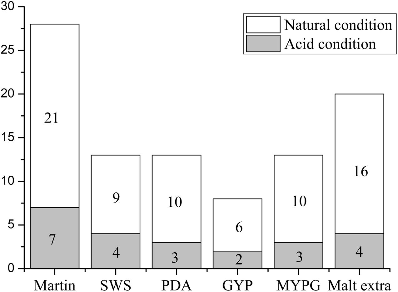
Figure 1. Numbers of fungi isolated from Thai mangrove samples using different media under acidic (pH 3.0) and natural (pH 5.0) conditions (95 fungi strains, 72 at pH 5.0 and 23 at pH 3.0). The horizontal axis represents the type of culture medium (SWS: peptone starch agar; PDA: potato dextrose agar; GYP: glucose yeast extract peptone; and MYPG: malt extract yeast extract peptone glucose), and the vertical axis represents the number of fungi isolates.
We investigated research references on fungi derived from the semi-mangrove plant H. tiliaceus from 2008 to 2020 (Li et al., 2008, 2010; Yan et al., 2010, 2012; Wang et al., 2012). Most results discussed the plant endophyte and their metabolites. The fungal genera included Aspergillus flavus, Penicillium commune, and Eurotium rubrum, among which A. flavus strain was isolated from the roots, and the others were isolated from flowers or stems. There is no reference reported on pH adaptability of these fungi from the soil around H. tiliaceus roots. So it is the first time to report the characteristics of acid-tolerant rhizosphere fungi derived from H. tiliaceus of Thailand mangroves. Mangrove soil is a special acidic geological environment, but there have been few reports of acid-tolerant fungi from this environment. Generally, a highly acidic environment is not conducive to microbial growth; however, our results showed more than a quarter of the fungi (23/95) showed better growth in the highly acidic medium (pH 3.0), which indicated that the microbial community in mangrove sediments associated with H. tiliaceus in Thailand has distinctive acidophilic properties.
To investigate the phylogenetic relationship of these mangrove fungi, the ITS sequences were compared using Clustal X software, the isolates with duplicated ITS sequences were excluded, and the remaining 41 strains were analyzed for phylogenetic relationships. According to the blast results compared with the sequences already registered in the NCBI gene databank, all the isolated fungi belonged to the Ascomycota phylum. These fungi showed 96–100% similarity to their closest relatives (Table 2). Based on the analysis of homology of the ITS rDNA gene, a phylogenetic relationship containing 41 representative fungi isolates was constructed by the neighbor-joining method (Figure 2). Considering the fact that the ITS phylogenetic analysis was not sufficient, different gene combinations were used for different genera. The β-tubulin gene was used for 33 isolates identified as Penicillium, Talaromyces, Aspergillus with the ITS genetic marker (Supplementary Figure 2). The phylogenetic relationship based on β-tubulin genes showed OUCMDZ-5245 and 5259 were clustered together with Talaromyces which were different from the ITS result Penicillium. Calmodulin and actin genes were used to analyze strain 5245 and 5259, respectively, and the results also showed that they all belonged to Talaromyces. At the level of genetic analysis, these two strains were considered more reliable as Talaromyces. Calmodulin and actin genes were used for analyzing OUCMDZ-5058, 5216, 5036, 5008, 5111 and 5083, 5040, 5048, respectively (Supplementary Figures 3, 4). Except for strain 5111, all the identification results were corresponded to the ITS rDNA analyzing results. The actin gene was used for identification of 5111, and the result showed that 5111 belonged to Allophoma which was the same as the result of ITS. Combined with all the genetic analysis results, all the identified fungi belonged to one phylum (Ascomycota), three classes (Eurotiomycetes, Dothideomycetes, and Sordariomycetes), four orders (Eurotiales, Capnodiales, Pleosporales, and Hypocreomycetidae) and seven genera including Penicillium (12 isolates), Aspergillus (10 isolates), Talaromyces (11), Cladosporium (4), Alternaria (2), Allophoma (1), and Trichoderma (1). Among them, 80.5% of the strains belonged to the order Eurotiales. Penicillium, Talaromyces, and Aspergillus were the dominant genera in this mangrove ecosystem and were extensively distributed.
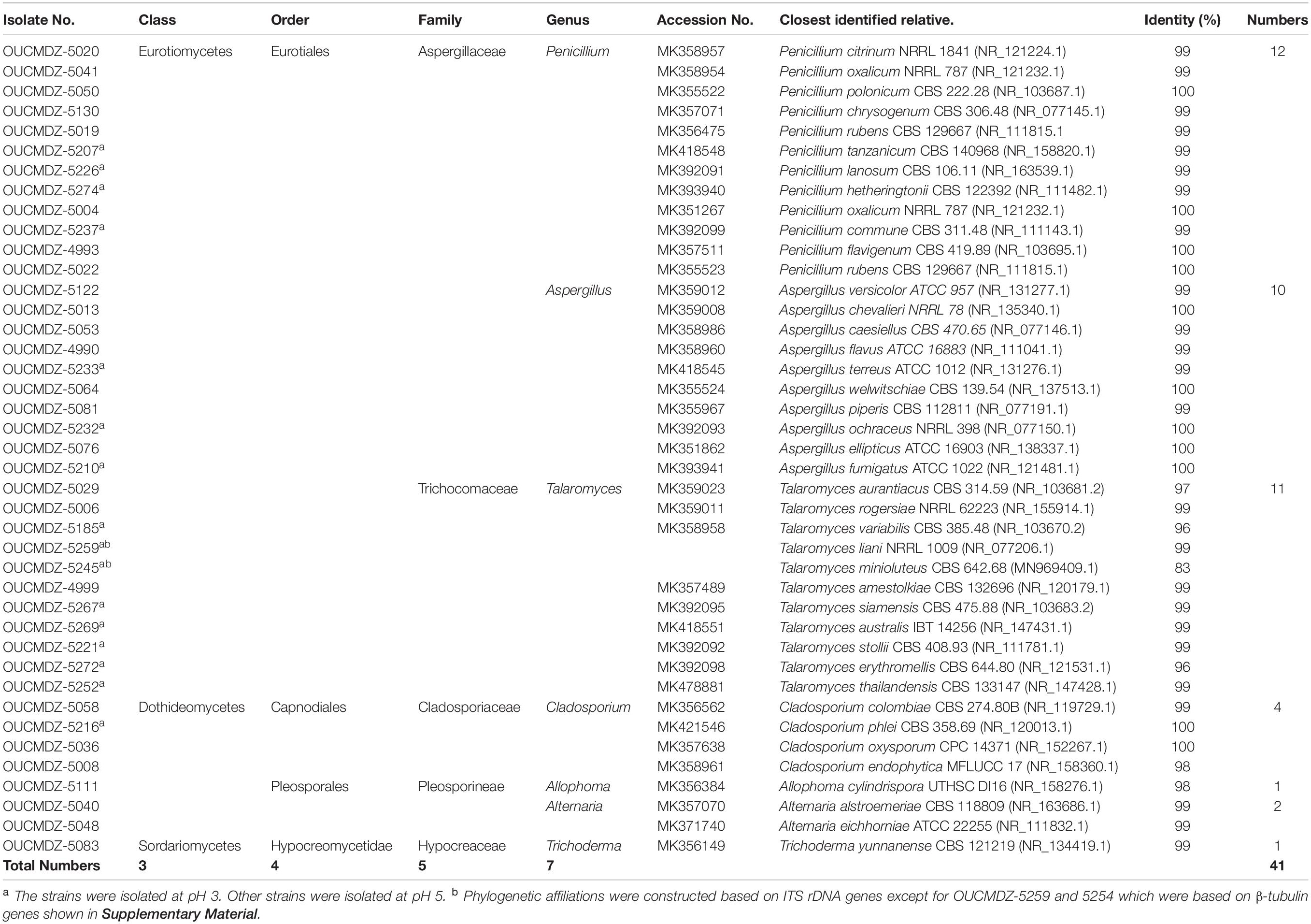
Table 2. Phylogenetic affiliations and the classification of 41 representative acid-tolerant fungal isolates from Thai mangrove samples.
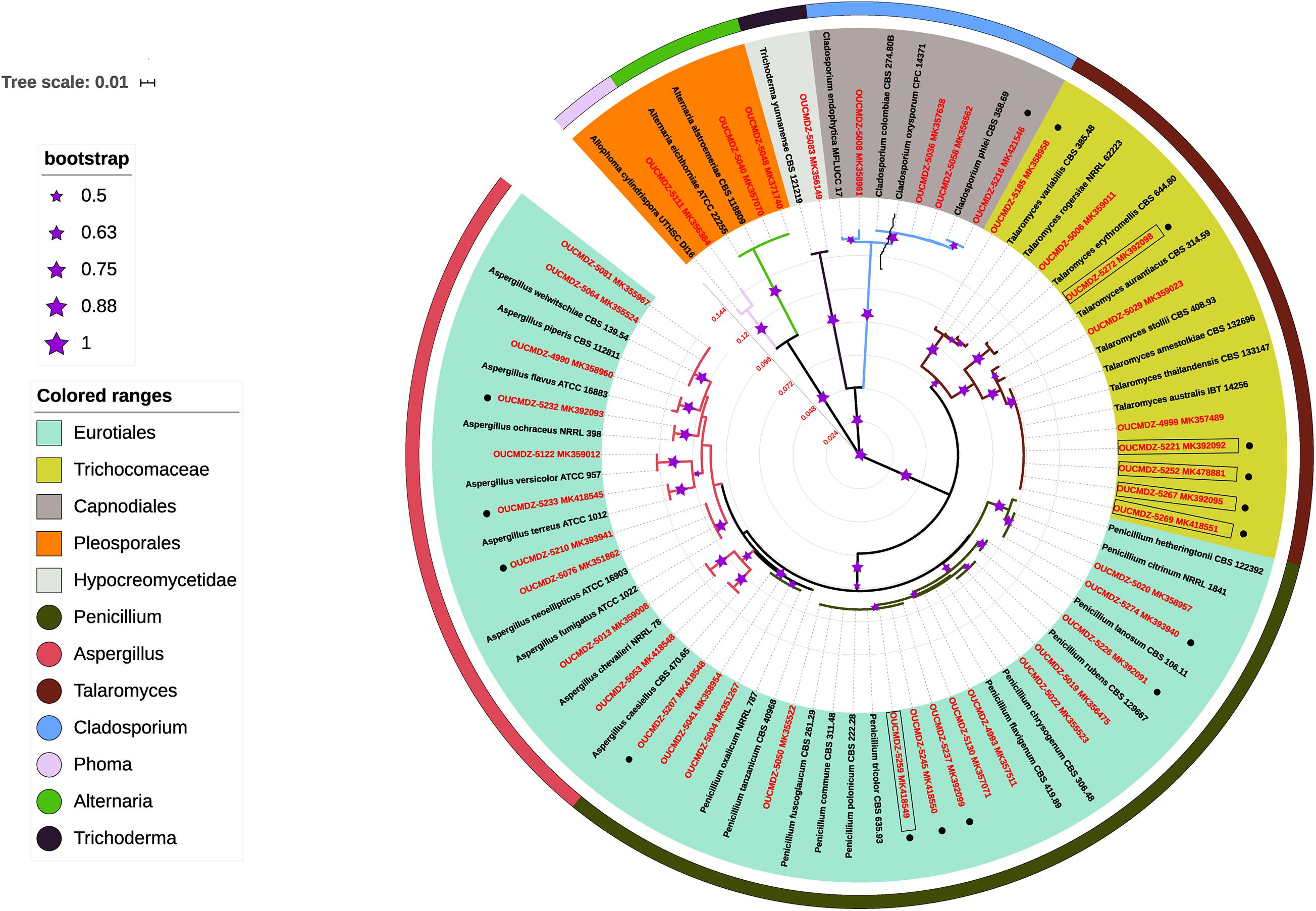
Figure 2. Phylogenetic relationship of 41 representative fungal isolates based on ITS rDNA gene sequences using the neighbor-joining method. The scale bar represents 0.01 substitutions per nucleotide. Different orders and genera are represented by different colors. The size of the asterisk indicates different bootstrap values from 1000 replicates (a larger asterisk means a higher credibility). The red label indicates the strains isolated from the experiment, and the black label indicates the reference strains in the database. Acidophilic fungi are represented by boxes. The strains obtained under pH 3.0 are indicated by black spots.
To understand the acid tolerance of these fungi, all the 41 strains obtained under (pH 3.0 and pH 5.0) were incubated under extremely acidic conditions (pH 2.0) and neutral conditions (pH 7), and the growth was observed. The results indicated that all of the 41 fungi strains could grow at pH 5.0, six (OUCMDZ-5221, 5252, 5259, 5267, 5269, and 5272) could not survive at pH 7.0 but grew well at pH 2.0, and two (OUCMDZ-4999 and 5111) could not survive at pH 2.0 or 3.0. The six isolates (OUCMDZ-5221, 5252, 5259, 5267, 5269, and 5272) can be characterized as acidophilic fungi that cannot survive in a neutral environment and have optimal growth at pH values between 2 and 5, and interestingly, all these six isolates belonged to the genus Talaromyces. These results came from the unrooted phylogenetic tree constructed with β-tubulin gene sequences (Figure 3).
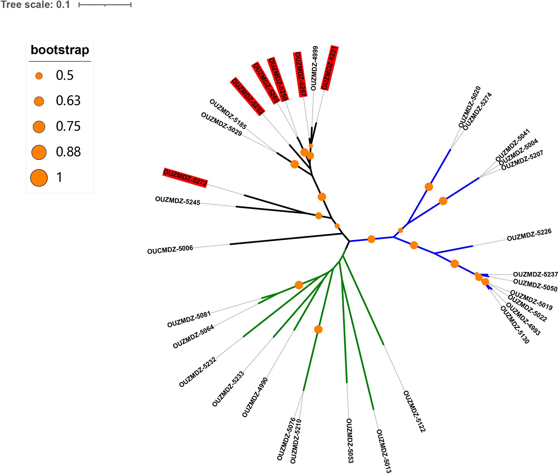
Figure 3. The unrooted phylogenetic tree constructed using all isolates based on β-tubulin gene sequences. The red colored background indicates acidophilic fungi. The blue, green, and black branches represent the genus Penicillium, Aspergillus, and Talaromyces, respectively. It can be seen that all the acidophilic isolates belonged to the genus Talaromyces.
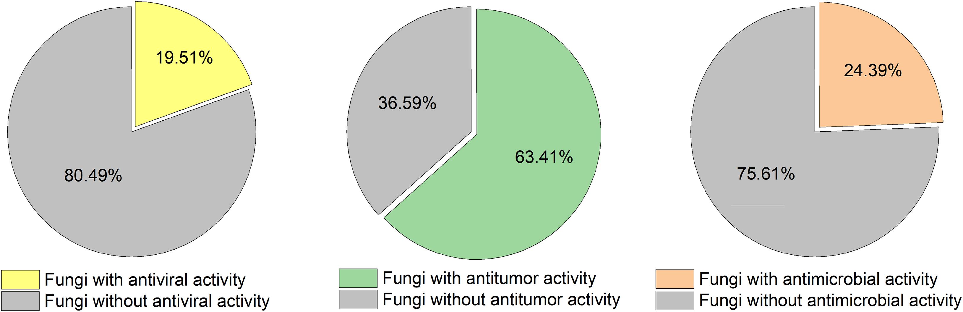
Figure 4. Percentage of bioactive acid-tolerant fungal strains isolated from Thai mangrove sediment.
All six acidophilic fungi were clustered in the branch of the genus Talaromyces. We summarized all the references on isolation and identification of Talaromyces since 2015 and found that no acidic culture conditions were involved in the isolation and culture process (including 34 new species of Talaromyces) (Adhikari et al., 2015; Visagie et al., 2015; Chen et al., 2016; Luo et al., 2016; Yilmaz et al., 2016a,b; Barbosa et al., 2018; Jiang et al., 2018; Rajeshkumar et al., 2019). However, in our experiments, all six acidophilic fungi isolates belonged to the genus Talaromyces. It was almost impossible to isolate these strains under neutral conditions. Although it was difficult to conclude whether the acidophilic fungi Talaromyces isolated by us were new species or not, these results revealed the acidophilic properties of the genus Talaromyces for the first time. This indicates that more samples of Talaromyces could be separated under acidic conditions.
The secondary metabolites of this genus have shown great potential in their chemical and bioactive diversity. Terpenoids, alkaloids, and polyketides with multiple bioactivities (including antibiotic, cytotoxic, and anti-HBV activity) have been reported as metabolites from Talaromyces (Hayashi et al., 2012; Kumar et al., 2013; Wu et al., 2015; Zhai et al., 2015; Fu et al., 2016; Guo et al., 2016; Xie et al., 2016; Zhao et al., 2016, 2019; Zhi et al., 2016; Cai et al., 2017; Küppers et al., 2017; Chen et al., 2018; Wang et al., 2018), with another review paper published in 2020 (Lan and Wu, 2020). Our investigation provides good indications for future studies on the metabolites of this genus, which require further exploration. The mangrove sediments in Thailand are the best source of acid-tolerant microbes for investigating the acidophilic properties and diverse metabolites of the genus Talaromyces.
Bioactive Diversity of Aciduric Fungi Derived From Thai Mangrove Sediment
Forty-one representative fungi were fermented in a PDB medium at pH 3.0 and/or pH 7.0 based on the results of the growth of the isolates in the acid resistance assessment (detailed in Supplementary Table 1). All of the EtOAc extracts were tested for antimicrobial activity against nine aquatic and eight human pathogenic microbes. Nearly a quarter of the samples showed inhibitory effects (Table 3). The fungal strain OUCMDZ-5130 displayed broad-spectrum inhibitory effects against C. albicans, C. glabrata, E. coli, V. alginolyticus, P. nigrifaciens, V. splendidus, S. aureus subsp. aureus Rosenbach, E. tarda, and B. cereus. The strains OUCMDZ-5081, 5210, 5233, and 5245 showed weak antibacterial activity against the aquatic pathogens P. nigrifaciens and V. alginolyticus, while the strains OUCMDZ-4993, 5020, and 5040 showed moderate antibacterial activity against the aquatic pathogen B. cereus. These fungi inhibited more aquatic pathogens than human pathogens.
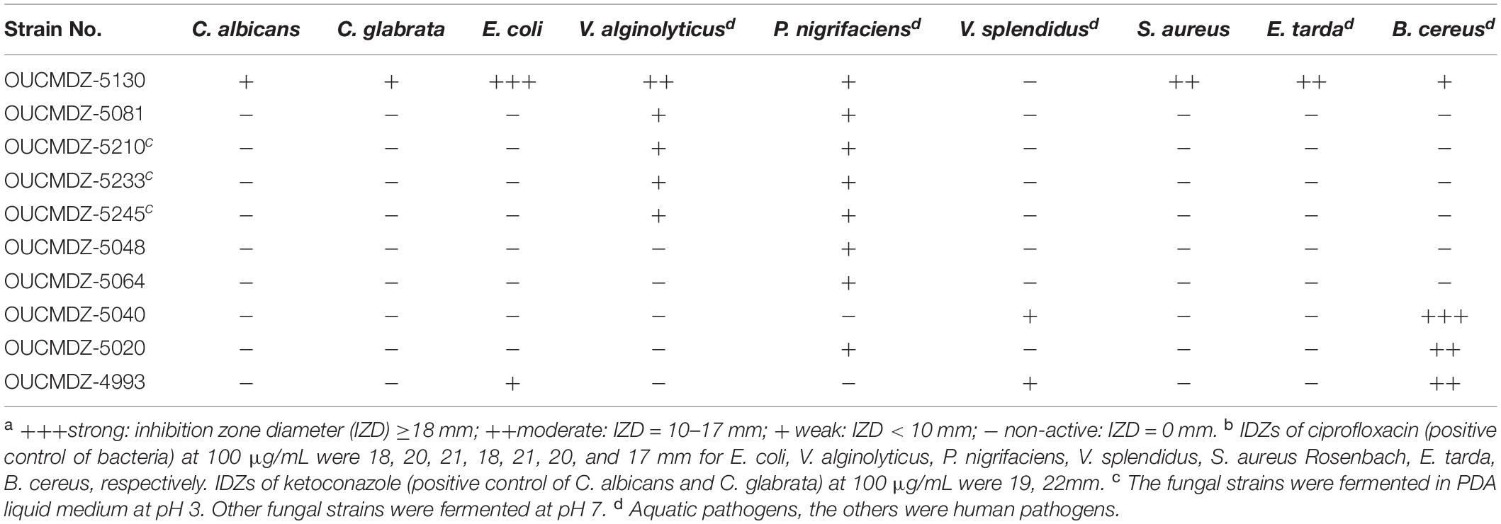
Table 3. The antimicrobial activity of acid-tolerant fungal EtOAc extracts at 1000 μg/mL a,b.
The extracts were also evaluated for cytotoxicity against A549 and MCF-7 tumor cell lines at a concentration of 100 μg/mL (Table 4). The positive control adriamycin (1 μM) showed 80 and 72% inhibition of A549 and MCF-7 cells, respectively. The strains, which produced extracts that showed more than 50% inhibition, were defined as active strains. The results showed that 26 of the fungal strains (63.4%) were active, 13 of which had been fermented at pH 3. Eight strains, OUCMDZ-5013, 5041, 5076, 5083, 5221, 5237, 5245, and 5274, showed more than 60% inhibition of both A549 and MCF-7 cell lines. The strains OUCMDZ-5048 and 5207 only showed inhibitory activity against A549 cells with 92 and 53% inhibition, respectively. The strains OUCMDZ-5053 and 5008 stimulated the proliferation of A549 cells and inhibited the growth of MCF-7 cells.
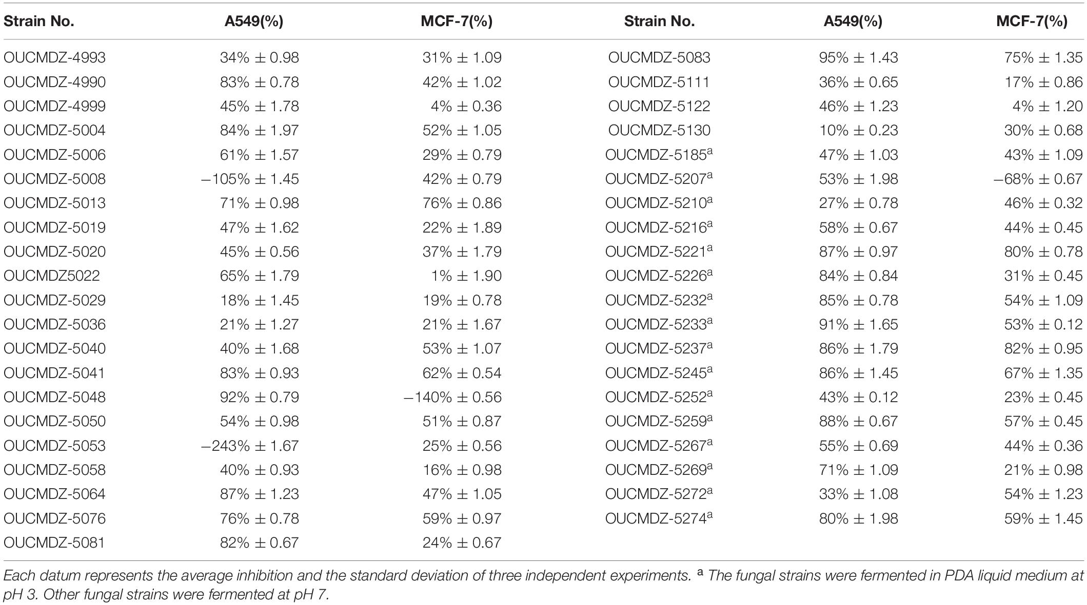
Table 4. Cytotoxicity of acid-tolerant fungal EtOAc extracts at 100 μg/mL.
Extracts of 41 fungi were evaluated for cytotoxicity against MDCK cell lines and the results showed that 28 strains (68.2%) were not cytotoxic at a concentration of 50 μg/mL. These non-cytotoxic strains were then selected to test for anti-H1N1 virus activity at the same concentration (Table 5). Eight strains, OUCMDZ-5008, 5013, 5022, 5029, 5040, 5048, 5083, and 5111, showed anti-H1N1 virus activity, among which OUCMDZ-5040 was the most active against the H1N1 virus with 42.1% inhibition, while the positive control, ribavirin, showed 47.0% inhibition at 50 μg/mL. Interestingly, all the strains with anti-H1N1 virus activity were fermented under neutral conditions (pH 7.0), which was different from the results relating to the anti-tumor activity (where 50.0% of the strains were fermented at pH 3.0).
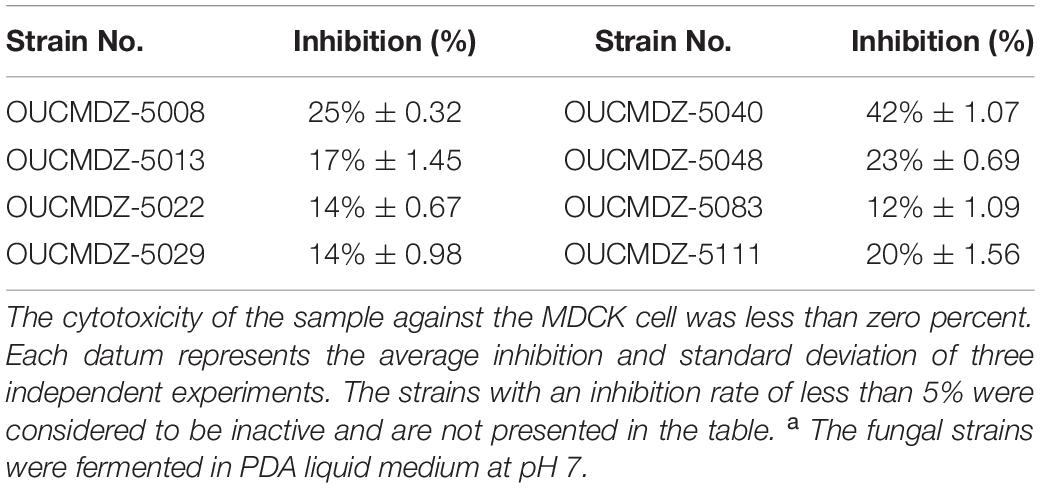
Table 5. Anti-H1N1 activities of acid-tolerant fungal EtOAc extracts at 50 μg/ml a.
The antimicrobial, cytotoxicity and antiviral activities of all the isolated aciduric and acidophilic fungal metabolites were evaluated. Nearly three-quarters of the fermentation extracts (26/41) showed cytotoxicity against A549 or/and MCF-7 cells, while less than a quarter of the isolates showed antimicrobial (10/41) or anti-H1N1 virus (8/41) activity (Figure 4). Most of the cytotoxic isolates were fermented under acidic conditions (pH 3), while all the isolates with anti-H1N1 virus activity were fermented under neutral conditions (pH 7.0). The metabolites of these acid-tolerant fungi did not show obvious antibacterial and antiviral activities but showed outstanding cytotoxic activities. These results suggest that an extremely acidic environment was not conducive to the growth of common microorganisms. However, these extreme conditions did not affect the survival of the acid-tolerant fungi, leading to the production of cytotoxic metabolites through population competition. These cytotoxic substances could potentially help mangrove plants to resist being eaten by other animals. More strains had inhibitory activity against aquatic pathogens than against human pathogens, which may be because the fungi exist in the rhizosphere environment of mangrove plants and protect the mangroves against pathogens.
The Metabolites From Penicillium sp. OUCMDZ-5207 at pH 3
The fungus strain OUCMDZ-5207 was identified as Penicillium according to the morphology and ITS sequences (Accession No. MK418548). No significant differences were observed in the growth or spore germination of OUCMDZ-5207 cultured at pH 3 and pH 7, but differences were found in the chemical diversity and the biomass under the acidic and neutral conditions (Figure 5). The EtOAc extracts fermented at pH 3 showed selective inhibitory activity against A549 cells with a 53% inhibition rate compared with a weak proliferative effect on MCF-7 cells. The metabolite yield at pH 3.0 (3.1240 g/L) far exceeded that at pH 7.0 (0.1236 g/L). Furthermore, compounds with retention times from 14 to 20 min were only produced at pH 3.0. Thus, strain OUCMDZ-5207 fermented at pH 3.0 was selected for chemical study, which resulted in the isolation and identification of three compounds that were only produced at pH 3.0, tetrahydroauroglaucin (1), flavoglaucin (2), and auroglaucin (3) (Figure 5; Miyake et al., 2009). Compounds 1–3 at a concentration of 10 μM showed cytotoxic activity against A549 and MCF-7 cells with 42 ± 0.96% and 53 ± 1.03%, 32 ± 0.45% and 27 ± 0.53%, and 79 ± 0.89% and 48 ± 1.09% inhibition, respectively. Compound 3 was the most active against A549 cells with an IC50 value of 5.67 μM (IC50 = 0.61 μM for the positive control, adriamycin). Compounds 1–3 have been previously reported to show good antimicrobial and antimalarial bioactivity, inhibition of LPS (Lipopolysaccharide)-induced NO release and opioid and cannabinoid receptor binding activity, and cytotoxicity against SF-268, MCF-7, NCI-H460, HeLa, and HepG-2 cells (Hamasaki et al., 1981; Miyake et al., 2009; Gao et al., 2011, 2012; Kim et al., 2014; Dzoyem et al., 2015; Liang et al., 2018; Elissawy et al., 2019).
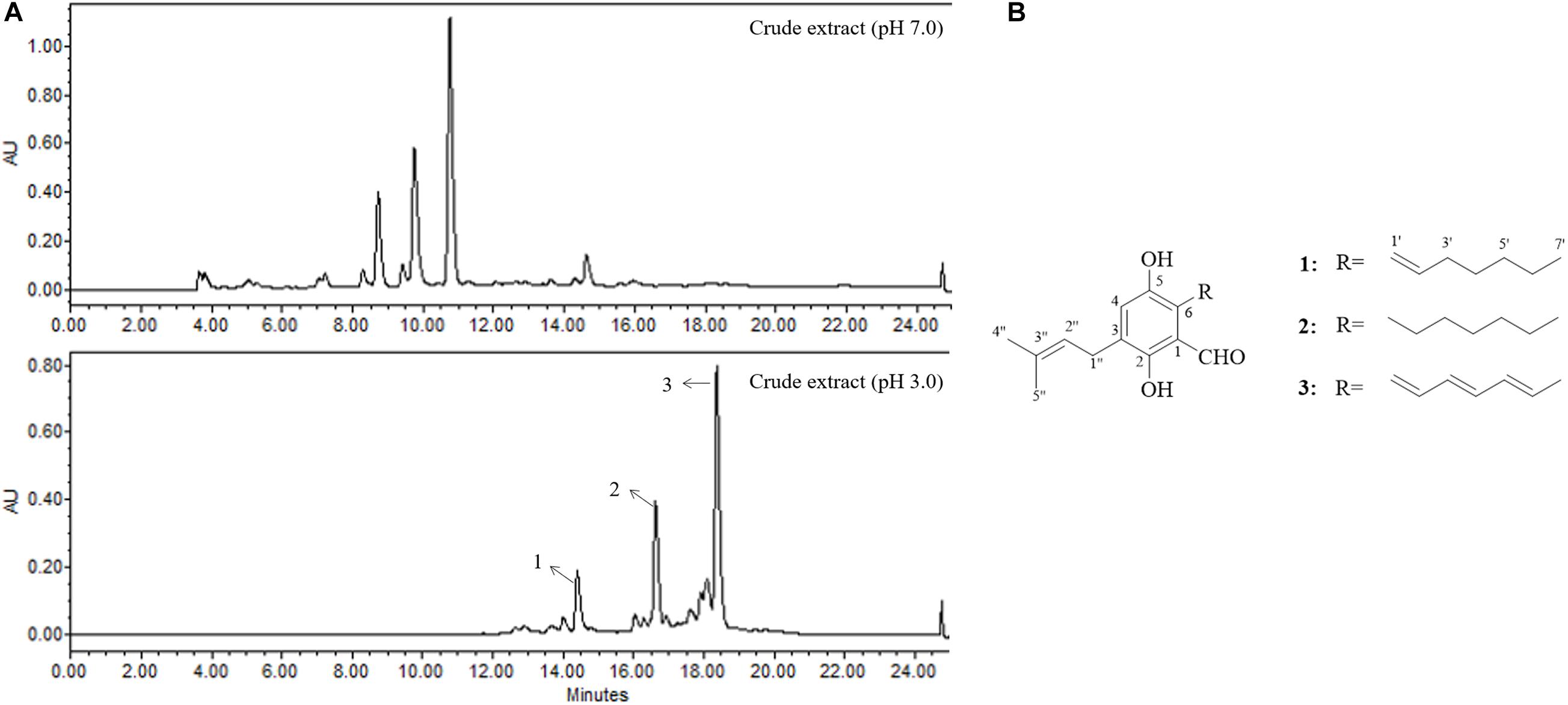
Figure 5. (A) The HPLC profile of secondary metabolites from OUCMDZ-5207 at pH 7.0 and pH 3.0 (HPLC gradient elution: 0–25 min, 10–100% CH3OH; UV wavelength: 300 nm; flow rate: 1 mL/min). (B) The chemical structures of compounds 1–3.
To the best of our knowledge, there have been no previous reports of compounds 1–3 outlining that they exhibit cytotoxicity against A549. In the present study, compound 3 demonstrated strong selective inhibition against A549. When the environmental pH changed, the fungi respond to the ambient pH and transmit pH signals to the nucleus through transcriptional regulators, thereby regulating a series of gene expressions including secondary metabolite gene clusters. It has also been reported that pH sensing in filamentous fungi relies mainly on the regulatory pathway PacC, a global regulator of fungi, which regulates the transcription of secondary metabolite-related genes, such as aflatoxin, cephalosporin, and penicillin (Brakhage, 2013). This is a complex regulation system and it was difficult to conclude if this regulation system was related to our experimental results. However, these results do suggest that acid stress can activate silent gene clusters to expand the diversity of secondary metabolites and this strategy will lead to potential medicinal discovery.
Conclusion
Our investigation indicated that mangroves in Thailand are rich in aciduric and acidophilic microbial resources. These acid-tolerant microorganisms are potential medicinal resources. Acid stress can activate silent gene clusters to produce secondary metabolites that cannot be obtained under neutral conditions, which could be a useful strategy for expanding the diversity of available metabolites for potential medicinal use.
Data Availability Statement
The datasets presented in this study can be found in online repositories. The names of the repository/repositories and accession number(s) can be found in Table 2.
Author Contributions
All authors listed have made a substantial, direct and intellectual contribution to the work, and approved it for publication.
Funding
This work was supported by grants from the National Natural Science Foundation of China (No. 81973198), the Key Research and Development Program of Shandong (No. 2018GSF118070), and the National Science and Technology Major Project for Significant New Drug Development (No. 2018ZX09735004).
Conflict of Interest
The authors declare that the research was conducted in the absence of any commercial or financial relationships that could be construed as a potential conflict of interest.
Supplementary Material
The Supplementary Material for this article can be found online at: https://www.frontiersin.org/articles/10.3389/fmicb.2020.609952/full#supplementary-material
References
Adhikari, M., Yadav, D. R., Kim, S., Um, Y. H., Kim, H. S., Lee, H. B., et al. (2015). Discovery of two new talaromyces species from crop field soil in korea. Mycobiology 43, 402–407. doi: 10.5941/MYCO.2015.43.4.402
Am-In, S., Limtong, S., Yongmanitchai, W., and Jindamorakot, S. (2011). Candida andamanensis sp. nov., Candida laemsonensis sp. nov. and Candida ranongensis sp. nov., anamorphic yeast species isolated from estuarine waters in a Thai mangrove forest. Int. J. Syst. Evol. Micr. 61, 454–461. doi: 10.1099/ijs.0.022038-0
Am-In, S., Yongmanitchai, W., and Limtong, S. (2008). Kluyveromyces siamensis sp. nov., an ascomycetous yeast isolated from water in a mangrove forest in Ranong province, Thailand. FEMS Yeast Res. 8, 823–828. doi: 10.1111/j.1567-1364.2008.00396.x
Atipan, S., Akio, T., Vorasan, S., and Suppasil, M. (2012). Mangrove sediment, a new source of potential biosurfactant-producing bacteria. Ann. Microbiol. 62, 1669–1679. doi: 10.1007/s13213-012-0424-9
Barbosa, R. N., Bezerra, J. D. P., Souza-Motta, C. M., Frisvad, J. C., Samson, R. A., Oliveira, N. T., et al. (2018). New Penicillium and Talaromyces species from honey, pollen and nests of stingless bees. Antonie Van Leeuwenhoek 111, 1883–1912. doi: 10.1007/s10482-018-1081-1
Brakhage, A. A. (2013). Regulation of fungal secondary metabolism. Nat. Rev. Microbiol. 11, 21–32. doi: 10.1038/nrmicro2916
Cai, R. L., Chen, S. H., Long, Y. H., Li, C. H., Huang, X. S., and She, Z. G. (2017). Depsidones from Talaromyces stipitatus SK-4, an endophytic fungus of the mangrove plant Acanthus ilicifolius. Phytochem. Lett. 20, 196–199. doi: 10.1016/j.phytol.2017.04.023
Carbone, I., and Kohn, L. M. (1999). A method for designing primer sets for speciation studies in filamentous ascomycetes. Mycologia 91, 553–556. doi: 10.1080/00275514.1999.12061051
Chen, A. J., Sun, B. D., Houbraken, J., Frisvad, J. C., Yilmaz, N., Zhou, Y. G., et al. (2016). New Talaromyces species from indoor environments in China. Stud. Mycol. 84, 119–144. doi: 10.1016/j.simyco.2016.11.003
Chen, S. H., Ding, M., Liu, W. Y., Huang, X. S., Liu, Z. M., Lu, Y. J., et al. (2018). Anti-inflammatory meroterpenoids from the mangrove endophytic fungus, Talaromyces amestolkiae, YX1. Phytochemistry 146, 8–15. doi: 10.1016/j.phytochem.2017.11.011
Deshmukh, R., Khardenavis, A. A., and Purohit, H. J. (2016). Diverse metabolic capacities of fungi for bioremediation. Indian J. Microbiol. 56, 247–264. doi: 10.1007/s12088-016-0584-6
Dzoyem, J. P., Lannang, A. M., Fouotsa, H., Mbazoa, C. D., Nkengfack, A. E., Sewald, N., et al. (2015). Anti-inflammatory activity of benzophenone and xanthone derivatives isolated from Garcinia (Clusiaceae) species. Phytochem. Lett. 14, 153–158. doi: 10.1016/j.phytol.2015.10.003
Elissawy, A. M., Ebada, S. S., Ashour, M. L., Ei-Neketi, M., Ebrahim, W., and Singab, A. B. (2019). New secondary metabolites from the mangrove-derived fungus Aspergillus sp. AV-2. Phytochem. Lett. 29, 1–5. doi: 10.1016/j.phytol.2018.10.014
Fan, Y. Q., Wang, Y., Fu, P., Arthit, C., Pawinee, P., and Zhu, W. M. (2018). Secopaxilline A, an indole-diterpenoid derivative from an aciduric Penicillium fungus, its identification and semisynthesis. Org. Chem. Front. 5, 2835–2839. doi: 10.1039/c8qo00756j
Fan, Y. Q., Wang, Y., Liu, P. P., Fu, P., Zhu, T. H., Wang, W., et al. (2013). Indole-diterpenoids with Anti-H1N1 activity from the aciduric fungus Penicillium camemberti OUCMDZ-1492. J. Nat. Prod. 76, 1328–1336. doi: 10.1021/np400304q
Fu, G. C., Yang, Z. D., Zhou, S. Y., Li, X. M., Yu, H. T., Yao, X. J., et al. (2016). Wortmannines A-C, three novel wortmannin derivatives with an unusual five-membered B ring from the endophytic fungus Talaromyces wortmannii LGT-4. Tetrahedron Lett. 57, 4608–4611. doi: 10.1016/j.tetlet.2016.09.004
Gao, J. T., León, F., Radwan, M. M., Dale, O. R., Husni, A. S., Manly, S. P., et al. (2011). Benzyl derivatives with in vitro binding affinity for human opioid and cannabinoid receptors from the fungus Eurotium repens. J. Nat. Prod. 74, 1636–1639. doi: 10.1021/np200147c
Gao, J. T., Radwan, M. M., León, F., Wang, X. N., Jacob, M. R., Tekwani, B. L., et al. (2012). Antimicrobial and antiprotozoal activities of secondary metabolites from the fungus Eurotium repens. Med. Chem. Res. 21, 3080–3086. doi: 10.1007/s00044-011-9798-7
Gardes, M., and Bruns, T. D. (1993). ITS primer with enhanced specificity for basidiomycetes application to the identification of mycorrizae and rusts. Mol. Ecol. 2, 113–118. doi: 10.1111/j.1365-294X.1993.tb00005.x
Glass, N. L., and Donaldson, G. C. (1995). Development of primer sets designed for use with the PCR to amplify conserved genes from filamentous ascomycetes. Appl. Environ. Microb. 61, 1323–1330. doi: 10.1128/aem.61.4.1323-1330.1995
Guo, J., Ran, H. M., Zeng, J., Liu, D., and Xin, Z. H. (2016). Tafuketide, a phylogeny-guided discovery of a new polyketide from Talaromyces funiculosus Salicorn 58. Appl. Microbiol. Biotechnol. 100, 5323–5338. doi: 10.1007/s00253-016-7311-4
Hamasaki, T., Kimura, Y., Hatsuda, Y., and Nagao, M. (1981). Structure of a new metabolite, dihydroauroglaucin, produced by Aspergillus chevalieri. Agric. Biol. Chem. 45, 313–314. doi: 10.1080/00021369.1981.10864508
Hayashi, H., Oka, Y., Kai, K., and Akiyama, K. (2012). New chrodrimanin congeners, chrodrimanins D-H, from YO-2 of Talaromyces sp. Biosci. Biotechnol. Biochem. 76, 1765–1768. doi: 10.1271/bbb.120365
H-Kittikun, A., Biscola, V., El-Ghaish, S., Jaffrès, E., Dousset, X., Pillot, G., et al. (2015). Bacteriocin-producing Enterococcus faecalis KT2W2G isolated from mangrove forests in southern Thailand: purification, characterization and safety evaluation. Food Control 54, 126–134. doi: 10.1016/j.foodcont.2014.12.037
Hujslová, M., Bystrianský, L., Benada, O., and Gryndler, M. (2019). Fungi, a neglected component of acidophilic biofilms: do they have a potential for biotechnology? Extremophiles 23, 267–275. doi: 10.1007/s00792-019-01085-9
Hwanhlem, N., Chobert, J. M., and H-Kittikun, A. (2014). Bacteriocin-producing lactic acid bacteria isolated from mangrove forests in southern Thailand as potential bio-control agents in food: isolation, screening and optimization. Food Control 41, 202–211. doi: 10.1016/j.foodcont.2014.01.021
Jiang, X., Yu, Z., Ruan, Y., and Wang, L. (2018). Three new species of Talaromyces sect. Talaromyces discovered from soil in China. Sci. Rep. 8:4932. doi: 10.1038/s41598-018-23370-x
Jin, Y. Z., Qin, S. D., Gao, H., Zhu, G. L., Wang, W., Zhu, W. M., et al. (2018). An anti-HBV anthraquinone from aciduric fungus Pennicillium sp. OUCMDZ-4736 under low pH stress. Extremophiles 22, 39–45. doi: 10.1007/s00792-017-0975-6
Johnson, D. B. (1998). Biodiversity and ecology of acidophilic microorganisms. FEMS Microbiol. Ecol. 27, 307–317. doi: 10.1111/j.1574-6941.1998.tb00547.x
Kim, K. S., Cui, X., Lee, D. S., Ko, W., Sohn, J. H., Yim, J. H., et al. (2014). Inhibitory effects of benzaldehyde derivatives from the marine fungus Eurotium sp. SF-5989 on inflammatory mediators via the induction of heme oxygenase-1 in lipopolysaccharide-stimulated RAW264.7 macrophages. Int. J. Mol. Sci. 15, 23749–23765. doi: 10.3390/ijms151223749
Kumar, M., Qadri, M., Sharma, P. R., Kumar, A., Andotra, S. S., Kaur, T., et al. (2013). Tubulin inhibitors from an endophytic fungus isolated from Cedrus deodara. J. Nat. Prod. 76, 194–199. doi: 10.1021/np3006666
Küppers, L., Ebrahim, W., El-Neketi, M., Özkaya, F. C., Mándi, A., Kurtán, T., et al. (2017). Lactones from the sponge-derived fungus Talaromyces rugulosus. Mar. Drugs 15:359. doi: 10.3390/md15110359
Lan, D., and Wu, B. (2020). Chemistry and bioactivities of secondary metabolites from the genus Talaromyces. Chem. Biodivers. 17, e2000229. doi: 10.1002/cbdv.202000229
Li, D. L., Li, X. M., Li, T. G., Dang, H. Y., Proksch, P., and Wang, B. G. (2008). Benzaldehyde derivatives from Eurotium rubrum, an endophytic fungus derived from the mangrove plant Hibiscus tiliaceus. Chem. Pharm. Bull. 56, 1282–1285. doi: 10.1248/cpb.56.1282
Li, D. L., Li, X. M., Proksch, P., and Wang, B. G. (2010). 7-O-Methylvariecolortide A, a new spirocyclic diketopiperazine alkaloid from a marine mangrove derived endophytic fungus, Eurotium rubrum. Nat. Prod. Commun. 5, 1583–1586. doi: 10.1177/1934578X1000501014
Liang, T. M., Fang, Y. W., Zheng, J. Y., and Shao, C. L. (2018). Secondary metabolites isolated from the gorgonian-derived fungus Aspergillus ruber and their antiviral activity. Chem. Nat. Compd. 54, 559–561. doi: 10.1007/s10600-018-2406-z
Limtong, S., Yongmanitchai, W., Kawasaki, H., and Seki, T. (2007). Candida thaimueangensis sp. nov., an anamorphic yeast species from estuarine water in a mangrove forest in Thailand. Int. J. Syst. Evol. Micr. 57, 650–653. doi: 10.1099/ijs.0.64698-0
Limtong, S., Yongmanitchai, W., Kawasaki, H., and Seki, T. (2008). Candida phangngensis sp. nov., an anamorphic yeast species in the Yarrowia clade, isolated from water in mangrove forests in Phang-Nga Province, Thailand. Int. J. Syst. Evol. Micr. 58, 515–519. doi: 10.1099/ijs.0.65506-0
Lin, Y. W., Wang, L. P., Wang, Y., Wang, W., Hao, J. J., and Zhu, W. M. (2015). Bioactive natural products of Aspergillus sp. OUCMDZ-1914, an aciduric fungus from the mangrove soils. Chin. J. Org. Chem. 35, 1955–1960. doi: 10.6023/cjoc201504007
Luo, Y., Lu, X., Bi, W., Liu, F., and Gao, W. (2016). Talaromyces rubrifaciens, a new species discovered from heating, ventilation and air conditioning systems in China. Mycologia 108, 773–779. doi: 10.3852/15-233
Miyake, Y., Ito, C., Itoigawa, M., and Osawa, T. (2009). Antioxidants produced by Eurotium herbariorum of filamentous fungi used for the manufacture of karebushi, dried bonito (Katsuobushi). Biosci. Biotechnol. Biochem. 73, 1323–1327. doi: 10.1271/bbb.80887
Mosmann, T. (1983). Rapid colorimetric assay for cellular growth and survival: application to proliferation and cytotoxicity assays. J. Immunol. Methods 65, 55–63. doi: 10.1016/0022-1759(83)90303-4
O’ Donnell, K., Nirenberg, H. I., Aoki, T., and Cigelnik, E. (2000). A Multigene phylogeny of the Gibberella fujikuroi species complex: detection of additional phylogenetically distinct species. Mycoscience 41, 61–78. doi: 10.1007/bf02464387
Peng, X. P., Wang, Y., Kunlai, S., Liu, P. P., Yin, X., and Zhu, W. M. (2011a). Cerebrosides and 2-Pyridone Alkaloids from the Halotolerant Fungus Penicillium chrysogenum grown in a hypersaline medium. J. Nat. Prod. 74, 1298–1302. doi: 10.1021/np1008976
Peng, X. P., Wang, Y., Liu, P. P., Hong, K., Chen, H., Yin, X., et al. (2011b). Aromatic compounds from the halotolerant fungal strain of Wallemia sebi PXP-89 in a hypersaline medium. Arch. Pharm. Res. 34, 907–912. doi: 10.1007/s12272-011-0607-0
Peng, X. P., Wang, Y., Zhu, G. L., and Zhu, W. M. (2018). Fatty acid derivatives from the halotolerant fungus Cladosporium cladosporioides. Magn. Reson. Chem. 56, 18–24. doi: 10.1002/mrc.4659
Qin, S. D., Wang, Y., Wang, W., and Zhu, W. M. (2016). Anti-H1N1-virus secondary metabolites from mangrove-derived aciduric fungus Penicillium sp. OUCMDZ-4736. Chin. J. Mar. Drugs 15, 21–28.
Rajeshkumar, K. C., Yilmaz, N., Marathe, S. D., and Seifert, K. A. (2019). Morphology and multigene phylogeny of Talaromyces amyrossmaniae, a new synnematous species belonging to the section Trachyspermi from India. Mycokeys 45, 41–56. doi: 10.3897/mycokeys.45.32549
Songsumanus, A., Tanasupawat, S., Igarashi, Y., and Kudo, T. (2013). Micromonospora maritima sp. nov., isolated from mangrove soil. Int. J. Syst. Evol. Micr. 63, 554–559. doi: 10.1099/ijs.0.039180-0
Stierle, A. A., Stierle, D. B., Decato, D., Priestley, N. D., Alverson, J. B., Hoody, J., et al. (2017). The berkeleylactones, antibiotic macrolides from fungal coculture. J. Nat. Prod. 80, 1150–1160. doi: 10.1021/acs.jnatprod.7b00133
Tamura, G., Suzuki, S., Takatsuki, A., Ando, K., and Arima, K. (1968). Ascochlorin, a new antibiotic, found by paper-disk agar-diffusion method. I. Isolation, biological and chemical properties of ascochlorin. (Studies on antiviral and antitumor antibiotics. I). J. Antibiot. 21, 539–544. doi: 10.7164/antibiotics.21.539
Visagie, C. M., Yilmaz, N., Frisvad, J. C., Houbraken, J., Seifert, K. A., Samson, R. A., et al. (2015). Five new Talaromyces species with ampulliform-like phialides and globose rough walled conidia resembling T. Verruculosus. Mycoscience 56, 486–502. doi: 10.1016/j.myc.2015.02.005
Wanapaisan, P., Laothamteep, N., Vejarano, F., Chakraborty, J., Shintani, M., Muangchinda, C., et al. (2018). Synergistic degradation of pyrene by five culturable bacteria in a mangrove sediment-derived bacterial consortium. J. Hazard. Mater. 342, 561–570. doi: 10.1016/j.jhazmat.2017.08.062
Wang, H., Lu, Z., Qu, H., Liu, P., Miao, C., Zhu, T., et al. (2012). Antimicrobial aflatoxins from the marine-derived fungus Aspergillus flavus 092008. Arch. Pharm. Res. 35, 1387–1392. doi: 10.1007/s12272-012-0808-1
Wang, H., Wang, Y., Liu, P. P., Wang, W., Fan, Y. Q., and Zhu, W. M. (2013). Purpurides B and C, two new sesquiterpene esters from the aciduric fungus Penicillium purpurogenum JS03-21. Chem. Biodivers. 10, 1185–1192. doi: 10.1002/cbdv.201200175
Wang, H., Wang, Y., Wang, W., Fu, P., Liu, P. P., and Zhu, W. M. (2011a). Anti-influenza virus polyketides from the acid-tolerant fungus Penicillium purpurogenum JS03-21. J. Nat. Prod. 74, 2014–2018. doi: 10.1021/np2004769
Wang, Y., Lu, Z. Y., Sun, K. L., and Zhu, W. M. (2011b). Effects of high salt stress on secondary metabolites production in the marine-derived fungus Spicaria elegans. Mar. Drugs 9, 535–542. doi: 10.3390/md9040535
Wang, Y., Zheng, J. K., Liu, P. P., Wang, W., and Zhu, W. M. (2011c). Three new compounds from Aspergillus terreus PT06-2 grown in a high salt medium. Mar. Drugs 9, 1368–1378. doi: 10.3390/md9081368
Wang, M. M., Wang, S. Y., Wang, W., Wang, Y., Wang, H., and Zhu, W. M. (2017). Inhibition effects of novel polyketide compound PPQ-B against influenza A virus replication by interfering with the cellular EGFR pathway. Antivir. Res. 143, 74–84. doi: 10.1016/j.antiviral.2017.04.007
Wang, W. J., Wan, X., Liu, J., Wang, J. P., Zhu, H. C., Chen, C. C., et al. (2018). Two new terpenoids from Talaromyces purpurogenus. Mar. Drugs 16:150. doi: 10.3390/md16050150
Wang, W. L., Wang, Y., Tao, H. W. T., Peng, X. P., Liu, P. P., and Zhu, W. M. (2009). Cerebrosides of the halotolerant fungus Alternaria raphanin isolated from a sea salt field. J. Nat. Prod. 72, 1695–1698. doi: 10.1021/np9002299
Wang, Y., Fan, Y. Q., Wang, Y. N., and Zhu, W. M. (2017). Research progress of natural products from aciduric microbes. Chin. J. Antibiot. 42, 255–260.
White, T. J., Bruns, T., Lee, S., and Taylor, J. (1990). “Amplification and direct sequencing of fungal ribosomal RNA genes for phylogenetics. PCR protocols: a guide to methods and applications,” in PCR Protocols. A Guide to Methods and Applications, eds M. A. Innis, D. H. Gelfand, J. J. Sninsky, and T. J. White (San Diego: Academic Press), 315–322.
Wilson, Z. E., and Brimble, M. A. (2009). Molecules derived from the extremes of life. Nat. Prod. Rep. 26, 44–71. doi: 10.1039/b800164m
Wongwongsee, W., Chareanpat, P., and Pinyakong, O. (2013). Abilities and genes for PAH biodegradation of bacteria isolated from mangrove sediments from the central of Thailand. Mar. Pollut. Bull. 74, 95–104. doi: 10.1016/j.marpolbul.2013.07.025
Wu, B., Ohlendorf, B., Oesker, V., Wiese, J., Malien, S., Schmaljohann, R., et al. (2015). Acetylcholinesterase inhibitors from a marine fungus Talaromyces sp. Strain LF458. Mar. Biotechnol. 17, 110–119. doi: 10.1007/s10126-014-9599-3
Xie, X. S., Fang, X. W., Huang, R., Zhang, S. P., Wei, H. X., and Wu, S. H. (2016). A new dimeric anthraquinone from endophytic Talaromyces sp. YE3016. Nat. Prod. Res. 30, 1706–1711. doi: 10.1080/14786419.2015.1136888
Yan, H., Gao, S., Li, C., Li, X., and Wang, B. (2010). Chemical constituents of a marine-derived endophytic fungus Penicillium commune G2M. Molecules 15, 3270–3275. doi: 10.3390/molecules15053270
Yan, H. J., Li, X. M., Li, C. S., and Wang, B. G. (2012). Alkaloid and anthraquinone derivatives produced by the marine-derived endophytic fungus Eurotium rubrum. Helv. Chim. Acta. 95, 163–168. doi: 10.1002/hlca.201100255
Yilmaz, N., López-Quintero, C. A., Vasco-Palacios, A. M., Frisvad, J. C., Theelen, B., Boekhout, T., et al. (2016a). Four novel Talaromyces species isolated from leaf litter from Colombian Amazon rain forests. Mycol. Prog. 15, 1041–1056. doi: 10.1007/s11557-016-1227-3
Yilmaz, N., Visagie, C. M., Frisvad, J. C., Houbraken, J., Jacobs, K., and Samson, R. A. (2016b). Taxonomic re-evaluation of species in Talaromyces section Islandici, using a polyphasic approach. Persoonia 36, 37–56. doi: 10.3767/003158516X688270
Zhai, M. M., Niu, H. T., Li, J., Xiao, H., Shi, Y. P., Di, D. L., et al. (2015). Talaromycolides A-C, novel phenyl-substituted phthalides isolated from the green chinese onion-derived fungus Talaromyces pinophilus AF-02. J. Agr. Food. Chem. 63, 9558–9564. doi: 10.1021/acs.jafc.5b04296
Zhao, J. W., Yang, Z. D., Zhou, S. Y., Yang, L. J., Sun, J. H., Yao, X. J., et al. (2019). Wortmannine F and G, two new pyranones from Talaromyces wortmannii LGT-4, the endophytic fungus of Tripterygium wilfordii. Phytochem. Lett. 29, 115–118. doi: 10.1016/j.phytol.2018.11.023
Zhao, Q. H., Yang, Z. D., Shu, Z. M., Wang, Y. G., and Wang, M. G. (2016). Secondary metabolites and biological activities of Talaromyces sp. LGT-2, an endophytic fungus from Tripterygium wilfordii. Iran J. Pharm. Res. 15, 453–457. doi: 10.22037/IJPR.2016.1873
Zheng, J. K., Wang, Y., Wang, J. F., Liu, P. P., Li, J., and Zhu, W. M. (2013). Antimicrobial ergosteroids and pyrrole derivatives from halotolerant Aspergillus flocculosus PT05-1 cultured in a hypersaline medium. Extremophiles 17, 963–971. doi: 10.1007/s00792-013-0578-9
Zheng, J. K., Xu, Z. H., Wang, Y., Hong, K., Liu, P. P., and Zhu, W. M. (2010). Cyclic Tripeptides from the halotolerant fungus Aspergillus sclerotiorum PT06-1. J. Nat. Prod. 73, 1133–1137. doi: 10.1021/np100198h
Zheng, J. K., Zhu, H. J., Hong, K., Wang, Y., Liu, P. P., Wang, X., et al. (2009). Novel cyclic hexapeptides from marine-derived fungus, Aspergillus sclerotiorum PT06-1. Org. Lett. 11, 5262–5265. doi: 10.1021/ol902197z
Keywords: acid-tolerant fungi, Thai mangrove sediment, genetic relationship, secondary metabolites, bioactive diversity
Citation: Gao H, Wang Y, Luo Q, Yang L, He X, Wu J, Kachanuban K, Wilaipun P, Zhu W and Wang Y (2021) Bioactive Metabolites From Acid-Tolerant Fungi in a Thai Mangrove Sediment. Front. Microbiol. 11:609952. doi: 10.3389/fmicb.2020.609952
Received: 24 September 2020; Accepted: 23 December 2020;
Published: 22 January 2021.
Edited by:
Runying Zeng, Third Institute of Oceanography, State Oceanic Administration, ChinaReviewed by:
Lea Atanasova, University of Natural Resources and Life Sciences, Vienna, AustriaSamantha Chandranath Karunarathna, Kunming Institute of Botany, China
Yonghong Liu, Chinese Academy of Sciences, China
Copyright © 2021 Gao, Wang, Luo, Yang, He, Wu, Kachanuban, Wilaipun, Zhu and Wang. This is an open-access article distributed under the terms of the Creative Commons Attribution License (CC BY). The use, distribution or reproduction in other forums is permitted, provided the original author(s) and the copyright owner(s) are credited and that the original publication in this journal is cited, in accordance with accepted academic practice. No use, distribution or reproduction is permitted which does not comply with these terms.
*Correspondence: Weiming Zhu, weimingzhu@ouc.edu.cn; Yi Wang, wangyi0213@ouc.edu.cn
†These authors have contributed equally to this work