 Robin A. Welsh
 Nianbin Song2†
 Scheherazade Sadegh-Nasseri
- 1Graduate Program in Immunology, Johns Hopkins School of Medicine, Baltimore, MD, United States
- 2Department of Biology, Krieger School of Arts & Sciences, Johns Hopkins University, Baltimore, MD, United States
- 3Department of Pathology, Johns Hopkins School of Medicine, Baltimore, MD, United States
Dendritic cells are the antigen presenting cells that process antigens effectively and prime the immune system, a characteristic that have gained them the spotlights in recent years. B cell antigen presentation, although less prominent, deserves equal attention. B cells select antigen experienced CD4 T cells to become memory and initiate an orchestrated genetic program that maintains memory CD4 T cells for life of the individual. Over years of research, we have demonstrated that low levels of antigens captured by B cells during the resolution of an infection render antigen experienced CD4 T cells into a quiescent/resting state. Our studies suggest that in the absence of antigen, the resting state associated with low-energy utilization and proliferation can help memory CD4 T cells to survive nearly throughout the lifetime of mice. In this review we would discuss the primary findings from our lab as well as others that highlight our understanding of B cell antigen presentation and the contributions of the MHC Class II accessory molecules to this outcome. We propose that the quiescence induced by the low levels of antigen presentation might be a mechanism necessary to regulate long-term survival of CD4 memory T cells and to prevent cross-reactivity to autoantigens, hence autoimmunity.
Dendritic Cells as Antigen Presenting Cells to Initiate a Primary Response
Initiation of an adaptive immune response begins with naïve T cells being activated by antigens presented on dendritic cells (DCs), a highly specialized professional antigen presenting cell (APC) (1, 2). As a frontline defender, DCs are key APCs bridging the gap between innate and adaptive immunity. Located primarily in peripheral tissues, immature DCs are well known for their ability to recognize and capture invading pathogens mainly through phagocytosis and micropinocytosis. Uptake of antigen (Ag) is closely followed by upregulation of MHC Class I, Class II and co-stimulatory molecules on the surface of DCs, as they lose the ability to perform macropinocytosis (3, 4). Mature DCs then migrate to draining lymph nodes where they present pathogen-derived epitopes to naïve CD4 and CD8 T cells (5). One unique characteristic of DCs is their ability to uptake Ag via phagocytosis and cross-present it on MHC Class I molecules. This makes DCs a perfect primary antigen presenter for initiation of an immune response (2). Moreover, it has also been noted that activation of immature DCs by various Toll-like receptor ligands (TLR3 and TLR9) transiently increases antigen specific micropinocytosis (6), which likely increases the ability of DCs to capture Ag within an inflammatory context. To date, research into what contributions DCs make to memory development has been limited and mainly focuses on memory CD8 T cells development (7–11). Use of Batf3 knock-out (KO) mice, which lack CD8a DCs responsible for cross-presentation, found no impact on primary CD4 T cell responses but drastically impaired CD8 responses (12). Likewise, work using Toxoplasma gondii showed a crucial role for CD4 T cells in protecting Batf3 KO mice from succumbing to T. gondii infection (13). Yet none of these studies using DC KO mice investigated a role for DCs in memory CD4 T cell development. Data from Dalai et al., however suggests that loss of DCs does not likely impact the formation of memory CD4 T cells as removal of CD11c+ DCs did not affect the development of quiescent memory CD4 T cells (14).
B Cells as APCs in Secondary Responses
B cells are another major professional APCs, which unlike DCs, take up antigens specifically by B cell receptor (BCR) (1). Upon interaction with a cognate Ag, the BCR-Ag complex would be internalized and shuttled to the specialized MHC class II enriched compartments (MIIC) for processing and presentation to the Ag-specific CD4 T cells (15). These CD4 T-B interactions provide essential activation signals to B cells for affinity maturation and differentiation into memory B, or antibody-secreting plasma B cells (16). The memory B cells generated from this T-B interaction have been found to also be important for CD4 T cell memory responses (17).
How B Cells and DCs Impact Memory T Cell Development
It is generally accepted that memory T cells differentiate after exposure to Ag followed by multiple rounds of proliferation (18–20). While characterization of memory T cells has been explored intensely, the onset of differentiation of Ag-experienced T cells into memory, and how APCs influence this process is less appreciated. Especially that in rare publications, it has been proposed that CD8 memory T cells may be generated upon asymmetric cell division, which precludes the need for interaction with antigen presenting cells (14, 21, 22). On another line of studies, CD8 memory T cell development and homeostasis has been reported to be mediated by IL-15Rα expressed by DCs and Macrophages (23, 24). It is also found that long-lasting CD8 memory can be achieved in the absence of CD4 T cells or B cells (25).
For CD4 Memory T development, however, TCR-pMHC interaction appears to drive CD4 Memory T development (14, 26–30). In this regard, Williams et al. found that lower levels of LCMV antigen density led to high functional avidity CD4 T memory differentiation, while higher levels of LCMV antigen density promoted both high avidity and low avidity CD4 T cells expansion (28). However, the authors did not explore whether DCs or B cells were the APCs to drive such differentiation. Studies addressing contributions of B cells to activation of naïve CD4 T cells has been inconclusive (31). Conversely, several investigations have reported that B cells play a critical role in regulating CD4 memory T development and differentiation (14, 17, 26, 30, 32–37). It is noteworthy that among these studies, both Chowdhury (17) and Misumi (35) found that absence of antigen specific B cells either from SCID mice without B cells or treatment of anti-CD20 mAb did not impact the priming of CD4 T cells in viral infection but impaired the development and effector function of memory CD4 T cells. By virtue of having antigen specific B Cell Receptors, B cells can recognize and internalize specific antigens, process, and present them to cognate CD4 T cells (15). As such, B cell antigen presentation adds a new and exciting dimension to our current knowledge.
The first clear demonstration that B cells play a role in memory CD4 T cell generation/differentiation came from Bradley and colleagues who reported B cell knockout mice did not develop memory CD4 T cells (32). Further studies have shown that loss of B cells adversely affects development of Tuberculosis (TB)-specific CD4 memory precursor effector cells (MPECs) in TB vaccinated B cell deficient mice (36). Because of the ability of B cells to produce antibodies that bind to Ag, it has been postulated that contribution of B cells to CD4 memory T cell development might be linked to Ag-Ab complexes. However, when this issue was specifically addressed by Whitmire et al., T cell responses to lymphocytic choriomeningitis virus (LCMV) infection, the team found that in contrast to B cell-deficient mice, membrane Ig expressing Tg mice retained functional Th cell memory, indicating that B cells selectively preserve CD4 T cell memory independently of immune complex formation (33).
To directly test if B cells were important for the development of CD4 T cell memory, Dalai et al. tested the specific interactions of various APCs with Ag experienced CD4 T cells (14). Using an ex vivo anergy assay, the group showed that only B cells, but not DCs, induced a resting state in Ag experienced CD4 T cells. Further in vivo characterization using an adoptive cell transfer assay further confirmed the ex vivo observations. Previous findings had demonstrated that sub-optimal levels of agonist peptides had induced a resting state in T cells in vitro, and in vivo (34, 38–44). Thus, the above observations that B cells, but not DCs, pulsed with low doses of Ag induced resting memory CD4 T cells confirmed prior findings that B cells are indispensable for memory CD4 T cell development/differentiation. In agreement with the above findings, B cell deficient mice did not develop quiescent CD4 memory T cells. However, when B cells were transferred to the B cell deficient mice, hyporesponsive CD4 memory T cells were developed. Importantly, B2 (B220+CD43+) follicular B cells, which have diverse BCR were identified as the cells that rendered CD4 memory T cells hyporesponsive (14). These finding were later supported by Keck et al, who found that B cells were required for both optimal expansion and T-bet expression in response to weak TCR stimulation and optimal generation of CD4 T memory (30).
Contribution of Ag Density Presented by Follicular B Cells to CD4 Memory T Cell Induction/Differentiation
Building upon those initial findings, Dalai et al. tested the effects of B cell presentation of peptide-MHC (pMHC) density on the induction of quiescent memory CD4 T cells. They used a clever strategy by recovering B cells from mice at various timepoints post immunization and transferring them into recipient mice harboring CD4 T memory precursor cells at 4-day intervals (26). This staggered timeframe allowed Dalai et al. to correlate the amount of pMHC presented by the B cells to the time post immunization; earlier time points displayed more pMHC, and later time points fewer pMHC. Interestingly, the group found that only B cells harvested between day 16-20 post OVA immunization induced resting hyporesponsive CD4 memory T cells. These findings supported the idea that CD4 memory T cells are signaled to a resting state by the presentation of a subthreshold numbers of pMHC. These conclusions were further expanded to HEL-specific B cells (45) HEL-specific B cells when used for induction of quiescence/resting state of Ag experienced T cells were more efficient in capturing the Ag and induced quiescence in Ag experienced CD4 T cells at much later time points, i.e., 41-48 days vs 16-20 days post immunization by non-specific B cells. In those experiments B cells immunized with protein antigens were transferred to mice that carried primed T cells at 4-day intervals. The rationale was to find out when during an immune response B cell presentation of pMHC reaches to the levels necessary for the induction of quiescence naturally, in vivo. It was quite gratifying to see that HEL-specific B cells had captured far more antigen so that the required densities of pMHC for inducing quiescence had reached 20 plus days later than the polyclonal B cells (26). Altogether, Dalai et al. established that: (1) B cells are the APCs responsible for rendering CD4 memory T cells the quiescent, and (2) low levels of pMHC presentation are the main driving force that signal CD4 T cells to enter a resting state (26).
More recently, we have explored how this state of anergy impacts both the longevity and function of CD4 T memory cells. Song et al. investigated gene expression dynamics in CD4 T memory cells at different stages post immunization representing activated, early memory, late memory, and long-term memory stages (46). OVA-specific DO11.10 T cells were adoptively transferred into naïve mice before infecting them with Vaccinia-OVA virus, followed by harvesting the CD44hiDO11.1pos T cells at different time points post immunization and subjecting their mRNA for gene expression analyses. Through this approach, the group was able to illustrate the gene expression dynamics occurring during CD4 T memory development up to almost 1 year. In agreement with findings of others (47–51), authors found that the OVA-specific CD4 memory T cells adopted a resting phenotype. Furthermore, the memory phenotype associated with multiple genetic programs regulating cellular proliferation, DNA repair, prevention of apoptosis, glucose, and lipid metabolism (Figure 1). Specifically, most genes regulating cellular proliferation and DNA repair response were found to be associated with p53 pathways, which highlights the importance of limiting cell proliferation and promoting DNA repair in long-lived CD4 Memory T cells. Also, of note was that like CD8 Memory T cells, genes regulating lipid metabolism were upregulated indicating that long-lived CD4 Memory T cells may also rely on lipid metabolism. However, unlike CD8 memory, the genes regulating lipid metabolism in CD4 T memory were found to be centered on regulating cellular cholesterol and ceramide levels, which could be related to the T cell signaling and prevention of apoptosis. Altogether, these programs play important roles in CD4 Memory T development and maintenance.
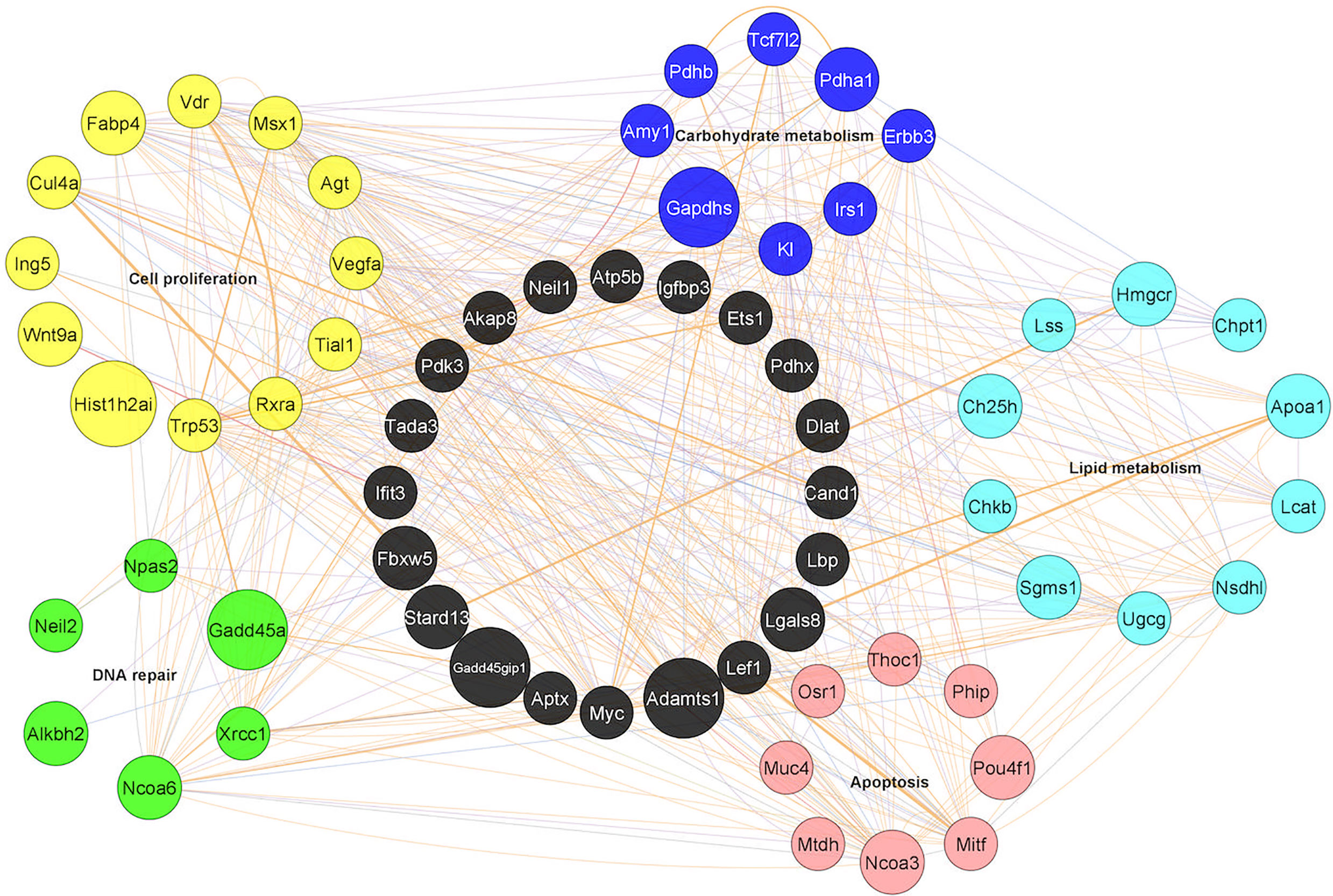
Figure 1 Gene networks in long-lived CD4 memory T cells. Five different gene programs were identified as dynamically regulated during memory CD4 T cell differentiation. The genes shown were from the long-lived memory CD4 cells 10.5 months post immunization as compared to naïve controls and are marked in different colors: Yellow: cell proliferation; Green: DNA repair; Red: Apoptosis; Light blue: Lipid metabolism; Dark blue: Carbohydrate metabolism; Black: Not identified in the five gene programs but served as connecting genes. Each line represents an interaction/co-expression of genes as identified by literature report.
The above genetic studies also revealed upregulated levels of CD99, CCR10 and Itga3 as potentially new surface markers for long-lived CD4 memory T cells. Importantly, the high expression levels of these new CD4 memory markers at the protein level were confirmed to hold true across different animal models and antigens. For example, CD99hi resting human CD4 T cells from flu vaccinated donors had much better proliferation responses than the CD99lo CD4 subsets to in vitro challenges, indicating that the gene expression programs found in murine CD4 memory T cells could also be applicable to human CD4 memory T cells. Overall speaking, this work indicated not only that the resting state of CD4 memory T cells was mediated by multiple genes and could be part of the reason for CD4 memory longevity, but also the surprising findings that the murine CD4 memory differentiation is regulated by genetic programs that evolve upwards of 6 months to fully appear.
Contribution of Class II Accessory Molecules in CD4 Memory Formation
The finding that proper development of CD4 T memory cells relies on quantitative differences in presentation of immunodominant epitopes by B cells, brings the focus to the potential roles that accessory molecules in antigen processing play in the selection of epitopes for binding to MHC Class II. It is demonstrated that as the main Class II peptide-editor, HLA-DM (human DM; murine H2-M) contributes to the selection of immunodominant epitopes by generating higher quantities of those epitopes (52–55). HLA-DO (human DO; murine H2-O), is a second accessory molecule, which requires DM for its expression; DO is mainly expressed in thymic epithelium and B cells (54–56). Both DM and DO contribute to T cell immunity in a significant way, because lymphocytes usually respond to a small portion of the potential determinants on a protein antigen, defined as ‘immunodominant’ (57). Immunodominant epitopes are the essential targets of the immune response against infectious diseases, cancer, autoimmune diseases, and allergy. Hence, deserve the attention devoted to the understanding of epitope selection and immunodominance. To better understand how each accessory molecule impacts immunodominant epitope selection, we must discuss each molecule individually.
Mechanism of DM in Finding the Immunodominant Determinants During Antigen Processing
It has been well established that the MHC II groove is flexible and requires a bound peptide to maintain its shape. Without a peptide, the MHC II groove would close and becomes inefficient in binding peptides (58–60). Thus, newly synthesized MHC II molecules bind to a domain of the Class II invariant chain (CLIP) that serves two functions; a) protects the groove from binding to peptide in the ER (61), and b) acts as a place-keeper, while another domain of Ii guides the complex to the specialized vesicular compartments filled with pathogen-derived antigenic peptides, MIIC. Within MIIC, DM is necessary to first dissociate CLIP to form a peptide-receptive conformation that can quickly scan unfolded exogenous proteins to find its suitable determinant (62). DM does this job by effectively dissociating any peptide sequences that do not fill in the pockets of the MHC II groove. Only when a sequence of antigenic determinant that would fit in the MHC II groove leading to formation of a compact folded conformation, the complex becomes resistant to DM-mediated dissociation (DM-resistant). Next, the proteases would trim the MHC II bound determinant. The proteases also cut the antigenic determinants that do not fit the groove, hence are susceptible to DM-mediated dissociation (DM-sensitive) and are dislodged by DM (63–71). The solution of the crystal structure of the DM/DR complex (72) using DR1/peptide complexes that enforced an open DR1 groove, revealed that DM would bind the P1 pocket of HLA-DR molecules tightly if empty, and would remain bound until a P1 filling peptide would bind the groove and induce closing of the groove, and displacing DM (72–74). The above findings were complemented by the measured thermodynamics of peptide binding to DR1, indicating that a greater entropic penalty, versus a smaller penalty, was associated with structural rigidity rather than with the flexibility of the pMHC complexes (75). These findings suggested that an overall dynamic MHC II conformation in addition to P1 pocket occupancy, determines susceptibility to DM-mediated peptide exchange and provides a molecular mechanism for DM to efficiently target poorly fitting pMHC II complexes and editing them for more stable ones. Hence, in addition to the removal of CLIP, DM helps in shaping epitope selection and immunodominance by producing a higher abundance of those determinants (62).
Different Models on How DO Fine-Tunes Antigenic Epitope Selection
DO also contributes to the selection of immunodominant epitopes, although understanding the contributions of DO to epitope selection has proven to be highly challenging (54–56, 76). In brief, our knowledge about DO can be distilled into two working hypotheses: (1) DO binds to DM to inhibit its activity, mainly removal of the CLIP peptide and, (2) DO differentially affects presentation of structurally diverse peptides and acts as a second accessory molecule working together with DM in fine tuning MHC II repertoire selection. Data in support of the former hypothesis mainly comes from studying over-expression of DO genes in cell lines, or dendritic cells (77, 78); Welsh, 2019 #13} and the recent mutagenesis and structural studies of DM/DO interactions (79, 80). The 3D structure of DM/DO showed that DO binds to DM at the same interface with which DM interacts with DR1 (74). Studies supporting the latter hypothesis came from biochemical (81) and biophysical studies demonstrating that DO only affected association kinetics of certain peptides to DR but, had no effect on the dissociation kinetics of any tested peptide/DR1 complexes (76, 82). The effects of DO on association kinetics directly correlated with peptide sensitivity to DM-mediated dissociation. DO reduced binding of peptides that formed DM-sensitive complexes with DR and enhanced the binding of peptides that formed DM-resistant complexes. In a nutshell, it was clearly shown that; i) DO works directly on DR1, and not by regulating the effect of DM, ii) DO can only bind the peptide-receptive MHC Class II, and iii) that this peptide-receptive conformation is generated by DM. Hence, authors proposed that DM and DO cooperate for a more effective epitope selection. Thus, in one model, DO would reduce presentation of immunodominant epitopes, whereas in the other, DO would increase the abundance of immunodominant epitopes.
Speculations for Future Research
The question of the potential contributions of DO and DM to memory CD4 T cell development is of most interest and is discussed below. A few characteristics of DO hint to its possible link to CD4 memory differentiation. First, DO is mainly expressed in B cells (81, 83, 84) and it enhances the presentation of immunodominant epitopes (56, 76). Next, it has been documented that successful entry of B cells into the germinal center (GC) requires high expression levels of pMHC (85–89). B cells enter GC and interact with CD4 T cells in search of proper signaling for affinity maturation. It is conceivable that CD4 T cells also receive signals from GC B cells for their own differentiation into resting memory T cells. One might say if high levels of pMHC equip B cells for entry into GC, how could B cells signal T cells to differentiate into resting memory, as this process requires suboptimal densities of pMHC presentation. An answer worth considering is that once B cells enter GC, their expression levels of DO and DM decreases, leading to a reduced level of pMHC II expression (90–92). As such, those GC B cells can interact with Ag-specific CD4 T cells in the Light Zone (LZ), selecting them to become memory precursor cells. In support of this argument, in an elegant study, Kim et al. have documented that memory CD4 T cells bear high affinity TCR for pMHC II (27), hence memory CD4 T cells are selected based on TCR affinity. One may predict that alterations in this controlled entry into the GC reaction could lead to faulty CD4 T cell memory development and possibly the development of increased autoreactivity.
Since biology tends to repeat itself, it would be interesting to compare the effects of pMHC numbers on APCs and their effects on CD8 memory T cell development. While as far as we know no studies has made such data available, in an exciting new study authors reported that in the absence of B cells CD8 T Cell memory formation was compromised, while CD8 effector function was enhanced. One might speculate that since CD4 T cells are essential for CD8 memory T cell development (93), perhaps their contributions to CD8 memory is mediated indirectly via CD4 memory T cells.
Future experimental evidence is needed to clarify the proposed relationship of these MHC II accessory molecules to the development and maintenance of CD4 memory T cells, and hopefully this review would prompt new research on the qualitative and quantitative antigen presentation on CD8 memory T cell development.
Author Contributions
All authors listed have made a substantial, direct, and intellectual contribution to the work and approved it for publication.
Funding
Supported by grants from NIAID, R01AI063764, R21AI101987, and R01AI120634, to SS-N.
Conflict of Interest
The authors declare that the research was conducted in the absence of any commercial or financial relationships that could be construed as a potential conflict of interest.
References
2. Itano AA, McSorley SJ, Reinhardt RL, Ehst BD, Ingulli E, Rudensky AY, et al. Distinct Dendritic Cell Populations Sequentially Present Antigen to CD4 T Cells and Stimulate Different Aspects of Cell-Mediated Immunity. Immunity (2003) 19:47–57. doi: 10.1016/S1074-7613(03)00175-4
3. Sallusto F, Cella M, Danieli C, Lanzavecchia A. Dendritic Cells Use Macropinocytosis and the Mannose Receptor to Concentrate Macromolecules in the Major Histocompatibility Complex Class II Compartment: Downregulation by Cytokines and Bacterial Products. J Exp Med (1995) 182:389–400. doi: 10.1084/jem.182.2.389
4. Lutz MB, Schuler G. Immature, Semi-Mature and Fully Mature Dendritic Cells: Which Signals Induce Tolerance or Immunity? Trends Immunol (2002) 23:445–9. doi: 10.1016/S1471-4906(02)02281-0
5. Chudnovskiy A, Pasqual G, Victora GD. Studying Interactions Between Dendritic Cells and T Cells In Vivo. Curr Opin Immunol (2019) 58:24–30. doi: 10.1016/j.coi.2019.02.002
6. West MA, Wallin RP, Matthews SP, Svensson HG, Zaru R, Ljunggren HG, et al. Enhanced Dendritic Cell Antigen Capture Via Toll-Like Receptor-Induced Actin Remodeling. Science (2004) 305:1153–7. doi: 10.1126/science.1099153
7. Zammit DJ, Lefrancois L. Dendritic Cell-T Cell Interactions in the Generation and Maintenance of CD8 T Cell Memory. Microbes Infect (2006) 8:1108–15. doi: 10.1016/j.micinf.2005.12.002
8. Zammit DJ, Cauley LS, Pham QM, Lefrancois L. Dendritic Cells Maximize the Memory CD8 T Cell Response to Infection. Immunity (2005) 22:561–70. doi: 10.1016/j.immuni.2005.03.005
9. Enamorado M, Khouili SC, Iborra S, Sancho D. Genealogy, Dendritic Cell Priming, and Differentiation of Tissue-Resident Memory Cd8(+) T Cells. Front Immunol (2018) 9:1751. doi: 10.3389/fimmu.2018.01751
10. Yu B, Zhang K, Milner JJ, Toma C, Chen R, Scott-Browne JP, et al. Epigenetic Landscapes Reveal Transcription Factors That Regulate CD8(+) T Cell Differentiation. Nat Immunol (2017) 18:573–82. doi: 10.1038/ni.3706
11. Johnnidis JB, Muroyama Y, Ngiow SF, Chen Z, Manne S, Cai Z, et al. Inhibitory Signaling Sustains a Distinct Early Memory CD8(+) T Cell Precursor That Is Resistant to DNA Damage. Sci Immunol (2021) 6:1–16. doi: 10.1126/sciimmunol.abe3702
12. Hildner K, Edelson BT, Purtha WE, Diamond M, Matsushita H, Kohyama M, et al. Batf3 Deficiency Reveals a Critical Role for CD8alpha+ Dendritic Cells in Cytotoxic T Cell Immunity. Science (2008) 322:1097–100. doi: 10.1126/science.1164206
13. Tussiwand R, Behnke MS, Kretzer NM, Grajales-Reyes GE, Murphy TL, Schreiber RD, et al. An Important Role for CD4(+) T Cells in Adaptive Immunity to Toxoplasma Gondii in Mice Lacking the Transcription Factor Batf3. mSphere (2020) 5:1–11. doi: 10.1128/mSphere.00634-20
14. Dalai SK, Mirshahidi S, Morrot A, Zavala F, Sadegh-Nasseri S. Anergy in Memory CD4+ T Cells is Induced by B Cells. J Immunol (2008) 181:3221–31. doi: 10.4049/jimmunol.181.5.3221
15. Adler LN, Jiang W, Bhamidipati K, Millican M, Macaubas C, Hung SC, et al. The Other Function: Class Ii-Restricted Antigen Presentation by B Cells. Front Immunol (2017) 8:319. doi: 10.3389/fimmu.2017.00319
16. Tarlinton D. B Cells Still Front and Centre in Immunology. Nat Rev Immunol (2019) 19:85–6. doi: 10.1038/s41577-018-0107-2
17. Chowdhury MG, Maeda K, Yasutomo K, Maekawa Y, Furukawa A, Azuma M, et al. Antigen-Specific B Cells Are Required for the Secondary Response of T Cells But Not for Their Priming. Eur J Immunol (1996) 26:1628–33. doi: 10.1002/eji.1830260733
18. Moulton VR, Farber DL. Committed to Memory: Lineage Choices for Activated T Cells. Trends Immunol (2006) 27:261–7. doi: 10.1016/j.it.2006.04.006
19. van Stipdonk MJ, Lemmens EE, Schoenberger SP. Naive CTLs Require a Single Brief Period of Antigenic Stimulation for Clonal Expansion and Differentiation. Nat Immunol (2001) 2:423–9. doi: 10.1038/87730
20. Hataye J, Moon JJ, Khoruts A, Reilly C, Jenkins MK. Naive and Memory CD4+ T Cell Survival Controlled by Clonal Abundance. Science (2006) 312:114–6. doi: 10.1126/science.1124228
21. Chang JT, Palanivel VR, Kinjyo I, Schambach F, Intlekofer AM, Banerjee A, et al. Asymmetric T Lymphocyte Division in the Initiation of Adaptive Immune Responses. Science (2007) 315:1687–91. doi: 10.1126/science.1139393
22. Pollizzi KN, Sun IH, Patel CH, Lo YC, Oh MH, Waickman AT, et al. Asymmetric Inheritance of mTORC1 Kinase Activity During Division Dictates CD8(+) T Cell Differentiation. Nat Immunol (2016) 17:704–11. doi: 10.1038/ni.3438
23. Stonier SW, Ma LJ, Castillo EF, Schluns KS. Dendritic Cells Drive Memory CD8 T-Cell Homeostasis Via IL-15 Transpresentation. Blood (2008) 112:4546–54. doi: 10.1182/blood-2008-05-156307
24. Mortier E, Advincula R, Kim L, Chmura S, Barrera J, Reizis B, et al. Macrophage- and Dendritic-Cell-Derived Interleukin-15 Receptor Alpha Supports Homeostasis of Distinct CD8+ T Cell Subsets. Immunity (2009) 31:811–22. doi: 10.1016/j.immuni.2009.09.017
25. Di Rosa F, Matzinger P. Long-Lasting CD8 T Cell Memory in the Absence of CD4 T Cells or B Cells. J Exp Med (1996) 183:2153–63.
26. Dalai SK, Khoruzhenko S, Drake CG, Jie CC, Sadegh-Nasseri S. Resolution o Infection Promotes a State of Dormancy and Long Survival of CD4 Memory T Cells. Immunol Cell Biol (2011) 89(8):870–81. doi: 10.1038/icb.2011.2
27. Kim C, Wilson T, Fischer KF, Williams MA. Sustained Interactions Between T Cell Receptors and Antigens Promote the Differentiation of CD4(+) Memory T Cells. Immunity (2013) 39:508–20. doi: 10.1016/j.immuni.2013.08.033
28. Williams MA, Ravkov EV, Bevan MJ. Rapid Culling of the CD4+ T Cell Repertoire in the Transition From Effector to Memory. Immunity (2008) 28:533–45. doi: 10.1016/j.immuni.2008.02.014
29. Rees W, Bender J, Teague TK, Kedl RM, Crawford F, Marrack P, et al. An Inverse Relationship Between T Cell Receptor Affinity and Antigen Dose During CD4(+) T Cell Responses In Vivo and In Vitro. Proc Natl Acad Sci USA (1999) 96:9781–6. doi: 10.1073/pnas.96.17.9781
30. Keck S, Schmaler M, Ganter S, Wyss L, Oberle S, Huseby ES, et al. Antigen Affinity and Antigen Dose Exert Distinct Influences on CD4 T-Cell Differentiation. Proc Natl Acad Sci USA (2014) 111:14852–7. doi: 10.1073/pnas.1403271111
31. Chen X, Jensen PE. MHC Class II Antigen Presentation and Immunological Abnormalities Due to Deficiency of MHC Class II and its Associated Genes. Exp Mol Pathol (2008) 85:40–4. doi: 10.1016/j.yexmp.2008.03.011
32. Linton PJ, Harbertson J, Bradley LM. A Critical Role for B Cells in the Development of Memory CD4 Cells. J Immunol (2000) 165:5558–65. doi: 10.4049/jimmunol.165.10.5558
33. Whitmire JK, Asano MS, Kaech SM, Sarkar S, Hannum LG, Shlomchik MJ, et al. Requirement of B Cells for Generating CD4+ T Cell Memory. J Immunol (2009) 182:1868–76. doi: 10.4049/jimmunol.0802501
34. Sadegh-Nasseri S, Dalai SK, Korb Ferris LC, Mirshahidi S. Suboptimal Engagement of the T-cell Receptor by a Variety of peptide-MHC Ligands Triggers T-cell Anergy. Immunology (2010) 129:1–7. doi: 10.1111/j.1365-2567.2009.03206.x
35. Misumi I, Whitmire JK. B Cell Depletion Curtails CD4+ T Cell Memory and Reduces Protection Against Disseminating Virus Infection. J Immunol (2014) 192:1597–608. doi: 10.4049/jimmunol.1302661
36. Dubois Cauwelaert N, Baldwin SL, Orr MT, Desbien AL, Gage E, Hofmeyer KA, et al. Antigen Presentation by B Cells Guides Programing of Memory CD4(+) T-Cell Responses to a TLR4-agonist Containing Vaccine in Mice. Eur J Immunol (2016) 46:2719–29. doi: 10.1002/eji.201646399
37. Pepper M, Jenkins MK. Origins of CD4(+) Effector and Central Memory T Cells. Nat Immunol (2011) 12:467–71. doi: 10.1038/ni.2038
38. Korb LC, Mirshahidi S, Ramyar K, Sadighi Akha AA, Sadegh-Nasseri S. Induction of T Cell Anergy by Low Numbers of Agonist Ligands. J Immunol (1999) 162:6401–9.
39. Ryan KR, Evavold BD. Persistence of Peptide-Induced CD4+ T Cell Anergy In Vitro. J Exp Med (1998) 187:89–96. doi: 10.1084/jem.187.1.89
40. Mirshahidi S, Ferris LC, Sadegh-Nasseri S. The Magnitude of TCR Engagement is a Critical Predictor of T Cell Anergy or Activation. J Immunol (2004) 172:5346–55. doi: 10.4049/jimmunol.172.9.5346
41. Ryan KR, McNeil LK, Dao C, Jensen PE, Evavold BD. Modification of Peptide Interaction With MHC Creates TCR Partial Agonists. Cell Immunol (2004) 227:70–8. doi: 10.1016/j.cellimm.2004.01.003
42. Ford ML, Evavold BD. Regulation of Polyclonal T Cell Responses by an MHC Anchor-Substituted Variant of Myelin Oligodendrocyte Glycoprotein 35-55. J Immunol (2003) 171:1247–54. doi: 10.4049/jimmunol.171.3.1247
43. Robertson JM, Evavold BD. Cutting Edge: Dueling TCRs: Peptide Antagonism of CD4+ T Cells With Dual Antigen Specificities. J Immunol (1999) 163:1750–4.
44. Mirshahidi S, Huang CT, Sadegh-Nasseri S. Anergy in Peripheral Memory CD4(+) T Cells Induced by Low Avidity Engagement of T Cell Receptor. J Exp Med (2001) 194:719–31. doi: 10.1084/jem.194.6.719
45. Garside P, Ingulli E, Merica RR, Johnson JG, Noelle RJ, Jenkins MK. Visualization of Specific B and T Lymphocyte Interactions in the Lymph Node. Science (1998) 281:96–9. doi: 10.1126/science.281.5373.96
46. Song N, Sengupta S, Khoruzhenko S, Welsh RA, Kim A, Kumar MR, et al. Multiple Genetic Programs Contribute to CD4 T Cell Memory Differentiation and Longevity by Maintaining T Cell Quiescence. Cell Immunol (2020) 357:104210. doi: 10.1016/j.cellimm.2020.104210
47. Tokoyoda K, Zehentmeier S, Hegazy AN, Albrecht I, Grun JR, Lohning M, et al. Professional Memory CD4+ T Lymphocytes Preferentially Reside and Rest in the Bone Marrow. Immunity (2009) 30:721–30. doi: 10.1016/j.immuni.2009.03.015
48. Prlic M, Bevan MJ. Immunology: A Metabolic Switch to Memory. Nature (2009) 460:41–2. doi: 10.1038/460041a
49. Pearce EL, Walsh MC, Cejas PJ, Harms GM, Shen H, Wang LS, et al. Enhancing CD8 T-Cell Memory by Modulating Fatty Acid Metabolism. Nature (2009) 460:103–7. doi: 10.1038/nature08097
50. Araki K, Turner AP, Shaffer VO, Gangappa S, Keller SA, Bachmann MF, et al. mTOR Regulates Memory CD8 T-Cell Differentiation. Nature (2009) 460:108–12. doi: 10.1038/nature08155
51. Waickman AT, Powell JD. mTOR, Metabolism, and the Regulation of T-cell Differentiation and Function. Immunol Rev (2012) 249:43–58. doi: 10.1111/j.1600-065X.2012.01152.x
52. Kim A, Sadegh-Nasseri S. Determinants of Immunodominance for CD4 T Cells. Curr Opin Immunol (2015) 34:9–15. doi: 10.1016/j.coi.2014.12.005
53. Kim C, Jay DC, Williams MA. Dynamic Functional Modulation of CD4+ T Cell Recall Responses is Dependent on the Inflammatory Environment of the Secondary Stimulus. PLoS Pathog (2014) 10:e1004137. doi: 10.1371/journal.ppat.1004137
54. Welsh RA, Song N, Foss CA, Boronina T, Cole RN, Sadegh-Nasseri S. Lack of the MHC Class II Chaperone H2-O Causes Susceptibility to Autoimmune Diseases. PLoS Biol (2020) 18:e3000590. doi: 10.1371/journal.pbio.3000590
55. Welsh R, Song N, Sadegh-Nasseri S. What to do With HLA-DO/H-2O Two Decades Later? Immunogenetics (2019) 71:189–96. doi: 10.1007/s00251-018-01097-3
56. Welsh RA, Sadegh-Nasseri S. The Love and Hate Relationship of HLA-DM/DO in the Selection of Immunodominant Epitopes. Curr Opin Immunol (2020) 64:117–23. doi: 10.1016/j.coi.2020.05.007
57. Sercarz EE, Lehmann PV, Ametani A, Benichou G, Miller A, Moudgil K. Dominance and Crypticity of T Cell Antigenic Determinants. Annu Rev Immunol (1993) 11:729–66. doi: 10.1146/annurev.iy.11.040193.003501
58. Natarajan SK, Assadi M, Sadegh-Nasseri S. Stable Peptide Binding to MHC Class II Molecule is Rapid and is Determined by a Receptive Conformation Shaped by Prior Association With Low Affinity Peptides. J Immunol (1999) 162:4030–6.
59. Sadegh-Nasseri S, Stern LJ, Wiley DC, Germain RN. MHC Class II Function Preserved by Low-Affinity Peptide Interactions Preceding Stable Binding. Nature (1994) 370:647–50. doi: 10.1038/370647a0
60. Rabinowitz JD, Vrljic M, Kasson PM, Liang MN, Busch R, Boniface JJ, et al. Formation of a Highly Peptide-Receptive State of Class II Mhc. Immunity (1998) 9:699–709. doi: 10.1016/S1074-7613(00)80667-6
61. Roche PA, Cresswell P. Invariant Chain Association With HLA-DR Molecules Inhibits Immunogenic Peptide Binding. Nature (1990) 345:615–8. doi: 10.1038/345615a0
62. Kim A, Hartman IZ, Poore B, Boronina T, Cole RN, Song N, et al. Divergent Paths for the Selection of Immunodominant Epitopes From Distinct Antigenic Sources. Nat Commun (2014) 5:5369. doi: 10.1038/ncomms6369
63. Chou CL, Sadegh-Nasseri S. Hla-DM Recognizes the Flexible Conformation of Major Histocompatibility Complex Class II. J Exp Med (2000) 192:1697–706. doi: 10.1084/jem.192.12.1697
64. Zarutskie JA, Busch R, Zavala-Ruiz Z, Rushe M, Mellins ED, Stern LJ. The Kinetic Basis of Peptide Exchange Catalysis by HLA-DM. Proc Natl Acad Sci USA (2001) 98:12450–5. doi: 10.1073/pnas.211439398
65. Belmares MP, Busch R, Mellins ED, McConnell HM. Formation of Two Peptide/MHC II Isomers is Catalyzed Differentially by HLA-DM. Biochemistry (2003) 42:838–47. doi: 10.1021/bi020466p
66. Pashine A, Busch R, Belmares MP, Munning JN, Doebele RC, Buckingham M, et al. Interaction of HLA-DR With an Acidic Face of HLA-DM Disrupts Sequence-Dependent Interactions With Peptides. Immunity (2003) 19:183–92. doi: 10.1016/S1074-7613(03)00200-0
67. Stratikos E, Wiley DC, Stern LJ. Enhanced Catalytic Action of HLA-DM on the Exchange of Peptides Lacking Backbone Hydrogen Bonds Between Their N-terminal Region and the MHC Class II Alpha-Chain. J Immunol (2004) 172:1109–17. doi: 10.4049/jimmunol.172.2.1109
68. Nicholson MJ, Moradi B, Seth NP, Xing X, Cuny GD, Stein RL, et al. Small Molecules That Enhance the Catalytic Efficiency of HLA-DM. J Immunol (2006) 176:4208–20. doi: 10.4049/jimmunol.176.7.4208
69. Chou CL, Mirshahidi S, Su KW, Kim A, Narayan K, Khoruzhenko S, et al. Short Peptide Sequences Mimic HLA-DM Functions. Mol Immunol (2008) 45:1935–43. doi: 10.1016/j.molimm.2007.10.033
70. Narayan K, Su KW, Chou CL, Khoruzhenko S, Sadegh-Nasseri S. Hla-DM Mediates Peptide Exchange by Interacting Transiently and Repeatedly With HLA-DR1. Mol Immunol (2009) 46:3157–62. doi: 10.1016/j.molimm.2009.07.001
71. Zhou Z, Callaway KA, Weber DA, Jensen PE. Cutting Edge: HLA-DM Functions Through a Mechanism That Does Not Require Specific Conserved Hydrogen Bonds in Class II MHC-Peptide Complexes. J Immunol (2009) 183:4187–91. doi: 10.4049/jimmunol.0901663
72. Anders AK, Call MJ, Schulze MS, Fowler KD, Schubert DA, Seth NP, et al. Hla-DM Captures Partially Empty HLA-DR Molecules for Catalyzed Removal of Peptide. Nat Immunol (2011) 12:54–61. doi: 10.1038/ni.1967
73. Painter CA, Negroni MP, Kellersberger KA, Zavala-Ruiz Z, Evans JE, Stern LJ. Conformational Lability in the Class II MHC 310 Helix and Adjacent Extended Strand Dictate HLA-DM Susceptibility and Peptide Exchange. Proc Natl Acad Sci U S A (2011) 108:19329–34. doi: 10.1073/pnas.1108074108
74. Pos W, Sethi DK, Call MJ, Schulze MS, Anders AK, Pyrdol J, et al. Crystal Structure of the HLA-DM-HLA-DR1 Complex Defines Mechanisms for Rapid Peptide Selection. Cell (2012) 151:1557–68. doi: 10.1016/j.cell.2012.11.025
75. Ferrante A, Templeton M, Hoffman M, Castellini MJ. The Thermodynamic Mechanism of Peptide-MHC Class II Complex Formation is a Determinant of Susceptibility to HLA-DM. J Immunol (2015) 195:1251–61. doi: 10.4049/jimmunol.1402367
76. Poluektov YO, Kim A, Hartman IZ, Sadegh-Nasseri S. Hla-DO as the Optimizer of Epitope Selection for MHC Class II Antigen Presentation. PLoS One (2013) 8:e71228. doi: 10.1371/journal.pone.0071228
77. Denzin LK, Cresswell P. Sibling Rivalry: Competition Between MHC Class II Family Members Inhibits Immunity. Nat Struct Mol Biol (2013) 20:7–10. doi: 10.1038/nsmb.2484
78. Nanaware PP, Jurewicz MM, Leszyk J, Shaffer SA, Stern LJ. Hla-DO Modulates the Diversity of the MHC-II Self-Peptidome. Mol Cell Proteomics (2018) 18(3):490–503 doi: 10.1074/mcp.RA118.000956
79. Yoon T, Macmillan H, Mortimer SE, Jiang W, Rinderknecht CH, Stern LJ, et al. Mapping the HLA-DO/HLA-DM Complex by FRET and Mutagenesis. Proc Natl Acad Sci USA (2012) 109:11276–81. doi: 10.1073/pnas.1113966109
80. Guce AI, Mortimer SE, Yoon T, Painter CA, Jiang W, Mellins ED, et al. Hla-DO Acts as a Substrate Mimic to Inhibit HLA-DM by a Competitive Mechanism. Nat Struct Mol Biol (2013) 20:90–8. doi: 10.1038/nsmb.2460
81. Kropshofer H, Vogt AB, Thery C, Armandola EA, Li BC, Moldenhauer G, et al. A Role for HLA-DO as a Co-Chaperone of HLA-DM in Peptide Loading of MHC Class II Molecules. EMBO J (1998) 17:2971–81. doi: 10.1093/emboj/17.11.2971
82. Poluektov YO, Kim A, Sadegh-Nasseri S. Hla-DO and Its Role in MHC Class II Antigen Presentation. Front Immunol (2013) 4:260. doi: 10.3389/fimmu.2013.00260
83. Liljedahl M, Winqvist O, Surh CD, Wong P, Ngo K, Teyton L, et al. Altered Antigen Presentation in Mice Lacking H2-O. Immunity (1998) 8:233–43. doi: 10.1016/S1074-7613(00)80475-6
84. Roucard C, Thomas C, Pasquier MA, Trowsdale J, Sotto JJ, Neefjes J, et al. In Vivo and In Vitro Modulation of HLA-DM and HLA-DO is Induced by B Lymphocyte Activation. J Immunol (2001) 167:6849–58. doi: 10.4049/jimmunol.167.12.6849
85. Yeh CH, Nojima T, Kuraoka M, Kelsoe G. Germinal Center Entry Not Selection of B Cells is Controlled by peptide-MHCII Complex Density. Nat Commun (2018) 9:928. doi: 10.1038/s41467-018-03382-x
86. Mesin L, Schiepers A, Ersching J, Barbulescu A, Cavazzoni CB, Angelini A, et al. Restricted Clonality and Limited Germinal Center Reentry Characterize Memory B Cell Reactivation by Boosting. Cell (2020) 180:92–106.e11. doi: 10.1016/j.cell.2019.11.032
87. Bannard O, McGowan SJ, Ersching J, Ishido S, Victora GD, Shin JS, et al. Ubiquitin-Mediated Fluctuations in MHC Class II Facilitate Efficient Germinal Center B Cell Responses. J Exp Med (2016) 213:993–1009. doi: 10.1084/jem.20151682
88. Shulman Z, Gitlin AD, Weinstein JS, Lainez B, Esplugues E, Flavell RA, et al. Dynamic Signaling by T Follicular Helper Cells During Germinal Center B Cell Selection. Science (2014) 345:1058–62. doi: 10.1126/science.1257861
89. Finney J, Kelsoe G. Ideal Vaccines: Balancing B Cell Recruitment and Differentiation. Immunity (2020) 53:473–5. doi: 10.1016/j.immuni.2020.08.008
90. Chalouni C, Banchereau J, Vogt AB, Pascual V, Davoust J. Human Germinal Center B Cells Differ From Naive and Memory B Cells by Their Aggregated MHC Class II-Rich Compartments Lacking HLA-do. Int Immunol (2003) 15:457–66. doi: 10.1093/intimm/dxg037
91. Glazier KS, Hake SB, Tobin HM, Chadburn A, Schattner EJ, Denzin LK. Germinal Center B Cells Regulate Their Capability to Present Antigen by Modulation of HLA-DO. J Exp Med (2002) 195:1063–9. doi: 10.1084/jem.20012059
92. Chen X, Laur O, Kambayashi T, Li S, Bray RA, Weber DA, et al. Regulated Expression of Human Histocompatibility Leukocyte Antigen (HLA)-DO During Antigen-Dependent and Antigen-Independent Phases of B Cell Development. J Exp Med (2002) 195:1053–62. doi: 10.1084/jem.20012066
Keywords: memory, CD4 lymphocyte, gene regulation, longevity, B cell Ag presentation, new CD4 memory markers, resting memory CD4+ T-cells
Citation: Welsh RA, Song N and Sadegh-Nasseri S (2021) How Does B Cell Antigen Presentation Affect Memory CD4 T Cell Differentiation and Longevity? Front. Immunol. 12:677036. doi: 10.3389/fimmu.2021.677036
Received: 07 March 2021; Accepted: 18 May 2021;
Published: 10 June 2021.
Edited by:
Eddie A. James, Benaroya Research Institute, United StatesReviewed by:
Bénédicte Manoury, Institut National de la Santé et de la Recherche Médicale (INSERM), FranceReinhard Obst, Ludwig Maximilian University of Munich, Germany
Copyright © 2021 Welsh, Song and Sadegh-Nasseri. This is an open-access article distributed under the terms of the Creative Commons Attribution License (CC BY). The use, distribution or reproduction in other forums is permitted, provided the original author(s) and the copyright owner(s) are credited and that the original publication in this journal is cited, in accordance with accepted academic practice. No use, distribution or reproduction is permitted which does not comply with these terms.
*Correspondence: Scheherazade Sadegh-Nasseri, ssadegh@jhmi.edu
†These authors have contributed equally to this work