Preparation of Complex Glycans From Natural Sources for Functional Study
 Qing Zhang
 Zhonghua Li
 Xuezheng Song
- Department of Biochemistry, Emory Comprehensive Glycomics Core, Emory University School of Medicine, Atlanta, GA, United States
One major barrier in glycoscience is the lack of diverse and biomedically relevant complex glycans in sufficient quantities for functional study. Complex glycans from natural sources serve as an important source of these glycans and an alternative to challenging chemoenzymatic synthesis. This review discusses preparation of complex glycans from several classes of glycoconjugates using both enzymatic and chemical release approaches. Novel technologies have been developed to advance the large-scale preparation of complex glycans from natural sources. We also highlight recent approaches and methods developed in functional and fluorescent tagging and high-performance liquid chromatography (HPLC) isolation of released glycans.
Introduction
Glycans, as one of the four major biological macromolecules in mammalian systems, are the most diverse and abundant biopolymers (Ohtsubo and Marth, 2006). Besides serving as structural support (such as cellulose) and energy storage (such as starch and glycogen), many glycans are covalently linked to proteins or lipids and play a wide variety of functional roles in physiological and pathophysiological states (Varki, 2017; Reily et al., 2019). Aberrations of glycan structures are associated with many diseases, including cancer, autoimmune, infectious, chronic inflammatory diseases, etc. (Reily et al., 2019).
Recently, glycoscience and functional glycomics have greatly advanced to systematically study the structure and function of glycans (Paulson et al., 2006; Cummings, 2009; Taniguchi et al., 2009; Smith and Cummings, 2013; Cummings and Pierce, 2014; Song et al., 2015). However, the functional study of glycans and glycoconjugates lags far behind those of proteins/peptides and nucleic acids. This is partially due to the fact that glycosylation is a post-translational modification, and the biosynthesis of glycans is not directly template-driven. Glycans are often highly branched structures as products of concerted reactions by glycosyltransferases and/or glycosidases. As a result, both high-throughput structural characterization (sequencing) and automated synthesis/expression are yet in the infant stage. Nevertheless, the importance of biological functions of glycans has been more and more recognized, driving significant interests to glycoscience study. Over the last decades, many new methods and technologies, such as those based on high-performance liquid chromatography (HPLC), mass spectrometry (MS), and LC-MS, have been developed to facilitate glycoscience study (Royle et al., 2008; Zaia, 2008; Doneanu et al., 2009; Ruhaak et al., 2010). Among those, the glycan microarray has proved to be very successful as a high-throughput screening tool for protein–glycan interactions. A glycan microarray is a presentation of a library of diverse glycan structures on a solid surface, such as microscope glass slides, for interrogation with fluorescently tagged glycan binding proteins (GBPs). As the biological functions of glycans are often realized through their specific interaction with GBPs, the glycan microarray has become extremely useful in elucidating ligand specificity of GBPs and generating biological hypothesis based on protein–glycan interactions (Fukui et al., 2002; Stowell et al., 2010; Song et al., 2014a, 2015; Smith et al., 2019). For a glycan microarray to be useful, the expansion of glycan libraries with more diverse and biomedically relevant structures is critical for advancing functional glycomics (Song et al., 2014a). The lack of more of these glycan structures for structural and functional study is a general problem for nearly all aspects of glycoscience. To address this problem, currently there are two main approaches to prepare glycans: chemical/chemo-enzymatic synthesis and isolation/separation of glycans from natural sources. Chemo-enzymatic approaches have been developed for the synthesis of structurally defined glycans in the last two decades (Koeller et al., 2000; Blixt and Razi, 2006; Boltje et al., 2009; Lepenies et al., 2010; Palcic, 2011; Schmaltz et al., 2011). A lot of effort and various synthetic methods have been introduced to make more complex glycans available (Wang et al., 2013, 2018; Chen, 2015; Li et al., 2015; Shivatare et al., 2016; Prudden et al., 2017; Zhang et al., 2017; Wen et al., 2018; Liu et al., 2019), and recently, two enzyme-mediated oligosaccharide synthesizers were reported to facilitate the synthetic progress (Zhang et al., 2018; Li et al., 2019). Despite many recent advancements in prototypic automated glycan synthesis, the synthesis of complex, highly branched glycan structures is still extremely challenging and can only be carried out in a number of noncommercialized laboratories. In addition, chemical/chemoenzymatic synthesis is target-driven, and the selection of biomedically relevant structures as synthetic targets relies on preliminary structural and functional analysis of natural glycome (Song et al., 2014a). On the other hand, the preparation of natural glycans has been traditionally carried out at μg scales for structural analysis. Because the biomedically relevant glycan structures often exist at low abundance and as heterogeneous glycoconjugates, the challenges to isolate sufficient quantities in high purity and define their structures are also high. Nevertheless, due to their higher potential biomedical relevance and lower technical barrier to access, we consider the production of natural glycan preparation for functional study to be an important and indispensable approach for glycoscience and functional glycomics.
In general, natural glycans occur in two categories: covalently attached to other biomolecules as glycoconjugates and free reducing glycans existing in organisms. The preparation of glycans from glycoconjugates requires the release of glycans first. Then glycans can be tagged, purified, and separated based on their physical and chemical properties. In this review, we discuss the diverse approaches for preparing different classes of nature glycans, including N-glycans, O-glycans, glycosphingolipids, glycosaminoglycans, glycosylphosphatidylinositol (GPI)-anchor glycans, and human milk oligosaccharides (HMOs). Glycans released from diverse natural glycoconjugates on cells or free glycans can be extracted, tagged, and purified to expand natural glycan libraries. These natural glycans can be printed onto glass slides as microarrays for functional glycomics study (Figure 1).
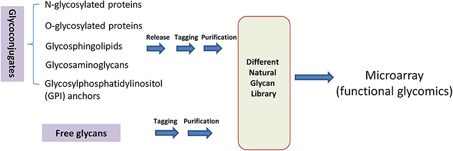
Figure 1. Preparation of natural glycans for functional glycomics.
N-Glycan Release From Natural Glycoproteins
As the most well-studied class of glycans until now, N-glycans can be cleaved off glycoproteins by several enzymes, such as Peptide-N-Glycosidases (PNGase) and endoglycosidases (Endo) (Figure 2). PNGase F is the most widely used enzyme to remove N-glycans from most N-linked glycoproteins and glycopeptides except core α3-fucosylated N-glycans, which are commonly found in plants and insects (Plummer et al., 1984; Tarentino et al., 1985; Tretter et al., 1991). PNGase A has broader substrate specificity and can cleave core α1–3-fucosylated N-glycan (Takahashi, 1977). Recently, PNGase F-II, acid-stable PNGase H+, and PNGase Yl from yeast are all reported to release core α3-fucosylated N-glycans (Du et al., 2015; Lee et al., 2015; Sun et al., 2015). PNGase Ar is able to release the unusual GalαFucα1,3-reducing terminal core from Caenorhabditis elegans (Yan et al., 2018). As another option for enzymatic N-glycan release, endoglycosidases are able to cleave the β1–4-linkage of the di-N-acetylchitobiose core, and such enzymes include Endo A, Endo H, Endo M, Endo D, and Endo S (Freeze and Kranz, 2008; Huang et al., 2012; Wang and Amin, 2014; Li et al., 2016). Although they cleave N-glycans at the same position, they have different substrate specificities related to the structures of the N-glycans (Fairbanks, 2017). Although it is not a focus of this review, it is worth noting that many mutants of endoglycosidases have been developed as synthases for N-glycopeptides and glycoproteins (Huang et al., 2012; Wang and Amin, 2014). The high cost of PNGases and endoglycosidases limits their application in large-scale preparation of N-glycans. Another enzymatic approach is using pronase to cleave peptide bonds and leave glycan-peptide linkages intact (Dodds et al., 2009; Song et al., 2009a; Lu et al., 2019). Pronase is much cheaper than PNGases and endoglycosidases, but its full digestion of glycoproteins to glycoamino acids is always a challenge and often difficult to reproduce.
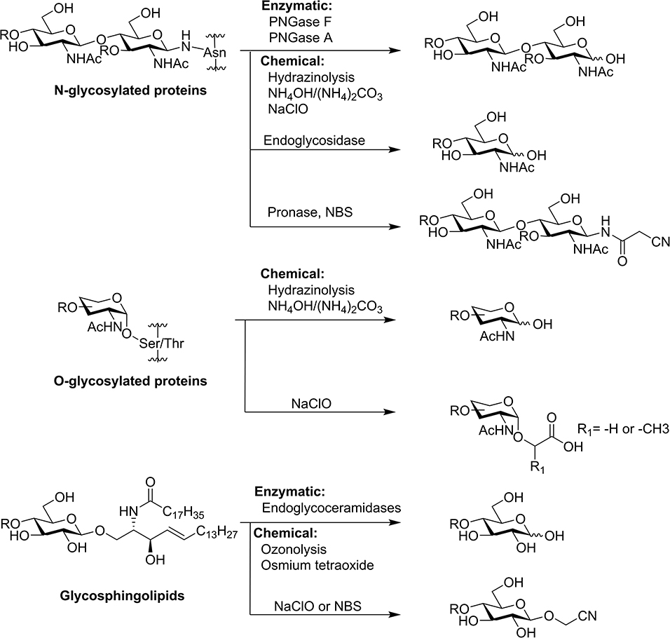
Figure 2. Common method to release glycans from glycoproteins and glycosphingolipids.
Chemical release approaches have provided an alternative to solve the high cost of enzymatic approaches in large preparation of N-glycans. Hydrazinolysis and ammonia/ammonium carbonate have been shown to release N-glycans from glycoproteins (Yosizawa et al., 1966; Huang et al., 2001; Nakakita et al., 2007). However, toxic reagents and/or harsh conditions are necessary, which is not amenable to large-scale preparation and may seriously affect the structural integrity of the released glycans. Under a set of optimized milder alkaline conditions, N-glycans without core α1–3-fucose can also be released by selective hydrolysis of N-glycopeptide (Yuan et al., 2014).
Recently, we reported two different chemical approaches for large-scale release of N-glycans. The first approach is a “chemoenzymatic” method to release N-glycans called threshing and trimming (TaT) (Song et al., 2014b). In the first threshing step, glycoproteins are treated with pronase to create a pool of N-glycoamino acids and glycopeptides with short peptide moieties. In the second trimming step, N-bromosuccinimide (NBS) is added to the mixture of glycoamino acids and glycopeptides to generate free-reducing glycans, nitriles, or aldehydes, depending on different reaction conditions. These products can be easily tagged with fluorescent tags for HPLC purification, MALDI-TOF-MS analysis, and functional study. The TaT approach releases N-glycans without using specialty enzymes, hazardous chemical reagents, and harsh reaction conditions; thus, it can be easily applied for relatively large-scale glycan preparation.
Inspired by the oxidative decarboxylation by NBS treatment, we explored other oxidative reagents and surprisingly discovered that sodium hypochlorite (NaClO) (household bleach) efficiently releases glycans from most classes of natural glycoconjugates (N-glycans, O-glycans, and GSLs) directly from cells, tissues, and organs (Song et al., 2016). In this oxidative release of natural glycan (ORNG) method, household bleach is added to homogenized natural materials (animal/plant tissues) and stirred for 15–30 min at room temperature. After acid precipitation, the free-reducing N-glycans in the supernatant are purified by chromatography techniques, including size exclusion, anion exchange, and hydrophobic/hydrophilic interaction. Purified glycans are ready for fluorescent tagging by reductive amination and separated into individual components by multidimensional HPLC. The ORNG approach is fast, easy to operate, and can be applied to multi-kilograms of natural materials to produce gram-scale natural complex glycans. In our most recent study, the ORNG approach was demonstrated as a complementary route for the preparation of multi-milligram quantities of purified high-mannose N-glycans (Zhu et al., 2018a).
O-Glycan Release From Natural Glycoproteins
Mucin-type O-GalNAc glycans, which attach to serine or threonine residues of proteins through an α-linkage, are the major O-glycans. Compared with N-glycans, which can be released from glycoproteins by several N-glycanases (Plummer et al., 1984; Tarentino et al., 1985; Plummer and Tarentino, 1991), there is a lack of effective general O-glycanase to release O-glycans. Natural O-glycans are traditionally released by chemical methods. The most commonly used method is reductive β-elimination using sodium hydroxide (NaOH) and sodium borohydride (NaBH4) (Carlson, 1966, 1968). Because the common 3-O-substituion at core GalNAc renders it susceptible toward a β-elimination-related peeling reaction after the release of O-glycan from the protein backbone by NaOH, in situ reduction of the reducing end by high-concentration NaBH4 is necessary. The reductive β-elimination converts the reducing end of O-glycan to alditols. Although it is useful for MS-based glycan profiling, it prevents further derivatization and functionalization for glycan purification and printing on a microarray. Several nonreductive β-elimination methods have been developed to keep the reducing end for further derivatization; (Patel et al., 1993; Chai et al., 1997; Huang et al., 2001; Merry et al., 2002; Miura et al., 2010; Yamada et al., 2010; Kozak et al., 2012) however, most of them are still based on base-catalyzed β-elimination, and “peeling” is nearly inevitable (Yu et al., 2010). Furthermore, even if an intact free-reducing end is generated, the following tagging step often generates open-ring O-glycans, which destroy the structural integrity of the O-glycans and, subsequently, may affect its functional study, such as the glycan recognition on a microarray (Prasanphanich et al., 2015). The regeneration of the natural α-O-linkage is significantly more challenging than that of the N-glycan linkage. A PMP-related releasing and tagging approach for O-glycans has also been developed by Wuhr's and Wang's groups (Wang et al., 2011; Zauner et al., 2012) using the combination of β-elimination followed by Michael addition, both of which are catalyzed by a strong base. However, the PMP or related tagged glycans are only suitable for glycomics analysis—not for further derivatization and functional screening on microarrays.
Interestingly, our novel ORNG method also can effectively release O-glycans from glycoproteins or tissues of organisms (Song et al., 2016). The release of O-glycans by ORNG is mechanistically different from all previously known methods. Instead of base-catalyzed elimination, sodium hypochlorite oxidatively degrades the protein backbone to generate O-glycan-acids containing glycolic acid (serine-linked) or lactic acid (threonine-linked) as aglycons in addition to a smaller fraction of free-reducing O-glycans. As a result, these glycolic/lactic acid–linked O-glycans to a great extent retain the structural integrities of the O-glycans as well as the α-O-linkage to the aglycon, preserving O-glycan recognition involving the linkage. In addition, compared to β-elimination, ORNG release is faster and the reaction condition is milder; thus, many labile functional groups, such as sulfation and O-acetylation, are uncompromised after NaClO treatment. More importantly, the released O-glycan acids can be easily labeled using a common amidation reaction with a florescent tag, such as mono-9-florenyl-methoxycarbonyl (mono-Fmoc) ethylenediamine for HPLC separation to prepare O-glycan libraries, and these mono-Fmoc tagged O-glycans can be deprotected by piperidine to expose the amino group for immobilization onto microarray slides for functional O-glycomics studies.
Unlike all the above release strategies, recently we have developed a novel technology termed cellular O-glycome reporter/amplification (CORA), which uses an O-glycan precursor (peracetylated benzyl-α-N-acetylgalactosamine, Ac3Bn-α-GalNAc) to amplify O-glycans in living cells and secretes free Bn-O-glycans into the cell media. The secreted Bn-O-glycans can be easily purified and analyzed by MS (Kudelka et al., 2016). CORA greatly enhances the sensitivity of MS analysis of O-glycome from living cells. However, the low UV absorption of the Bn group makes the isolation of these glycans using HPLC challenging. In order to overcome this limit, we have recently designed and synthesized many Ac3Bn-α-GalNAc derivatives as CORA precursors to replace Ac3Bn-α-GalNAc. These new CORA precursors include many function groups, such as the fluorescence group and bioorthogonal reactive groups (Zhang et al., 2019), allowing O-glycans produced by CORA to be tagged, separated, and purified by chromatography for functional study. Preparative CORA using these derivatives as precursors is currently under investigation, and we believe this method could become a promising approach for preparation of O-glycans (Figure 3).
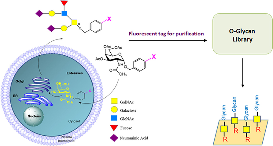
Figure 3. CORA method for preparation of O-glycans by living cells. Ac3Bn-α-GalNAc derivative can enter the cell, be deacetylated to form a Bn-α-GalNAc derivative, and then be extended by glycosyltransferases in the O-glycosylation pathway in Golgi. The Bn-O-glycan derivatives are secreted to cell media. The fluorescently labeled O-glycans can be purified to prepare O-glycan libraries for functional O-glycome study.
Glycan Release From Glycosphingolipids
Glycosphingolipids (GSLs) are amphipathic glycoconjugates widely distributed on the cell surfaces. Although exoglycosidases and endoglycosidases are only able to cleave the glycan moieties from GSLs (Li and Li, 1999), endoglycoceramidases are found to release entire glycans from GSLs (Ishibashi et al., 2007; Li et al., 2009; Albrecht et al., 2016). However, the enzymes are expensive and specific to certain GSL structures, preventing their wide application in larger scale glycan preparation from GSLs.
Traditional chemical methods utilize ozonolysis or osmium tetraoxide to oxidize the C=C double bond in the sphingosine moiety, followed by base-catalyzed β-elimination (Wiegandt and Baschang, 1965; Hakomori, 1966). In order to prevent the potential adverse effect of base treatment on glycan structural integrities, we have developed several approaches to release glycans from GSLs for functional study through glycan microarray preparation using covalent immobilization. The first approach takes advantage of the aldehyde group generated by ozone treatment of GSLs, which can be directly coupled with functional and fluorescent tags by reductive amination. This approach preserves a significant portion of the lipid moiety and may benefit functional studies requiring the lipid component (Song et al., 2011). The second approach is to heat ozonized GSLs gently under neutral pH, which interestingly releases free-reducing glycans fairly efficiently (Song et al., 2012). Both of these methods still require ozone to oxidize the C=C double bond to initiate the reaction and can only be applied to purified GSLs. In our most recent ORNG approach, we found that in addition to N- and O-glycans, NaClO can also release glycans as cyanomethyl glycosides from GSLs—apparently through the oxidative degradation of the lipid moiety at the polar head group (Song et al., 2016). The ORNG approach can be applied not only to gangliosides purified by organic solvent extraction, but also directly to aqueous homogenized brain tissue (Song et al., 2016). The ability to release GSL-glycans without involving organic solvent extraction significantly reduced the complexity of GSL-glycan preparation and is essential to larger scale glycan production. Interestingly, although NBS can also release glycan nitriles from gangliosides at 65°C, this reaction does not work directly on homogenized brain tissue.
Glycan Release From Glycosaminoglycans and GPI-Anchors
Glycosaminoglycans (GAGs) are linear polydisperse heteropolysaccharides, consisting of up to 1,000 repetitive disaccharide units (Murata et al., 1985; Jackson et al., 1991). Heparin, a highly sulfated form of heparan sulfate (HS) glycosaminoglycans, has been shown to possess important biological functions that vary according to its fine structure (Liu et al., 2009). Heparin has widespread clinical use as an intravenous anticoagulant with more than 100,000 kg produced annually worldwide (Liu et al., 2009). Commercial heparin is currently produced from animal tissues, such as porcine intestine and beef lung (Bhaskar et al., 2012). The methods used for commercial preparation of heparin involve five basic steps: (1) preparation of tissue, (2) extraction of heparin from tissue, (3) recovery of raw heparin, (4) purification of heparin, and (5) recovery of purified heparin (Linhardt and Gunay, 1999). While being similar, the heparins derived from different animal sources have diverse structures that relate to different functional activities, such as AT- and thrombin-binding affinities (Liu et al., 2009). A worldwide health crisis in 2007, associated with contamination of several heparin batches, reportedly resulted in more than 200 deaths alone in the United States (Liu et al., 2009; Turnbull, 2011). Low-molecular weight heparins (LMWH, MW avg <8 kDa) are subcutaneously administered, have a longer half-life than unfractionated heparin, and can be prepared with different structures by different depolymerization methods, including oxidation, deaminative degradation, and β-elimination (Linhardt and Gunay, 1999).
With the improvement in chemical and chemo-enzymatic methods, the synthetic scale of GAGs has reached gram scale, and the automated solid-phase synthesis of chondroitin sulfate GAGs is available (Eller et al., 2013; Mende et al., 2016; Xu et al., 2017), which enable facilitated access to functional and biological study of GAGs. Although glycan microarray analysis of natural GAG oligomers have been reported for more than 10 years (Noti et al., 2006; Park et al., 2008), large-scale GAG microarrays for general screening of GAG-binding proteins are only reported in a synthetic approach (Yang et al., 2017; Zhang et al., 2017; Zong et al., 2017). The high heterogeneity of the sulfation patterns of the GAG chains make the isolation of homogeneous GAG oligomers, structural characterization, and chemical/enzymatic synthesis a challenging task. Nevertheless, with the recent progress in HPLC analysis and separation, preparation of a more comprehensive natural GAG glycan library for functional study with GAG-binding proteins will become possible in the near future.
GPI-anchor proteins play critical roles in numerous biological processes, such as cell recognition and interaction (He et al., 1987; Takeda and Kinoshita, 1995; Paulick and Bertozzi, 2008). Because the first total synthesis of an intact GPI anchor was in 1991 (Murakata and Ogawa, 1991), convergent chemical and chemo-enzymatic strategies for GPI synthesis were developed, and more than 30 GPIs were isolated and characterized (Wu et al., 2008; Yu and Guo, 2009; Swarts and Guo, 2010; Guo, 2013). AN effective strategy of labeling of cell-surface GPIs and GPI-anchored proteins was developed for biological studies (Lu et al., 2015). However, natural-sourced GPI anchor preparation for functional study is not well studied yet, presumably due to the lack of well-defined enzymatic and chemical release methods and low abundance of GPI-anchors in cells.
Preparation of Human Milk Oligosaccharides
Human milk oligosaccharides (HMOs), occurring as free-reducing glycans, are the third major component of human milk after lactose and lipids and are known to play important roles benefiting infant health (Chen, 2015). HMOs are extended from lactose by a collection of glycosyltransferases, adding N-acetyl-glucosamine, galactose, fucose, and neuraminic acid (Jenness, 1979). More than a hundred different HMO structures have been identified and elucidated (Zopf et al., 1978; Prieto and Smith, 1985; Smith et al., 1985; Jensen et al., 1995). Due to its high abundance in human milk (5–15 g/L) as free-reducing glycan without the need to release from other biomolecules, large-scale isolation and separation of HMOs have been practiced for many years. In an early study, individual HMOs were isolated directly by size exclusive, anion-exchange, and paper chromatography without being derivatized (Kobata et al., 1969; Donald and Feeney, 1988). More recently, with the wide use of HPLC isolation and MS analysis, tagging of HMOs by functional and/or fluorescent groups for separation and further functional study is more common. We have applied our bifunctional fluorescent tag AEAB to HMO isolation and fractionation (Song et al., 2009b). Isolated glycans can be directly printed on a microarray for functional screening with various GBPs and viruses (Yu et al., 2012). With more complex HMO structures becoming available for functional study, we expect further elucidation of their functions through interaction with the infant microbiome.
Functional and Fluorescent Tagging of Released Glycans
After being released from natural sources, glycans existing in the heterogeneous mixture need to be separated for analysis or for preparation of pure glycans. Due to the lack of an exploitable chromophore in natural glycans and the anomeric mutual rotation at the reducing end, it's a challenge to monitor glycans during HPLC separation. The preparation of glycan microarrays also requires that the glycans are derivatized with functional groups, such as an amino group. Therefore, it is important to install functional and fluorescent tags on the released glycans for easier and more efficient separation and solid phase immobilization afterward.
Reductive amination of free-reducing glycans with fluorescent amines has long been used for the HPLC profiling of glycans (Figure 4). 2-aminopyridine (2-AP), 2-aminobenzamide (2-AB), 2-aminobenzoic acid, or anthranilic acid (2-AA) are commonly use fluorescent amines (Hase et al., 1978; Bigge et al., 1995; Anumula, 2014). However, these small fluorescent amines lack a functional group for efficient solid phase immobilization or covalent derivatization. With an aromatic amino group, a homobifunctional tag, 2,6-diaminopyridine (DAP) conjugated glycans can be immobilized onto activated surfaces for microarray preparation (Xia et al., 2005; Song et al., 2008). To efficiently immobilize precious natural glycans, we developed a novel heterobifunctional tag, 2-amino-N-(2-aminoethyl)benzamide (AEAB), which contains both arylamine and alkylamine (Song et al., 2009b). The aromatic amine selectively reacts with the free-reducing end of released glycans by reductive amination while the alkylamine is used for efficient solid-phase immobilization onto both NHS and epoxy-activated glass slides.

Figure 4. Several typical methods of fluorescent tagging of released glycans for functional study preparation.
One inherent problem with commonly used reductive amination is breaking the reducing end ring structure, affecting the glycan structural integrity. To address this drawback, new methods and linkers have been reported, such as 2-amino-methyl-N,O-hydroxyethyl (AMNO) (Bohorov et al., 2006) and N-Fmoc-3-(methoxyamino)propylamine (F-MAPA) (Wei et al., 2019). We also developed a procedure to prepare HMO-AEAB conjugates with an intact reducing end ring structure (Yu et al., 2012). More recently, we have designed a new tag, O-benzylhydroxylamine (BHA), which can be easily and efficiently installed on HMOs and keep the glycan structure integrity (Zhang et al., 2020). By Pd/C-catalyzed hydrogenation, free HMO can be easily regenerated from HMO-BHA.
Compared to nonderivatized glycans, the installation of a fluorescent tag to released glycans often increases the sensitivity of MS analysis. Although premethylation is considered a necessary step for detailed sequencing by MS (Ashline et al., 2014), the conjugated tags often generate structural complexity during permethylation. Therefore, we developed a facile and mild method using NBS to remove tags of aminated glycans, which regenerates free-reducing glycans for permethylation (Song et al., 2013). This method can be efficiently applied to all types of tags installed through reductive amination, including 2-AP, 2-AB, 2-AA, and AEAB.
Because of the easy installation and removal, the 9-fluorenylmethoxycarbonyl (Fmoc) group is widely used as an amino-protecting group in organic chemistry, especially in peptide synthesis. After being installed on released glycans, the fluorescent Fmoc group can greatly enhance sensitivity of HPLC to tagged glycans (Kamoda et al., 2005; Song et al., 2009a; Yamada et al., 2013; Lu et al., 2019). It can also serve as an affinity tag due to the hydrophobicity. The amino group can be easily regenerated for solid-phase immobilization in microarray printing (Kamoda et al., 2005; Yamada et al., 2013; Wei et al., 2019). We have successfully installed an Fmoc tag on the released glycan from natural O-glycanconjugates and glycosyphingolipids in our ORNG method (Song et al., 2016).
HPLC Separation of Glycans for Functional Study
Over the years, various HPLC methods have been commonly used for glycan purification, including hydrophilic interaction liquid chromatography (HILIC), high-performance anion-exchange chromatography (HPAEC), and reversed-phase chromatography (Ruhaak et al., 2010; Nagy et al., 2017). HILIC mode HPLC is an efficient technique for separation of unprotected saccharides (Fu et al., 2010; Melmer et al., 2011; Wan et al., 2015) while reversed-phase chromatography is suitable for hydrophobic saccharides (Rajakylä, 1986; El Rassi, 1995; Dallabernardina et al., 2016). HPAEC is usually used for negatively charged unprotected carbohydrates (Rohrer et al., 2013, 2016). Porous graphitized carbon (PGC) as a unique stationary phase combining both hydrophobic and anionic interactions separates glycans based very well on their isomeric structures under reverse-phase elution conditions (Fan et al., 1994; Itoh et al., 2002; Ruhaak et al., 2009; West et al., 2010; Lie and Pedersen, 2018). Because the glycans obtained from biological sources are often complex mixtures, multidimensional HPLC is necessary to separate them into individual glycans with significant purity (Nagy et al., 2017). We have successfully applied multidimensional HPLC to isolate an individual glycan library for microarray study (Song et al., 2009b; Yu et al., 2012).
Most of the HPLC separation methods are designed for analytical glycomics using small samples, which does not generate significant quantities of glycans for detailed functional study. There have been a few examples in which a more significant amount of starting materials are used to generate a sufficient amount glycans for NMR study (Green et al., 1988; Da Silva et al., 1995). However, no real preparative-scale HPLC separations have been tacked previously, presumably due to the unavailability of a large amount of released glycans. With gram-scale glycans from a natural source are available because of the ORNG technique, development of preparative-scale purification becomes practical and provides an effective route to address the lack of glycans for functional study. We have reported isolation of high mannose N-glycans from soy proteins and egg yolks by a preparative scale multidimensional HPLC method (Zhu et al., 2018a,b). However, even after multidimensional HPLC, some fractions are still mixtures of isomers that are very difficult to separate even on analytical columns. To address this problem, recycled HPLC could be a good solution (Alley et al., 2013; Sidana and Joshi, 2013). Most recently, we have reported a simple and affordable closed-loop recycled HPLC method for separation of complex glycans in the preparative scale. It was successfully applied to reverse-phase chromatography, HILIC, and sizes using size-exclusion chromatography (SEC) (Zhu et al., 2020).
Conclusions
With a highly diverse structure, natural glycans are likely more biologically relevant for functional study. Here, we have summarized the preparation of several classes of complex glycans from glycoconjugates. Both enzymatic and chemical approaches have been discussed, and each method has its own advantages and should be carefully selected based on the specific goal of individual study. When a large amount of natural glycans are desired, chemical approaches, especially the new ORNG approach provides a good alternative to chemoenzymatic synthesis. The ORNG approach is able to quickly release up to grams of glycans from several major classes of glycoconjugates using affordable chemical reagents (household bleach), a mild reaction condition, and a simple operation. Nevertheless, more preparation methods are still in demand, especially for O-glycans, GAGs, and GPI-anchors. The novel CORA method provides a potential new route toward O-glycans if preparative scale can be achieved.
Author Contributions
QZ, ZL, and XS wrote and edited the manuscript. All authors contributed to the article and approved the submitted version.
Funding
This work was supported by NIH Common Fund Glycoscience (U01GM116254, U01CA207821) and partially by STTR grants R41GM122139, SBIR R43GM131534, and R43GM133252. It was also partially supported by Emory Comprehensive Glycomics Core (ECGC), which is subsidized by the Emory University School of Medicine and is one of the Emory Integrated Core Facilities.
Conflict of Interest
XS is a co-founder of NatGlycan LLC, which commercializes the ORNG process.
The remaining authors declare that the research was conducted in the absence of any commercial or financial relationships that could be construed as a potential conflict of interest.
References
Albrecht, S., Vainauskas, S., Stöckmann, H., McManus, C., Taron, C. H., and Rudd, P. M. (2016). Comprehensive profiling of glycosphingolipid glycans using a novel broad specificity endoglycoceramidase in a high-throughput workflow. Anal. Chem. 88, 4795–4802. doi: 10.1021/acs.analchem.6b00259
Alley, W. R. Jr., Mann, B. F., Hruska, V., and Novotny, M. V. (2013). Isolation and purification of glycoconjugates from complex biological sources by recycling high-performance liquid chromatography. Anal. Chem. 85, 10408–10416. doi: 10.1021/ac4023814
Anumula, K. R. (2014). Single tag for total carbohydrate analysis. Anal. Biochem. 457, 31–37. doi: 10.1016/j.ab.2014.04.019
Ashline, D. J., Yu, Y., Lasanajak, Y., Song, X., Hu, L., Ramani, S., et al. (2014). Structural characterization by multistage mass spectrometry (MSn) of human milk glycans recognized by human rotaviruses. Mol. Cell. Proteom. 13, 2961–2974. doi: 10.1074/mcp.M114.039925
Bhaskar, U., Sterner, E., Hickey, A. M., Onishi, A., Zhang, F., Dordick, J. S., et al. (2012). Engineering of routes to heparin and related polysaccharides. Appl. Microbiol. Biotechnol. 93, 1–16. doi: 10.1007/s00253-011-3641-4
Bigge, J., Patel, T., Bruce, J., Goulding, P., Charles, S., and Parekh, R. (1995). Nonselective and efficient fluorescent labeling of glycans using 2-amino benzamide and anthranilic acid. Anal. Biochem. 230, 229–238. doi: 10.1006/abio.1995.1468
Blixt, O., and Razi, N. (2006). Chemoenzymatic synthesis of glycan libraries. Methods Enzymol. 415, 137–153. doi: 10.1016/S0076-6879(06)15009-0
Bohorov, O., Andersson-Sand, H., Hoffmann, J., and Blixt, O. (2006). Arraying glycomics: a novel bi-functional spacer for one-step microscale derivatization of free reducing glycans. Glycobiology 16, 21C−27C. doi: 10.1093/glycob/cwl044
Boltje, T. J., Buskas, T., and Boons, G.-J. (2009). Opportunities and challenges in synthetic oligosaccharide and glycoconjugate research. Nat. Chem. 1:611. doi: 10.1038/nchem.399
Carlson, D. M. (1966). Oligosaccharides isolated from pig submaxillary mucin. J. Biol. Chem. 241, 2984–2986.
Carlson, D. M. (1968). Structures and immunochemical properties of oligosaccharides isolated from pig submaxillary mucins. J. Biol. Chem. 243, 616–626.
Chai, W., Feizi, T., Yuen, C. T., and Lawson, A. M. (1997). Nonreductive release of O-linked oligosaccharides from mucin glycoproteins for structure/function assignments as neoglycolipids: application in the detection of novel ligands for E-selectin. Glycobiology 7, 861–872. doi: 10.1093/glycob/7.6.861
Chen, X. (2015). Human milk oligosaccharides (HMOS): structure, function, and enzyme-catalyzed synthesis. Adv. Carbohydr. Chem. Biochem. 72, 113–190. doi: 10.1016/bs.accb.2015.08.002
Cummings, R. D. (2009). The repertoire of glycan determinants in the human glycome. Mol. bioSyst. 5, 1087–1104. doi: 10.1039/b907931a
Cummings, R. D., and Pierce, J. M. (2014). The challenge and promise of glycomics. Chem. Biol. 21, 1–15. doi: 10.1016/j.chembiol.2013.12.010
Da Silva, M. L. C., Stubbs, H. J., Tamura, T., and Rice, K. G. (1995). 1H NMR characterization of a hen ovalbumin tyrosinamide N-linked oligosaccharide library. Arch. Biochem. Biophys. 318, 465–475. doi: 10.1006/abbi.1995.1255
Dallabernardina, P., Schuhmacher, F., Seeberger, P. H., and Pfrengle, F. (2016). Automated glycan assembly of xyloglucan oligosaccharides. Org. Biomol. Chem. 14, 309–313. doi: 10.1039/C5OB02226F
Dodds, E. D., Seipert, R. R., Clowers, B. H., German, J. B., and Lebrilla, C. B. (2009). Analytical performance of immobilized pronase for glycopeptide footprinting and implications for surpassing reductionist glycoproteomics. J. Proteome Res. 8, 502–512. doi: 10.1021/pr800708h
Donald, A. S., and Feeney, J. (1988). Separation of human milk oligosaccharides by recycling chromatography. First isolation of lacto-N-neo-difucohexaose II and 3′-galactosyllactose from this source. Carbohydr. Res. 178, 79–91. doi: 10.1016/0008-6215(88)80103-4
Doneanu, C. E., Chen, W., and Gebler, J. C. (2009). Analysis of oligosaccharides derived from heparin by ion-pair reversed-phase chromatography/mass spectrometry. Anal. Chem. 81, 3485–3499. doi: 10.1021/ac802770r
Du, Y. M., Xia, T., Gu, X. Q., Wang, T., Ma, H. Y., Voglmeir, J., et al. (2015). Rapid sample preparation methodology for plant N-glycan analysis using acid-stable PNGase H+. J. Agric. Food Chem. 63, 10550–10555. doi: 10.1021/acs.jafc.5b03633
El Rassi, Z. (1995). Reversed-phase and hydrophobic interaction chromatography of carbohydrates and glycoconjugates. J. Chromatogr. Libr. 58, 41–101. doi: 10.1016/S0301-4770(08)60507-2
Eller, S., Collot, M., Yin, J., Hahm, H. S., and Seeberger, P. H. (2013). Automated solid-phase synthesis of chondroitin sulfate glycosaminoglycans. Angew. Chem. Int. Ed. 52, 5858–5861. doi: 10.1002/anie.201210132
Fairbanks, A. J. (2017). The ENGases: versatile biocatalysts for the production of homogeneous N-linked glycopeptides and glycoproteins. Chem. Soc. Rev. 46, 5128–5146. doi: 10.1039/C6CS00897F
Fan, J.-Q., Kondo, A., Kato, I., and Lee, Y. C. (1994). High-performance liquid chromatography of glycopeptides and oligosaccharides on graphitized carbon columns. Anal. Biochem. 219, 224–229. doi: 10.1006/abio.1994.1261
Freeze, H. H., and Kranz, C. (2008). Endoglycosidase and glycoamidase release of N-linked glycans. Curr. Protoc. Immunol. 83:8.15. doi: 10.1002/0471142735.im0815s83
Fu, Q., Liang, T., Zhang, X., Du, Y., Guo, Z., and Liang, X. (2010). Carbohydrate separation by hydrophilic interaction liquid chromatography on a ‘click'maltose column. Carbohydr. Res. 345, 2690–2697. doi: 10.1016/j.carres.2010.09.033
Fukui, S., Feizi, T., Galustian, C., Lawson, A. M., and Chai, W. (2002). Oligosaccharide microarrays for high-throughput detection and specificity assignments of carbohydrate-protein interactions. Nat. Biotechnol. 20:1011. doi: 10.1038/nbt735
Green, E. D., Adelt, G., Baenziger, J. U., Wilson, S., and Van Halbeek, H. (1988). The asparagine-linked oligosaccharides on bovine fetuin. Structural analysis of N-glycanase-released oligosaccharides by 500-megahertz proton NMR spectroscopy. J. Biol. Chem. 263, 18253–18268.
Guo, Z. (2013). Synthetic studies of glycosylphosphatidylinositol (GPI) anchors and GPI-anchored peptides, glycopeptides, and proteins. Curr. Org. Synth. 10, 366–383. doi: 10.2174/1570179411310030003
Hakomori, S.-I. (1966). Release of carbohydrates from sphingoglycolipid by osmium-catalyzed periodate oxidation followed by treatment with mild alkali. J. Lipid Res. 7, 789–792.
Hase, S., Ikenaka, T., and Matsushima, Y. (1978). Structure analyses of oligosaccharides by tagging of the reducing end sugars with a fluorescent compound. Biochem. Biophys. Res. Commun. 85, 257–263. doi: 10.1016/S0006-291X(78)80037-0
He, H.-T., Finne, J., and Goridis, C. (1987). Biosynthesis, membrane association, and release of N-CAM-120, a phosphatidylinositol-linked form of the neural cell adhesion molecule. J. Cell Biol. 105, 2489–2500. doi: 10.1083/jcb.105.6.2489
Huang, W., Giddens, J., Fan, S.-Q., Toonstra, C., and Wang, L.-X. (2012). Chemoenzymatic glycoengineering of intact IgG antibodies for gain of functions. J. Am. Chem. Soc. 134, 12308–12318. doi: 10.1021/ja3051266
Huang, Y., Mechref, Y., and Novotny, M. V. (2001). Microscale nonreductive release of O-linked glycans for subsequent analysis through MALDI mass spectrometry and capillary electrophoresis. Anal. Chem. 73, 6063–6069. doi: 10.1021/ac015534c
Ishibashi, Y., Nakasone, T., Kiyohara, M., Horibata, Y., Sakaguchi, K., Hijikata, A., et al. (2007). A novel endoglycoceramidase hydrolyzes oligogalactosylceramides to produce galactooligosaccharides and ceramides. J. Biol. Chem. 282, 11386–11396. doi: 10.1074/jbc.M608445200
Itoh, S., Kawasaki, N., Ohta, M., Hyuga, M., Hyuga, S., and Hayakawa, T. (2002). Simultaneous microanalysis of N-linked oligosaccharides in a glycoprotein using microbore graphitized carbon column liquid chromatography-mass spectrometry. J. Chromatogr. A 968, 89–100. doi: 10.1016/S0021-9673(02)00951-2
Jackson, R. L., Busch, S. J., and Cardin, A. D. (1991). Glycosaminoglycans: molecular properties, protein interactions, and role in physiological processes. Physiol. Rev. 71, 481–539. doi: 10.1152/physrev.1991.71.2.481
Jensen, R. G., Blanc, B., and Patton, S. (1995). Particulate Constituents in Human and Bovine Milks. Handbook of Milk Composition. New York, NY: Academic Press.
Kamoda, S., Nakano, M., Ishikawa, R., Suzuki, S., and Kakehi, K. (2005). Rapid and sensitive screening of N-Glycans as 9-fluorenylmethyl derivatives by high-performance liquid chromatography: a method which can recover free oligosaccharides after analysis. J. Proteome Res. 4, 146–152. doi: 10.1021/pr049825o
Kobata, A., Ginsburg, V., and Tsuda, M. (1969). Oligosaccharides of human milk: I. Isolation and characterization. Arch. Biochem. Biophys. 130, 509–513. doi: 10.1016/0003-9861(69)90063-0
Koeller, K. M., Smith, M. E., and Wong, C.-H. (2000). Chemoenzymatic synthesis of PSGL-1 glycopeptides: sulfation on tyrosine affects glycosyltransferase-catalyzed synthesis of the O-glycan. Bioorg. Med. Chem. 8, 1017–1025. doi: 10.1016/S0968-0896(00)00041-9
Kozak, R. P., Royle, L., Gardner, R. A., Fernandes, D. L., and Wuhrer, M. (2012). Suppression of peeling during the release of O-glycans by hydrazinolysis. Anal. Biochem. 423, 119–128. doi: 10.1016/j.ab.2012.01.002
Kudelka, M. R., Antonopoulos, A., Wang, Y., Duong, D. M., Song, X., Seyfried, N. T., et al. (2016). Cellular O-glycome reporter/amplification to explore O-glycans of living cells. Nat. Methods 13, 81–86. doi: 10.1038/nmeth.3675
Lee, K. J., Gil, J. Y., Kim, S.-Y., Kwon, O., Ko, K., Kim, D.-I., et al. (2015). Molecular characterization of acidic peptide: N-glycanase from the dimorphic yeast Yarrowia lipolytica. J. Biochem. 157, 35–43. doi: 10.1093/jb/mvu051
Lepenies, B., Yin, J., and Seeberger, P. H. (2010). Applications of synthetic carbohydrates to chemical biology. Curr. Opin. Chem. Biol. 14, 404–411. doi: 10.1016/j.cbpa.2010.02.016
Li, L., Liu, Y., Ma, C., Qu, J., Calderon, A. D., Wu, B., et al. (2015). Efficient chemoenzymatic synthesis of an N-glycan isomer library. Chem. Sci. 6, 5652–5661. doi: 10.1039/C5SC02025E
Li, T., Liu, L., Wei, N., Yang, J.-Y., Chapla, D. G., Moremen, K. W., et al. (2019). An automated platform for the enzyme-mediated assembly of complex oligosaccharides. Nat. Chem. 11, 229–236. doi: 10.1038/s41557-019-0219-8
Li, T., Tong, X., Yang, Q., Giddens, J. P., and Wang, L.-X. (2016). Glycosynthase mutants of endoglycosidase S2 show potent transglycosylation activity and remarkably relaxed substrate specificity for antibody glycosylation remodeling. J. Biol. Chem. 291, 16508–16518. doi: 10.1074/jbc.M116.738765
Li, Y.-T., Chou, C.-W., Li, S.-C., Kobayashi, U., Ishibashi, Y.-H., and Ito, M. (2009). Preparation of homogenous oligosaccharide chains from glycosphingolipids. Glycoconjug. J. 26:929. doi: 10.1007/s10719-008-9125-9
Li, Y.-T., and Li, S.-C. (1999). Enzymatic hydrolysis of glycosphingolipids. Anal. Biochem. 273, 1–11. doi: 10.1006/abio.1999.4191
Lie, A., and Pedersen, L. H. (2018). Resolution and signal-to-noise in analysis of carbohydrate isomers by graphitized carbon chromatography with charged aerosol detection. J. Chromatogr. A 1567, 147–154. doi: 10.1016/j.chroma.2018.06.068
Linhardt, R. J., and Gunay, N. S. (1999). Production and chemical processing of low molecular weight heparins. Semin. Thromb. Hemost. 25 (Suppl. 3), 5–16.
Liu, H., Zhang, Z., and Linhardt, R. J. (2009). Lessons learned from the contamination of heparin. Nat. Product Rep. 26, 313–321. doi: 10.1039/b819896a
Liu, L., Prudden, A. R., Capicciotti, C. J., Bosman, G. P., Yang, J.-Y., Chapla, D. G., et al. (2019). Streamlining the chemoenzymatic synthesis of complex N-glycans by a stop and go strategy. Nat. Chem. 11, 161–169. doi: 10.1038/s41557-018-0188-3
Lu, L., Gao, J., and Guo, Z. (2015). Labeling cell surface GPIs and GPI-anchored proteins through metabolic engineering with artificial inositol derivatives. Angew. Chem. Int. Ed. 54, 9679–9682. doi: 10.1002/anie.201503814
Lu, Y., Li, C., Wei, M., Jia, Y., Song, J., Zhang, Y., et al. (2019). Release, separation, and recovery of monomeric reducing N-glycans with pronase E combined with 9-chloromethyl chloroformate and glycosylasparaginase. Biochemistry 58, 1120–1130. doi: 10.1021/acs.biochem.8b01224
Melmer, M., Stangler, T., Premstaller, A., and Lindner, W. (2011). Comparison of hydrophilic-interaction, reversed-phase and porous graphitic carbon chromatography for glycan analysis. J. Chromatogr. A 1218, 118–123. doi: 10.1016/j.chroma.2010.10.122
Mende, M., Bednarek, C., Wawryszyn, M., Sauter, P., Biskup, M. B., Schepers, U., et al. (2016). Chemical synthesis of glycosaminoglycans. Chem. Rev. 116, 8193–8255. doi: 10.1021/acs.chemrev.6b00010
Merry, A. H., Neville, D. C., Royle, L., Matthews, B., Harvey, D. J., Dwek, R. A., et al. (2002). Recovery of intact 2-aminobenzamide-labeled O-glycans released from glycoproteins by hydrazinolysis. Anal. Biochem. 304, 91–99. doi: 10.1006/abio.2002.5620
Miura, Y., Kato, K., Takegawa, Y., Kurogochi, M., Furukawa, J., Shinohara, Y., et al. (2010). Glycoblotting-assisted O-glycomics: ammonium carbamate allows for highly efficient o-glycan release from glycoproteins. Anal. Chem. 82, 10021–10029. doi: 10.1021/ac101599p
Murakata, C., and Ogawa, T. (1991). A total synthesis of GPI anchor of Trypanosoma brucei. Tetrahedron Lett. 32, 671–674. doi: 10.1016/S0040-4039(00)74856-8
Murata, K., Ochiai, Y., and Akashio, K. (1985). Polydispersity of acidic glycosaminoglycan components in human liver and the changes at different stages in liver cirrhosis. Gastroenterology 89, 1248–1257. doi: 10.1016/0016-5085(85)90640-7
Nagy, G., Peng, T., and Pohl, N. L. (2017). Recent liquid chromatographic approaches and developments for the separation and purification of carbohydrates. Anal. Methods 9, 3579–3593. doi: 10.1039/C7AY01094J
Nakakita, S.-I., Sumiyoshi, W., Miyanishi, N., and Hirabayashi, J. (2007). A practical approach to N-glycan production by hydrazinolysis using hydrazine monohydrate. Biochem. Biophys. Res. Commun. 362, 639–645. doi: 10.1016/j.bbrc.2007.08.032
Noti, C., de Paz, J. L., Polito, L., and Seeberger, P. H. (2006). Preparation and use of microarrays containing synthetic heparin oligosaccharides for the rapid analysis of heparin–protein interactions. Chem. Eur. J. 12, 8664–8686. doi: 10.1002/chem.200601103
Ohtsubo, K., and Marth, J. D. (2006). Glycosylation in cellular mechanisms of health and disease. Cell 126, 855–867. doi: 10.1016/j.cell.2006.08.019
Palcic, M. M. (2011). Glycosyltransferases as biocatalysts. Curr. Opin. Chem. Biol. 15, 226–233. doi: 10.1016/j.cbpa.2010.11.022
Park, T.-J., Lee, M.-Y., Dordick, J. S., and Linhardt, R. J. (2008). Signal amplification of target protein on heparin glycan microarray. Anal. Biochem. 383, 116–121. doi: 10.1016/j.ab.2008.07.037
Patel, T., Bruce, J., Merry, A., Bigge, C., Wormald, M., Jaques, A., et al. (1993). Use of hydrazine to release in intact and unreduced form both N- and O-linked oligosaccharides from glycoproteins. Biochemistry 32, 679–693. doi: 10.1021/bi00053a037
Paulick, M. G., and Bertozzi, C. R. (2008). The glycosylphosphatidylinositol anchor: a complex membrane-anchoring structure for proteins. Biochemistry 47, 6991–7000. doi: 10.1021/bi8006324
Paulson, J. C., Blixt, O., and Collins, B. E. (2006). Sweet spots in functional glycomics. Nat. Chem. Biol. 2, 238–248. doi: 10.1038/nchembio785
Plummer, T. H. Jr., Elder, J. H., Alexander, S., Phelan, A. W., and Tarentino, A. L. (1984). Demonstration of peptide:N-glycosidase F activity in endo-beta-N-acetylglucosaminidase F preparations. J. Biol. Chem. 259, 10700–10704.
Plummer, T. H. Jr., and Tarentino, A. L. (1991). Purification of the oligosaccharide-cleaving enzymes of Flavobacterium meningosepticum. Glycobiology 1, 257–263. doi: 10.1093/glycob/1.3.257
Prasanphanich, N. S., Song, X., Heimburg-Molinaro, J., Luyai, A. E., Lasanajak, Y., Cutler, C. E., et al. (2015). Intact reducing glycan promotes the specific immune response to lacto-N-neotetraose-BSA neoglycoconjugates. Bioconjug. Chem. 26, 559–571. doi: 10.1021/acs.bioconjchem.5b00036
Prieto, P. A., and Smith, D. F. (1985). A new ganglioside in human meconium detected by antiserum against the human milk sialyloligosaccharide, LS-tetrasaccharide b. Arch. Biochem. Biophys. 241, 281–289. doi: 10.1016/0003-9861(85)90384-4
Prudden, A. R., Liu, L., Capicciotti, C. J., Wolfert, M. A., Wang, S., Gao, Z., et al. (2017). Synthesis of asymmetrical multiantennary human milk oligosaccharides. Proc. Natl. Acad. Sci. U.S.A. 114, 6954–6959. doi: 10.1073/pnas.1701785114
Rajakylä, E. (1986). Use of reversed-phase chromatography in carbohydrate analysis. J. Chromatogr. A 353, 1–12. doi: 10.1016/S0021-9673(01)87070-9
Reily, C., Stewart, T. J., Renfrow, M. B., and Novak, J. (2019). Glycosylation in health and disease. Nat. Rev. Nephrol. 15, 346–366. doi: 10.1038/s41581-019-0129-4
Rohrer, J., Basumallick, L., and Hurum, D. (2013). High-performance anion-exchange chromatography with pulsed amperometric detection for carbohydrate analysis of glycoproteins. Biochemistry 78, 697–709. doi: 10.1134/S000629791307002X
Rohrer, J. S., Basumallick, L., and Hurum, D. C. (2016). Profiling N-linked oligosaccharides from IgG by high-performance anion-exchange chromatography with pulsed amperometric detection. Glycobiology 26, 582–591. doi: 10.1093/glycob/cww006
Royle, L., Campbell, M. P., Radcliffe, C. M., White, D. M., Harvey, D. J., Abrahams, J. L., et al. (2008). HPLC-based analysis of serum N-glycans on a 96-well plate platform with dedicated database software. Anal. Biochem. 376, 1–12. doi: 10.1016/j.ab.2007.12.012
Ruhaak, L. R., Deelder, A. M., and Wuhrer, M. (2009). Oligosaccharide analysis by graphitized carbon liquid chromatography-mass spectrometry. Anal. Bioanal. Chem. 394, 163–174. doi: 10.1007/s00216-009-2664-5
Ruhaak, L. R., Zauner, G., Huhn, C., Bruggink, C., Deelder, A. M., and Wuhrer, M. (2010). Glycan labeling strategies and their use in identification and quantification. Anal. Bioanal. Chem. 397, 3457–3481. doi: 10.1007/s00216-010-3532-z
Schmaltz, R. M., Hanson, S. R., and Wong, C.-H. (2011). Enzymes in the synthesis of glycoconjugates. Chem. Rev. 111, 4259–4307. doi: 10.1021/cr200113w
Shivatare, S. S., Chang, S.-H., Tsai, T.-I., Tseng, S. Y., Shivatare, V. S., Lin, Y.-S., et al. (2016). Modular synthesis of N-glycans and arrays for the hetero-ligand binding analysis of HIV antibodies. Nat. Chem. 8:338. doi: 10.1038/nchem.2463
Sidana, J., and Joshi, L. K. (2013). Recycle HPLC: a powerful tool for the purification of natural products. Chromatogr. Res. Int. 2013:509812. doi: 10.1155/2013/509812
Smith, D. F., and Cummings, R. D. (2013). Application of microarrays for deciphering the structure and function of the human glycome. Mol. Cell. Proteome. 12, 902–912. doi: 10.1074/mcp.R112.027110
Smith, D. F., Cummings, R. D., and Song, X. (2019). History and future of shotgun glycomics. Biochem. Soc. Transact. 47, 1–11. doi: 10.1042/BST20170487
Smith, D. F., Prieto, P. A., and Torres, B. V. (1985). Rabbit antibodies against the human milk sialyloligosaccharide alditol of LS-tetrasaccharide a (NeuAcα2-3Galβ1-3GlcNAcβ1-3Galβ1-4GlcOH). Arch. Biochem. Biophys. 241, 298–303. doi: 10.1016/0003-9861(85)90386-8
Song, X., Heimburg-Molinaro, J., Cummings, R. D., and Smith, D. F. (2014a). Chemistry of natural glycan microarrays. Curr. Opin. Chem. Biol. 18, 70–77. doi: 10.1016/j.cbpa.2014.01.001
Song, X., Heimburg-Molinaro, J., Smith, D. F., and Cummings, R. D. (2015). Glycan microarrays of fluorescently-tagged natural glycans. Glycoconjug. J. 32, 465–473. doi: 10.1007/s10719-015-9584-8
Song, X., Johns, B. A., Ju, H., Lasanajak, Y., Zhao, C., Smith, D. F., et al. (2013). Novel cleavage of reductively aminated glycan-tags by N-bromosuccinimide to regenerate free, reducing glycans. ACS Chem. Biol. 8, 2478–2483. doi: 10.1021/cb400513k
Song, X., Ju, H., Lasanajak, Y., Kudelka, M. R., Smith, D. F., and Cummings, R. D. (2016). Oxidative release of natural glycans for functional glycomics. Nat. Methods 13:528. doi: 10.1038/nmeth.3861
Song, X., Ju, H., Zhao, C., and Lasanajak, Y. (2014b). Novel strategy to release and tag N-glycans for functional glycomics. Bioconjug. Chem. 25, 1881–1887. doi: 10.1021/bc500366v
Song, X., Lasanajak, Y., Rivera-Marrero, C., Luyai, A., Willard, M., Smith, D. F., et al. (2009a). Generation of a natural glycan microarray using 9-fluorenylmethyl chloroformate (FmocCl) as a cleavable fluorescent tag. Anal. Biochem. 395, 151–160. doi: 10.1016/j.ab.2009.08.024
Song, X., Lasanajak, Y., Xia, B., Heimburg-Molinaro, J., Rhea, J. M., Ju, H., et al. (2011). Shotgun glycomics: a microarray strategy for functional glycomics. Nat. Methods 8:85. doi: 10.1038/nmeth.1540
Song, X., Smith, D. F., and Cummings, R. D. (2012). Nonenzymatic release of free reducing glycans from glycosphingolipids. Anal. Biochem. 429, 82–87. doi: 10.1016/j.ab.2012.06.029
Song, X., Xia, B., Lasanajak, Y., Smith, D. F., and Cummings, R. D. (2008). Quantifiable fluorescent glycan microarrays. Glycoconjug. J. 25, 15–25. doi: 10.1007/s10719-007-9066-8
Song, X., Xia, B., Stowell, S. R., Lasanajak, Y., Smith, D. F., and Cummings, R. D. (2009b). Novel fluorescent glycan microarray strategy reveals ligands for galectins. Chem. Biol. 16, 36–47. doi: 10.1016/j.chembiol.2008.11.004
Stowell, S. R., Arthur, C. M., Dias-Baruffi, M., Rodrigues, L. C., Gourdine, J.-P., Heimburg-Molinaro, J., et al. (2010). Innate immune lectins kill bacteria expressing blood group antigen. Nat. Med. 16, 295–301. doi: 10.1038/nm.2103
Sun, G., Yu, X., Bao, C., Wang, L., Li, M., Gan, J., et al. (2015). Identification and characterization of a novel prokaryotic peptide N-glycosidase from elizabethkingia meningoseptica. J. Biol. Chem. 290, 7452–7462. doi: 10.1074/jbc.M114.605493
Swarts, B. M., and Guo, Z. (2010). Synthesis of a glycosylphosphatidylinositol anchor bearing unsaturated lipid chains. J. Am. Chem. Soc. 132, 6648–6650. doi: 10.1021/ja1009037
Takahashi, N. (1977). Demonstration of a new amidase acting on glycopeptides. Biochem. Biophys. Res. Commun. 76, 1194–1201. doi: 10.1016/0006-291X(77)90982-2
Takeda, J., and Kinoshita, T. (1995). GPI-anchor biosynthesis. Trends Biochem. Sci. 20, 367–371. doi: 10.1016/S0968-0004(00)89078-7
Taniguchi, N., Hancock, W., Lubman, D. M., and Rudd, P. M. (2009). The second golden age of glycomics: from functional glycomics to clinical applications. J. Proteome Res. 8, 425–426. doi: 10.1021/pr801057j
Tarentino, A. L., Gomez, C. M., and Plummer, T. H. Jr. (1985). Deglycosylation of asparagine-linked glycans by peptide: N-glycosidase. F. Biochemistry 24, 4665–4671. doi: 10.1021/bi00338a028
Tretter, V., Altmann, F., and März, L. (1991). Peptide-N4-(N-acetyl-β-glucosaminyl) asparagine amidase F cannot release glycans with fucose attached α1 → 3 to the asparagine-linked N-acetylglucosamine residue. Eur. J. Biochem. 199, 647–652. doi: 10.1111/j.1432-1033.1991.tb16166.x
Turnbull, J. E. (2011). Getting the farm out of pharma for heparin production. Science 334, 462–463. doi: 10.1126/science.1211605
Wan, H., Sheng, Q., Zhong, H., Guo, X., Fu, Q., Liu, Y., et al. (2015). Evaluation of a silicon oxynitride hydrophilic interaction liquid chromatography column in saccharide and glycoside separations. J. Separ. Sci. 38, 1271–1276. doi: 10.1002/jssc.201401413
Wang, C., Fan, W., Zhang, P., Wang, Z., and Huang, L. (2011). One-pot nonreductive O-glycan release and labeling with 1-phenyl-3-methyl-5-pyrazolone followed by ESI-MS analysis. Proteomics 11, 4229–4242. doi: 10.1002/pmic.201000677
Wang, L.-X., and Amin, M. N. (2014). Chemical and chemoenzymatic synthesis of glycoproteins for deciphering functions. Chem. Biol. 21, 51–66. doi: 10.1016/j.chembiol.2014.01.001
Wang, S., Zhang, Q., Chen, C., Guo, Y., Gadi, M. R., Yu, J., et al. (2018). Facile chemoenzymatic synthesis of O-mannosyl glycans. Angew. Chem. Int. Ed. 57, 9268–9273. doi: 10.1002/anie.201803536
Wang, Z., Chinoy, Z. S., Ambre, S. G., Peng, W., McBride, R., de Vries, R. P., et al. (2013). A general strategy for the chemoenzymatic synthesis of asymmetrically branched N-glycans. Science 341, 379–383. doi: 10.1126/science.1236231
Wei, M., McKitrick, T. R., Mehta, A. Y., Gao, C., Jia, N., McQuillan, A. M., et al. (2019). Novel reversible fluorescent glycan linker for functional glycomics. Bioconjug. Chem. 30, 2897–2908. doi: 10.1021/acs.bioconjchem.9b00613
Wen, L., Edmunds, G., Gibbons, C., Zhang, J., Gadi, M. R., Zhu, H., et al. (2018). Toward automated enzymatic synthesis of oligosaccharides. Chem. Rev. 118, 8151–8187. doi: 10.1021/acs.chemrev.8b00066
West, C., Elfakir, C., and Lafosse, M. (2010). Porous graphitic carbon: a versatile stationary phase for liquid chromatography. J. Chromatogr. A 1217, 3201–3216. doi: 10.1016/j.chroma.2009.09.052
Wiegandt, H., and Baschang, G. (1965). Die gewinnung des zuckeranteiles der glykosphingolipide durch ozonolyse und fragmentierung. Zeitschrift Naturforschung B 20, 164–166. doi: 10.1515/znb-1965-0215
Wu, X., Shen, Z., Zeng, X., Lang, S., Palmer, M., and Guo, Z. (2008). Synthesis and biological evaluation of sperm CD52 GPI anchor and related derivatives as binding receptors of pore-forming CAMP factor. Carbohydr. Res. 343, 1718–1729. doi: 10.1016/j.carres.2008.03.033
Xia, B., Kawar, Z. S., Ju, T., Alvarez, R. A., Sachdev, G. P., and Cummings, R. D. (2005). Versatile fluorescent derivatization of glycans for glycomic analysis. Nat. Methods 2, 845–850. doi: 10.1038/nmeth808
Xu, Y., Chandarajoti, K., Zhang, X., Pagadala, V., Dou, W., Hoppensteadt, D. M., et al. (2017). Synthetic oligosaccharides can replace animal-sourced low–molecular weight heparins. Sci. Transl. Med. 9:eaan5954. doi: 10.1126/scitranslmed.aan5954
Yamada, K., Hirabayashi, J., and Kakehi, K. (2013). Analysis of O-glycans as 9-fluorenylmethyl derivatives and its application to the studies on glycan array. Anal. Chem. 85, 3325–33. doi: 10.1021/ac303771q
Yamada, K., Hyodo, S., Kinoshita, M., Hayakawa, T., and Kakehi, K. (2010). Hyphenated technique for releasing and MALDI MS analysis of O-glycans in mucin-type glycoprotein samples. Anal. Chem. 82, 7436–7443. doi: 10.1021/ac101581n
Yan, S., Vanbeselaere, J., Jin, C., Blaukopf, M., Wöls, F., Wilson, I. B., et al. (2018). Core richness of N-glycans of Caenorhabditis elegans: a case study on chemical and enzymatic release. Anal. Chem. 90, 928–935. doi: 10.1021/acs.analchem.7b03898
Yang, J., Hsieh, P.-H., Liu, X., Zhou, W., Zhang, X., Zhao, J., et al. (2017). Construction and characterisation of a heparan sulphate heptasaccharide microarray. Chem. Commun. 53, 1743–1746. doi: 10.1039/C6CC08204A
Yosizawa, Z., Sato, T., and Schmid, K. (1966). Hydrazinolysis of α1-acid glycoprotein. Biochim. Biophys. Acta Gen. Subj. 121, 417–420. doi: 10.1016/0304-4165(66)90134-6
Yu, F., and Guo, Z. (2009). Efficient syntheses of chiral myo-inositol derivatives—key intermediates in glycosylphosphatidylinositol (GPI) syntheses. Bioorg. Med. Chem. Lett. 19, 3852–3855. doi: 10.1016/j.bmcl.2009.03.151
Yu, G., Zhang, Y., Zhang, Z., Song, L., Wang, P., and Chai, W. (2010). Effect and limitation of excess ammonium on the release of O-glycans in reducing forms from glycoproteins under mild alkaline conditions for glycomic and functional analysis. Anal. Chem. 82, 9534–9542. doi: 10.1021/ac102300r
Yu, Y., Mishra, S., Song, X., Lasanajak, Y., Bradley, K. C., Tappert, M. M., et al. (2012). Functional glycomic analysis of human milk glycans reveals the presence of virus receptors and embryonic stem cell biomarkers. J. Biol. Chem. 287, 44784–44799. doi: 10.1074/jbc.M112.425819
Yuan, J., Wang, C., Sun, Y., Huang, L., and Wang, Z. (2014). Nonreductive chemical release of intact N-glycans for subsequent labeling and analysis by mass spectrometry. Anal. Biochem. 462, 1–9. doi: 10.1016/j.ab.2014.05.029
Zaia, J. (2008). Mass spectrometry and the emerging field of glycomics. Chem. Biol. 15, 881–892. doi: 10.1016/j.chembiol.2008.07.016
Zauner, G., Koeleman, C. A., Deelder, A. M., and Wuhrer, M. (2012). Mass spectrometric O-glycan analysis after combined O-glycan release by beta-elimination and 1-phenyl-3-methyl-5-pyrazolone labeling. Biochim. Biophys. Acta 1820, 1420–1428. doi: 10.1016/j.bbagen.2011.07.004
Zhang, J., Chen, C., Gadi, M. R., Gibbons, C., Guo, Y., Cao, X., et al. (2018). Machine-driven enzymatic oligosaccharide synthesis by using a peptide synthesizer. Angew. Chem. 130, 16880–16884. doi: 10.1002/ange.201810661
Zhang, Q., Li, Z., Chernova, T., Saikam, V., Cummings, R., Song, X., et al. (2019). Synthesis and characterization of versatile O-glycan precursors for cellular O-glycomics. ACS Synth. Biol. 8, 2507–2513. doi: 10.1021/acssynbio.9b00168
Zhang, X., Pagadala, V., Jester, H. M., Lim, A. M., Pham, T. Q., Goulas, A. M. P., et al. (2017). Chemoenzymatic synthesis of heparan sulfate and heparin oligosaccharides and NMR analysis: paving the way to a diverse library for glycobiologists. Chem. Sci. 8, 7932–7940. doi: 10.1039/C7SC03541A
Zhang, Y., Zhu, Y., Lasanajak, Y., Smith, D. F., and Song, X. (2020). O-Benzylhydroxylamine (BHA) as a cleavable tag for isolation and purification of reducing glycans. SLAS Technol. 2472630319898150. doi: 10.1177/2472630319898150. [Epub ahead of print].
Zhu, Y., Bowen, T. J., and Song, X. (2020). Preparative scale purification of natural glycans by closed-loop recycle HPLC. Anal. Biochem. 599:113702. doi: 10.1016/j.ab.2020.113702
Zhu, Y., Liu, X., Zhang, Y., Wang, Z., Lasanajak, Y., and Song, X. (2018b). Anthranilic acid as a versatile fluorescent tag and linker for functional glycomics. Bioconjug. Chem. 29, 3847–3855. doi: 10.1021/acs.bioconjchem.8b00678
Zhu, Y., Yan, M., Lasanajak, Y., Smith, D. F., and Song, X. (2018a). Large scale preparation of high mannose and paucimannose N-glycans from soybean proteins by oxidative release of natural glycans (ORNG). Carbohydr. Res. 464, 19–27. doi: 10.1016/j.carres.2018.05.002
Zong, C., Venot, A., Li, X., Lu, W., Xiao, W., Wilkes, J.-S. L., et al. (2017). Heparan sulfate microarray reveals that heparan sulfate–protein binding exhibits different ligand requirements. J. Am. Chem. Soc. 139, 9534–9543. doi: 10.1021/jacs.7b01399
Keywords: natural glycans, CORA, oxidative release, HPLC, tagging, large scale preparation
Citation: Zhang Q, Li Z and Song X (2020) Preparation of Complex Glycans From Natural Sources for Functional Study. Front. Chem. 8:508. doi: 10.3389/fchem.2020.00508
Received: 06 April 2020; Accepted: 18 May 2020;
Published: 03 July 2020.
Edited by:
Yanmei Li, Tsinghua University, ChinaCopyright © 2020 Zhang, Li and Song. This is an open-access article distributed under the terms of the Creative Commons Attribution License (CC BY). The use, distribution or reproduction in other forums is permitted, provided the original author(s) and the copyright owner(s) are credited and that the original publication in this journal is cited, in accordance with accepted academic practice. No use, distribution or reproduction is permitted which does not comply with these terms.
*Correspondence: Xuezheng Song, xsong2@emory.edu