
Location detection of key areas in medical images based on Haar-like fusion contour feature learning
Abstract
BACKGROUND:
Key area location is an important content of medical image processing and an important detail of auxiliary medical diagnosis.
OBJECTIVE:
In this paper, a prior knowledge fusion method based on Haar-like feature and contour feature is proposed to locate and detect key areas in medical images.
METHOD:
For the image to be processed, six Haar-like features and five contour features are extracted respectively. The improvement of Haar-like feature extraction template better adapts to the complexity of regional structure of medical images. The design of the contour feature extraction process fully reflects the consideration of feature invariance. The two features, together with prior knowledge, are fed into their respective decision makers and final fusers as the basis for determining and locating key regions.
RESULTS:
The experimental results show that the proposed method has excellent performance in locating key regions of medical images on MRI. When the capacity of the database increases from 10 to 200, the accuracy of locating the key areas of the image to be processed still reaches more than 90%.
CONCLUSION:
The proposed method realizes the accurate location of the key areas of medical images, which is of great significance for the auxiliary medical diagnosis.
1.Introduction
With the rapid development of information technology and medical imaging, medical imaging technology provides an important basis for clinical diagnosis [1]. Medical images can make people more intuitive and delicate in understanding the internal structure of the human body and case information, make doctors more clear and comprehensive in observing the internal organs of the human body, and greatly improve the accuracy and efficiency of the doctor’s diagnosis of patients’ condition [2].
Among many medical imaging technologies, the key area location technology of medical image is very important [3]. According to the different characteristics of different regions in the patient’s body structure, this technique will differentiate the regions that need to be diagnosed [4]. At the same time, the target area and background area are marked for doctors to assist doctors in making correct diagnosis. However, due to the limitations of medical imaging technology, low contrast, high noise and disorderly distribution of pixel values among organs of medical images make the location effect of key areas of medical images unsatisfactory [5]. Therefore, the location of key areas in medical images has attracted the attention of many scholars and researchers.
In the research process of regional location of medical image, segmentation and location methods based on feature and prior model are gradually formed [6]. Among them, feature-based segmentation and localization methods are mainly based on the characteristics of medical images, such as gray-scale features, texture features, changes in the magnitude of different regions of the image, and so on [7, 8]. These methods can be further divided into contour-based localization and region-based localization. A priori model-based localization method is robust when applied to images with complex organizational structures, which is one of the important characteristics different from image feature-based localization methods. Therefore, the location method based on a priori model is a hot research topic in the field of medical image location. The basic principle of location method based on priori model is to establish a standard model which can be used to segment new medical images on the basis of many years of research experience and rich pathological knowledge of medical experts. Compared with the method based on image features, the speed and accuracy of the method based on priori model are improved considerably [9, 10].
Tsai proposes an active contour model localization method, which converts the image localization and segmentation problem into solving the energy functional minimum problem. This method provides a new idea for medical image localization and provides a theoretical basis for subsequent medical image key area localization [11]. Han improved the active contour model by solving Euler-Lagrange equation under partial differential equation, which made the segmentation result more accurate and stable [12]. Bing introduced the level set method into active contour model and proposed a geometric active contour model based on geometric feature parameters, which was applied in medical image segmentation [13]. Based on the active contour model, Rezakhah builds the active shape model using the prior information of training samples. The model trains the shape and gray value of marker points, searches by adjusting the relevant parameters of marker points, makes the shape of segmentation iterate repeatedly and adjust constantly, and finally achieves the optimal state, that is, the result of regional location [14].
On the basis of previous research work, this paper combines feature-based and prior knowledge-based methods. It not only utilizes regional features, but also uses training and learning of prior knowledge to propose a new method for locating key areas in medical images, in order to achieve better positioning accuracy.
2.The proposed method
In order to improve the accuracy and reliability of key region location in medical images, a new location detection method is proposed in this paper, which combines the idea of feature-based method and prior knowledge-based method. In this method, improved Haar-like feature and contour feature will be used at the same time.
2.1Haar-like feature extraction
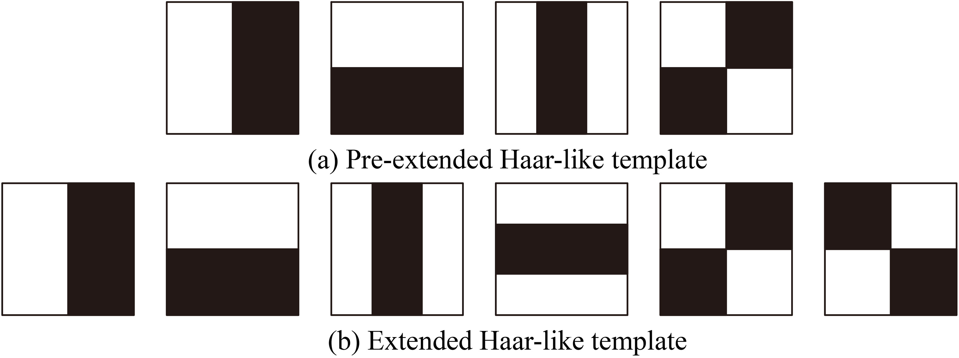
Haar-like feature is widely used in regional location recognition. Haar-like feature template first defines a rectangular area in the image, and describes the feature value by calculating the difference of the sum of the gray values of the adjacent pixels in the rectangular area. This method can clearly reflect the local gray changes of the detection area, and it can be used as the training feature of the classifier to significantly improve the image. Detection performance of labeled area. The original Haar-like feature template exists in the form of four rectangles and calculates the eigenvalues in the rectangular region. However, the form of feature areas in medical images is more complex. For this reason, this paper further expands the Haar-like template style. The Haar-like template before and after the expansion is shown in Fig. 1. In this figure it can be seen that the extended Haar-like template has better adaptability to the complexity of different regions in medical images.
Figure 1.
Comparison of Haar-like templates before and after expansion.
When using Haar-like template for feature calculation, the large amount of computation will affect the overall real-time performance of the algorithm. Therefore, the cumulative sum method based on integral graphs is used to simplify the calculation. The specific process is as follows: First, the horizontal search is carried out in the template, and
(1)
(2)
For different templates that may appear in Fig. 1, the pixel values of a four-pixel region are calculated as follows:
(3)
The Haar-like features of any scale can be calculated in a constant time by using the integral graph method. At the same time, the eigenvalues calculated by the integral graph method are not related to the size and shape of the image, but only to the image integral graph. Therefore, the eigenvalues at any point can be obtained by simple addition and subtraction operations, which effectively guarantees the real-time performance of the algorithm.
When extracting visual features from the Haar-like feature extension template, the rectangular template is scanned in the positive sample image, and the Haar-like feature values of the edge of the target are obtained according to the corresponding calculation method of the template. If the rectangular template is scanned in the negative sample image area, the detection window with edge characteristics is different from the Haar-like eigenvalue obtained by the detection window with non-edge characteristics. Therefore, this method can quantify the edge features and achieve the purpose of distinguishing edges from non-edges.
In the practical application of medical image, Haar-like feature is extracted from a sample image, and its number is calculated as follows:
(4)
Here,
After the above detection, six types of Haar-like features corresponding to the six templates in Fig. 1b can be obtained, which are marked as
2.2Contour feature extraction considering invariance
In the research of key region localization of medical image, it is customary to treat the image containing the target object as a positive sample and the image not containing the target object as a negative sample. Therefore, the problem of target region detection can be understood as detecting whether part of the image window in the whole image contains the target object.
In fact, the error-free detection of target region is to determine the contour of target region. This requires comparing the attitude information of each region in the region detection, which includes position, scale and direction information.
Considering the invariance of attitude information, this paper sets five contour feature parameters, including two position features, one direction feature and two scale features.
Combining these five contour features, the criteria for detecting the positive samples of the areas to be detected in medical images are as follows:
(5)
Here,
2.3Regional location detection based on two kinds of feature fusion learning
After the previous processing, six Haar-like features and five contour features can be extracted from each region. Some medical images with key regions are learned and trained as prior knowledge, and then unknown image regions are classified, detected and located.
Figure 2.
The block diagram of the key area location method proposed in this paper.
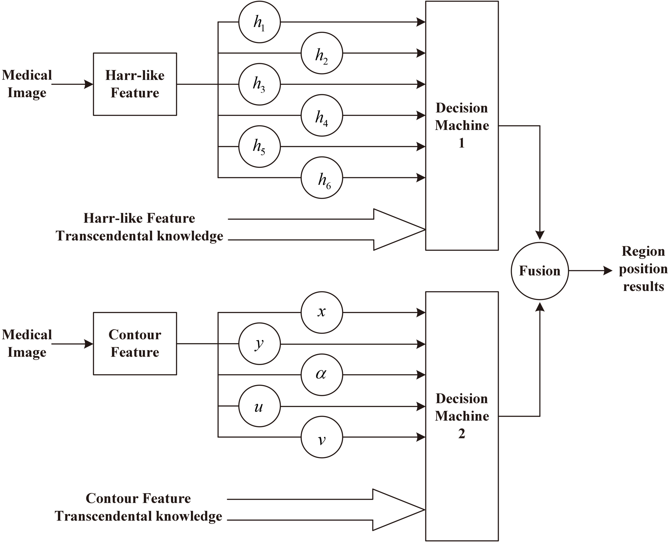
As can be seen from Fig. 2, in the process of detecting the key area, this paper extracts two kinds of features from the detecting image, namely, Haar-like features and contour features, and then combines the prior knowledge of each type of feature to form their own detection results. Then, through the fusion of the two kinds of detection results, a pairing can be formed.
Combining it with the above block diagram, the flowchart of the key region location method for medical image proposed in this paper is as follows: The first step is to extract Haar-like features and contour features from medical images to be processed. The second step is to incorporate the six Haar-like features of medical images and the prior knowledge of the Haar-like features stored in the algorithm into decision maker 1. In the third step, according to the detection result of decision maker 1, it is incorporated into the fuser. In the fourth step, the five contour features of medical images and the prior knowledge of contour features stored in the algorithm are incorporated into decision maker 2. In the fifth step, according to the detection result of decision maker 2, it is incorporated into the fuser. In the sixth step, according to the synthetical detection result of the fuser, the detection result of locating the key areas in the processed image is output.
3.Experimental results and analysis
3.1Experimental configuration
The whole experiment is carried out in the laboratory under its own conditions. The computer configuration of the algorithm is: CPU is 8-core 8-threaded Core Processor, 4.9 GHz main frequency, memory size is 32 GB, and the size of the independent graphics card is 5 GB, and the size of the hard disk is 1 TB.
In the process of experiment, the programming and compiling of the program are all carried out under the environment of MATLAB. The medical image data needed for the experiment is from the cardiac MRI data set, in which all medical images are in the format of MRI. In order to form an intuitive comparison with the method in this paper, the key area location method based on traditional Haar-like feature and the key area location method based on prior knowledge are selected as the comparison method of this method.
In the experiment, the detection parameters are allocated for the Haar-like feature decision maker and the contour feature decision maker. The results are shown in Table 1.
Table 1
Parameter configuration for twelve decision makers
| Feature | Parameters | Value | |
|---|---|---|---|
| 1 |
|
| 0.1 |
| 2 |
|
| 0.1 |
| 3 |
|
| 0.05 |
| 4 |
|
| 0.05 |
| 5 |
|
| 0.1 |
| 6 |
|
| 0.1 |
| 7 |
|
| 2 pixels |
| 8 |
|
| 4 pixels |
| 9 |
|
|
|
| 10 |
|
| 4 |
| 11 |
|
| 8 |
Figure 3.
Location results of key regions found in the remaining 24 cardiac MRI images.

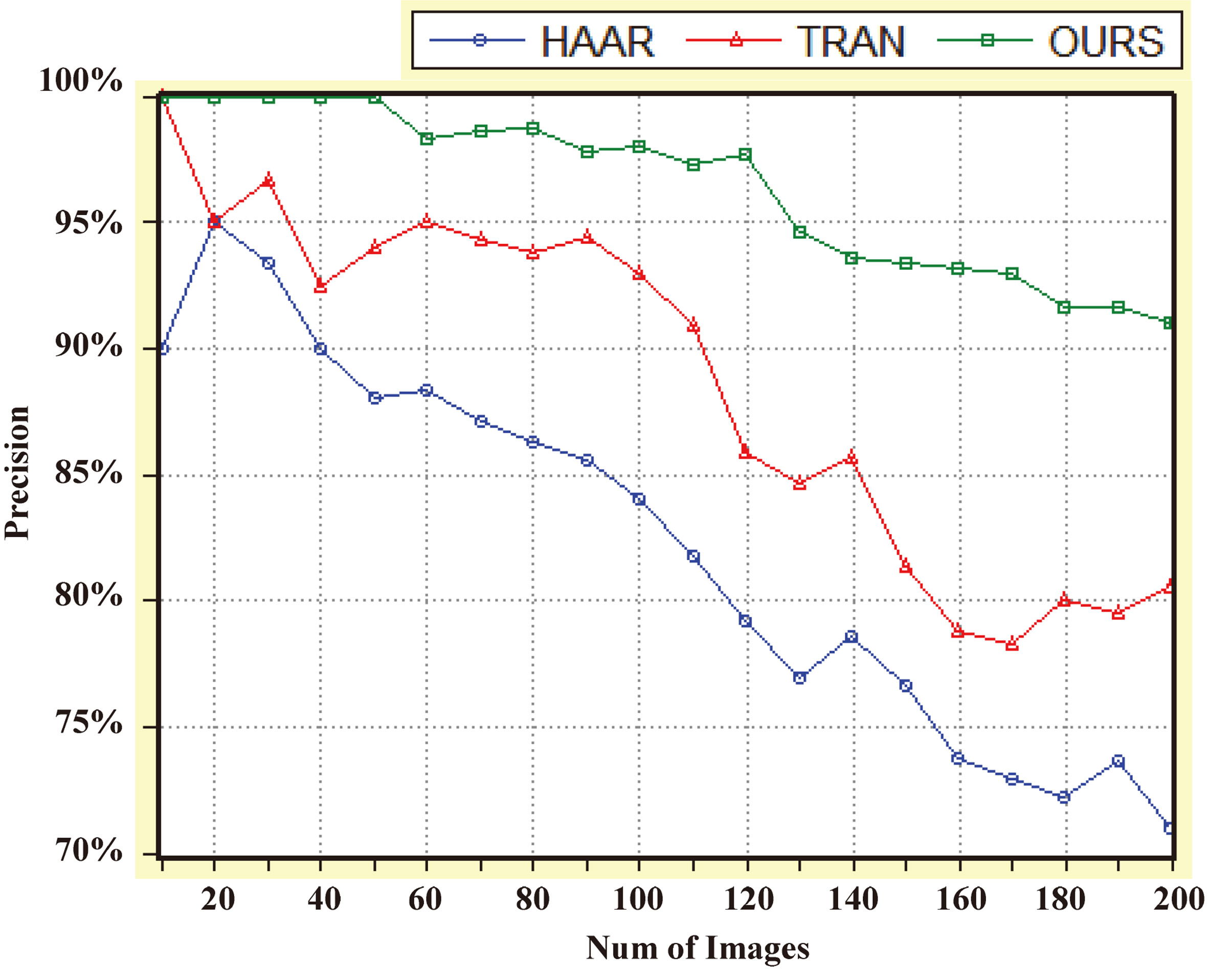
Figure 4.
Comparison curves of positioning accuracy of the three methods.
The method is trained by fusing these prior knowledge, combining the Haar-like features and contour features of each region in each image to be processed, and detecting them in two decision makers and one fuser. Finally, the key feature localization results corresponding to the remaining 12 cardiac MRI images are obtained, as shown in the Fig. 3.
From the experimental results it can be seen that although the cardiac MRI images of different patients are different and the angles in the process of MRI imaging are also different, the methods in this paper can accurately locate the key areas, which fully illustrates that the method has been achieved by using two features and fusing prior knowledge. It has a satisfactory positioning effect.
3.2Comparisons with other methods
In order to compare the performance of the two reference methods, the cardiac MRI data set was further expanded to 200 medical image data sets, including cardiac MRI, brain MRI, chest MRI and spinal MRI.
In the process of comparison, we mainly study how to increase the data capacity of the database step by step, and how to locate the key areas accurately and time. The incremental step of the database capacity is 10 pieces. The curves corresponding to the results are shown in Fig. 4.
From the comparison results of the curves in Fig. 4, we can see that the positioning accuracy of the three methods decreases with the increase of the retrieval image amplitude. Among them, the positioning accuracy of Haar feature-based positioning method decreases the most, while our method keeps the best positioning accuracy. When the number of retrieved images increases to 200, the positioning accuracy of our method is still above 90%.
4.Conclusion
The structure of medical images is complex, and there are many interference information in the target area. In order to improve the accuracy of location and detection of key areas in medical images, a new method is proposed in this paper. In this method, both Haar-like features and contour features are used. By improving the processing, Haar-like template is increased to 6, which has better adaptability to the complexity of medical structure and achieves better solution speed by combining integral graph. Two position features, one angle feature and two scale features are used to form a comprehensive expression of contour features, which takes into account the invariant attributes of key areas in different perspectives. A two-feature fusion location detection method based on prior knowledge is constructed. Haar-like features and contour features are combined with the corresponding prior knowledge into their respective decision makers, and then the final decision results are obtained through the processing of the fusion. Experiments are carried out on the medical image data set of MRI. The experimental results show that the positioning accuracy of the proposed method for key areas is significantly higher than that of the two reference methods. When the database capacity continues to expand, the positioning accuracy for key areas remains above 90%.
Acknowledgments
This study was supported by the National Natural Science Foundation of China (no. 61774107), the University Nursing Program for Young Scholars with Creative Talents in Heilongjiang Province (no. UNPYSCT-2017086), the Fundamental Research Foundation for Universities of Heilongjiang Province (no. LGYC2018JQ014), and the Heilongjiang Postdoctoral Scientific Research Developmental Fund.
Conflict of interest
None to report.
References
[1] | Banuaji A, Cha H-K. A 15-V bidirectional ultrasound interface analog front-end IC for medical imaging using standard CMOS technology. IEEE Transactions on Circuits & Systems II Express Briefs. (2017) ; 61: (8): 604-608. |
[2] | Lei H, Peng Z, Everding B, et al. A comparative study of deformable contour methods on medical image segmentation. Image & Vision Computing. (2008) ; 26: (2): 141-163. |
[3] | Heimann T, Meinzer H-P. Statistical shape models for 3D medical image segmentation: A review. Medical Image Analysis. (2009) ; 13: (4): 543-563. |
[4] | Ng EYK. A special section on development of new clinical procedures and techniques in modern medicine by medical imaging technology. Journal of Medical Imaging & Health Informatics. (2017) ; 7: (4): 840-843. |
[5] | Bjrn K. Evaluation of open source medical imaging software: A case study on health technology student learning experience. Procedia Computer Science. (2017) ; 121: : 724-731. |
[6] | Chen X, Udupa JK, Bagci U, et al. Medical image segmentation by combining graph cuts and oriented active appearance models. IEEE Transactions on Image Processing A Publication of the IEEE Signal Processing Society. (2012) ; 21: (4): 2035. |
[7] | Alenazy AB, Well RG, Terrence TD. New solid state cadmium-zinc-telluride technology for cardiac single photon emission computed tomographic myocardial perfusion imaging. Expert Review of Medical Devices. (2017) ; 14: (3): 213-229. |
[8] | Wright CL, Binzel K, Zhang J, et al. Advanced functional tumor imaging and precision nuclear medicine enabled by digital PET technologies. Contrast Media & Molecular Imaging. (2017) ; 17: (5): 1-7. |
[9] | Zhang DQ, Chen S-C. A novel kernelized fuzzy C-means algorithm with application in medical image segmentation. Artificial Intelligence in Medicine. (2004) ; 32: (1): 37-50. |
[10] | Aishwarya N, Bennila TC. A novel multimodal medical image fusion using sparse representation and modified spatial frequency. International Journal of Imaging Systems & Technology. (2018) ; 12: (6): 315-322. |
[11] | Tsai A, Wells W, Tempany C, et al. Mutual information in coupled multi-shape model for medical image segmentation. Medical Image Analysis. (2004) ; 8: (4): 429-445. |
[12] | Han S, Huang Y. Medical imaging technology shock and volatility of macro economics: Analysis using a three-sector dynamical stochastic general equilibrium REC model. Journal of X-ray Science and Technology. (2017) ; (2-3): 1-12. |
[13] | Bing NL, Chui CK, Chang S, et al. Integrating spatial fuzzy clustering with level set methods for automated medical image segmentation. Computers in Biology & Medicine. (2011) ; 41: (1): 1-10. |
[14] | Rezakhah A. Integrating spatial fuzzy clustering with level set methods for improving medical imaging segmentation. Computers in Biology & Medicine. (2014) ; 41: (1): 1-10. |

