IMR Press / FBL / Volume 28 / Issue 6 / DOI: 10.31083/j.fbl2806130
Open Access Original Research
A Comprehensive Analysis of Prognostic Indicators in Serous Ovarian Cancer Based on Leukocyte Migration and Immune Microenvironment
Show Less
1 Department of Radiation Oncology, Clinical Oncology School of Fujian Medical University, Fujian Cancer Hospital, 350011 Fuzhou, Fujian, China
2 Department of Gynecology, Clinical Oncology School of Fujian Medical University, Fujian Cancer Hospital, 350011 Fuzhou, Fujian, China
3 Department of Ultrasound, Clinical Oncology School of Fujian Medical University, Fujian Cancer Hospital, 350011 Fuzhou, Fujian, China
*Correspondence: 407062881@qq.com (Lina Tang); sufangqiu@fjmu.edu.cn (Sufang Qiu); 1379423879@qq.com (Qin Xu)
These authors contributed equally.
Front. Biosci. (Landmark Ed) 2023, 28(6), 130; https://doi.org/10.31083/j.fbl2806130
Submitted: 27 November 2022 | Revised: 27 January 2023 | Accepted: 10 February 2023 | Published: 30 June 2023
(This article belongs to the Special Issue Diagnostic, Prognostic, and Therapeutic Biomarkers for Ovarian Cancer)
Copyright: © 2023 The Author(s). Published by IMR Press.
This is an open access article under the CC BY 4.0 license.
Abstract

Background: High-grade serous ovarian cancer (HGSOC) treatment is facing clinical challenges. The tumor immune microenvironment (TME) has recently been shown to perform a critical function in the prediction of clinical outcomes as well as the effectiveness of treatment. Leukocyte migration is enhanced in malignant tumors and promotes immunity. However, its role in how to underlie the migration of immune cells into the TME remains to be further explained in HGSOC. 
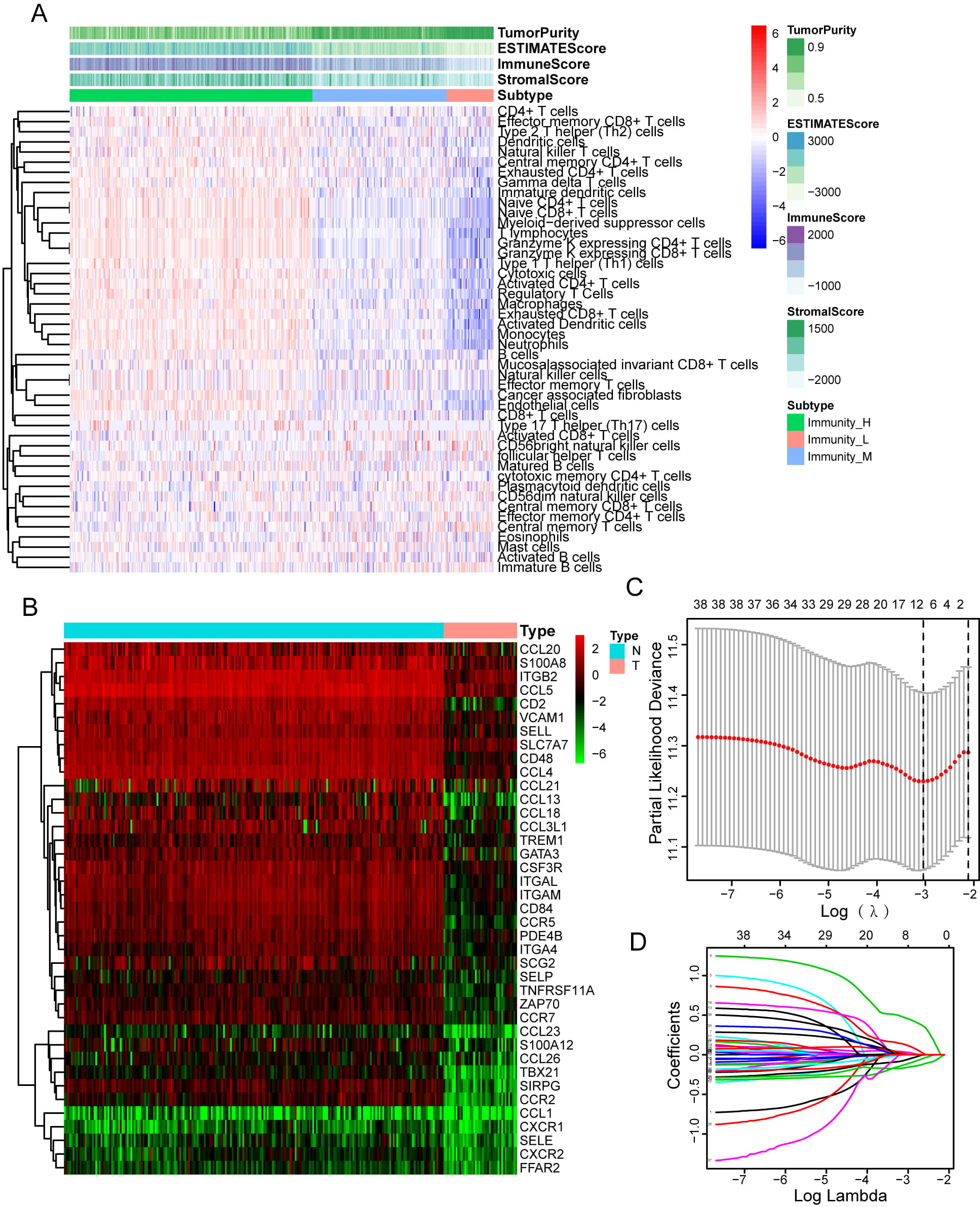
Methods: We built a prognostic multigene signature with leukocyte migration-related differentially expressed genes (LMDGs), which is associated with TME by single-sample gene set enrichment analysis (ssGSEA), in the The Cancer Genome Atlas (TCGA) cohort. Furthermore, we systematically correlated risk signature with immunological characteris-tics in TME, mutational profiles of HGSOC, and potential value in predicting efficacy of platinum-based chemotherapy and immunotherapy. Screening of the most important prognostic factor among risk signatures by Friends analysis, and immunofluorescence was employed to examine both the expression of CD2 as well as its relationship with CD8 and PD-1. Results: LMDGs-related prognostic model showed good prediction performance. Patients who had high-risk scores exhibited significantly reduced progression-free survival (PFS) and overall survival (OS) than those with low-risk scores, according to the results of the survival analysis (p < 0.001). In the TCGA cohort, the risk signature was found to have independent prognostic sig-nificance for HGSOC (HR =1.829, 95% CI = 1.460–2.290, p < 0.001) and validated in the Gene Expression Omnibus (GEO) cohort. Samples with high-risk scores had lower levels of CD8+ T cells infiltration. The low-risk signature shapes an inflamed TME in HGSOC. Furthermore, immune therapy might be effective for the low-risk subtype of HGSOC patients (p < 0.001). Friends analysis revealed that CD2 was the most important prognostic gene among risk signatures. Real-time quantitative PCR analysis showed the expression of CD2 was greater in tumor cells as opposed to normal ovarian cells. CD8, PD-1, and CD2 were shown to be co-localized in HGSOC tissues, according to immunofluorescence analyses. CD2 was significantly correlated with CD8 (r = 0.47). Conclusions: Our study identified and validated a promising LMDGs signature associated with inflamed TME, which might offer some prospective clinical implications for the treatment of SOC. CD2 might be a novel biomarker to predict immune efficacy.

Keywords
leukocyte migration
HGSOC
inflamed TME
CD2
TCGA
Funding
2020CX0101/Joint Funds for the National Clinical Key Specialty Construction Program
2020J011126/Natural Science Foundation of Fujian Province
2020Y2012/Fujian Provincial Clinical Research Center for Cancer Radiotherapy and Immunotherapy
Figures
Fig. 1.
Share
Back to top