
More Information
Submitted: 28 November 2019 | Approved: 03 January 2020 | Published: 06 January 2020
How to cite this article: Erdheim J, Koeck H. A case study on Erdheim ‐ Chester Disease. Arch Case Rep. 2019; 4: 001-003.
DOI: 10.29328/journal.acr.1001027
Copyright License: © 2020 Erdheim J, et al. This is an open access article distributed under the Creative Commons Attribution License, which permits unrestricted use, distribution, and reproduction in any medium, provided the original work is properly cited.
Keywords: Pathology; Erdheim - Chester Disease; Erdheim gsell syndrom; Idiopathic medionecrosis; Ernst Freund; Henry Jaffe; Jakob Erdheim Institute Vienna

A case study on Erdheim ‐ Chester Disease
Jakob Erdheim and Harald Koeck*
Vienna 1070, Zieglergasse 31/16/455, Austria
*Address for Correspondence: Harald Koeck, Vienna 1070, Zieglergasse 31/16/455, Austria, Tel: +43 664 4118467; Email: haraldkoeck@yahoo.de
A case study on Jakob Erdheim-Chester disease.
Jakob Erdheim, pathologist, collector, scientist and educator was born in 1874 in Galicia and received his medical degree from the University of Vienna in 1900. He became interested in pathology and joined the Pathology Institute of the Municipal Hospital (Lainz) of Vienna (Figure 1).
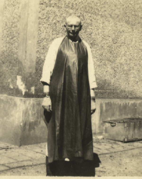
Figure 1: IMAGE kuba.jpg
In 1923, he became Director of that Institution and with great commitment. He performed thousands of autopsies and trained hundreds of students in pathology [1]. Among his revering students were Fritz Schajowicz, Henry Jaffe, Heinrich Karpas, Leo Low-Beer, Ernst Freund and Fuller Albright. His capacity as an investigator is reflected in his remarkable, studies of hyperparathyroidism, acromegaly, Paget’s disease, pituitary gland abnormalities, action of growth hormone and a variety of pathologic entities (Figure 2).
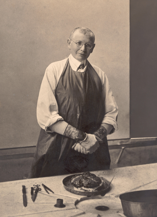
Figure 2: IMAGE jacob with brain.jpg
Jakob Erdheim was a reclusive pathologist. He lived in a room in his Hospital, he was not married and was twice hospitalized, once for tuberculosis once and later for typhoid fever. He recovered from both but concealed his illnesses from his colleagues and students. According to Dr. Sharon Romm, in 1937, while alone in his hospital residence room Jakob Erdheim suddenly died (67) of previous unrecognized cardiac disease [2,3].
There is some doubt as to this cause of his death although allegedly an autopsy disclosed the occlusion of his left coronary artery [4]. A few months prior to his death Erdheim sent his entire collection of pathologic material concealed in a rug to the United States to Ernst Freund who was working with Henry Jaffe at the Hospital for Joint Diseases at the time. When Freund died, he willed the collection to Jaffe; who in turn, when he died, willed it to Henry Mankin. Erdheim was Jewish and the reason for his desire to send this extraordinary collection of pathology away may have been related to the Viennese Anschluss, during which many people of various faiths were killed by the Nazis (Figure 3).
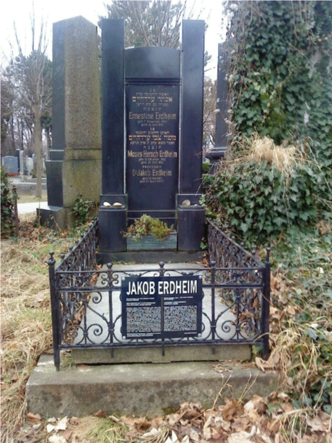
Figure 3: Austia, Vienna - Central Cemetery, Gate 1 or 11, Old Jewish Cemetery Grave 53B/ R28. IMAGE DSC00760.JPG.
Erdheim-Chester Disease
Erdheim-Chester disease is one of the most rarest and most mysterious clinical entities. In 1930 this disease was described by William Chester in an article in Virchow’s Archives. At the time Chester was a pupil of the famous Viennese pathologist and educator Jakob Erdheim. Henry Jaffe, a colleague and great admirer of Erdheim, subsequently named the entity, Erdheim-Chester disease. The disease is characterized by development of xanthogranulomatous material in histiocytes in multiple sites including the skin, heart, breast, marrow, kidney, lungs, abdomen, eyes, brain, pituitary gland and bones. To some extent the histologic character of the disease resembles Langerhans cell histiocytosis but differs in terms of the age at onset, the biochemical characteristics, a striking difference in the nature of the bone lesions and the degree of neurologic involvement [5]. The disease is so rare that there have been very limited opportunities for research as to causation, but thus far, there is no evidence for an infectious, genetic or biologic cause (Figures 4-6).
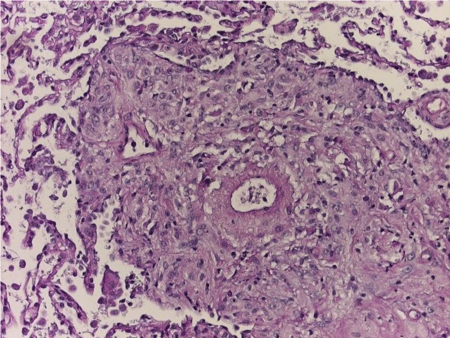
Figure 4: IMAGE BILD 0001.JPG.
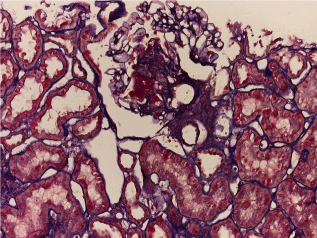
Figure 5: IMAGE BILD 0001.JPG.
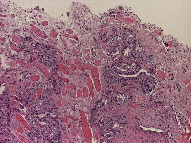
Figure 6: IMAGE BILD 0001.JPG.
Erdheim Gsell Syndrom
The variants of aortic lesions were revealed in autopsy material: intimal tear with the following dissection which extends along the aorta to variable distances - in most cases into iliac arteries. The morphologic changes, associated with were: anuclear zones, elastolysis, thinning, dyschromia and fragmentation of elastic fibers, focal hyperelastosis. Such changes were revealed in all parts of the aortic wall, independently of the site of rupture.
Idiopathic medionecrosis
Here one see an aortic and cardiac specimen with a disorder, which was first described by Jakob Erdheim. It is the socalled idiopathic medionecrosis “Erdheim-Gsell”. In this aortic specimen one can see that the inner layer of the aorta, the vascular wall has dissected interrupted and separated into its various different layers. The tunica media is exposed because part of the tunica intima has flapped back (Figure 7).
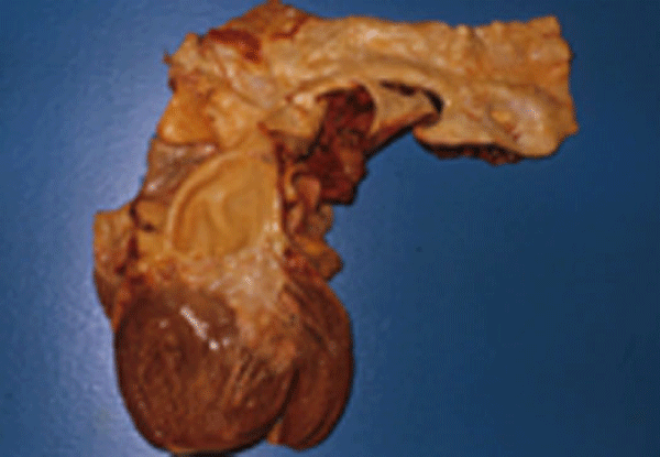
Figure 7: IMAGE Medianekro.jpg.
The cause of this disease is a degeneration of the connective tissue or collagen and elastic fibers in the wall of the aortic. One apparently sees macroscopic scattered, aortic sclerotic looking plagues in the aortic intima lining, as a substance partially designed grey white, glassy - partially liquid.
The heart is dilated, that is the heart chambers are effectively enlarged because of the change in the aorta the heart muscles have an increased volumetric load and therefore is an increase of pressure which leads to continuous hypertension. This results in a further enlargement of the heart chambers. In the heart section, one can recognize this well here in the aortic arch which is shaped like a roman arch or bow [6]. The trapezius muscle and papillary muscles have laminated because of the overload caused by the increased pressure.
One also sees that below the point of dissection, the aorta above the heart valves has bluged. This is called Aortic Neurysma or Aneuyrrisma.
Jakob Erdheim Institute Vienna
The Jakob Erdheim Institute - Chair Univ. Prof. Walter Ulrich MD - is one of the largest pathological institutes in Vienna. About 1.750 autopsy services, 31.000 histopathogical, 8.500 cytological and 80.000 microbiological examinations are carried out here per year. These methods are part complemented by modern molecular biological techniques [7]. Pathology is therefore not only relevant to post-mortem diagnosis but also to the quality of clinical diagnosis. The findings collected here form the basis for the diagnosis and therapy in clinical medicine, being the essential aspects of quality assurance in a hospital [8] (Figures 8,9).
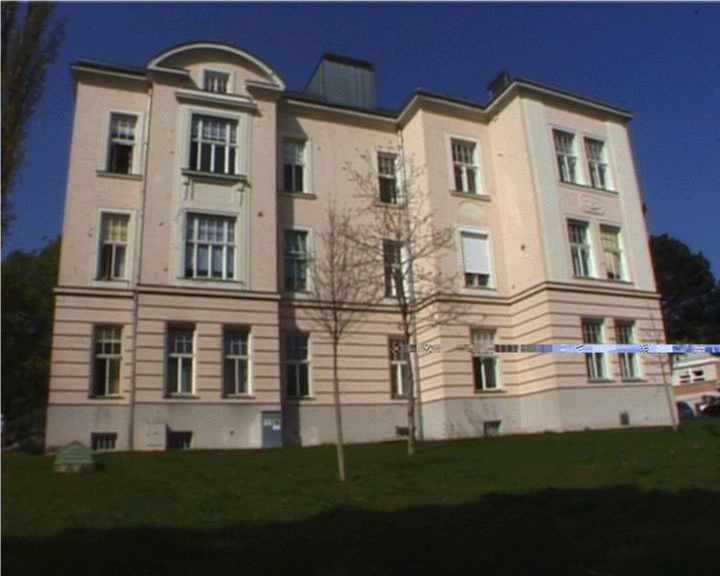
Figure 8: IMAGE FotoPatho_54677.jpg.
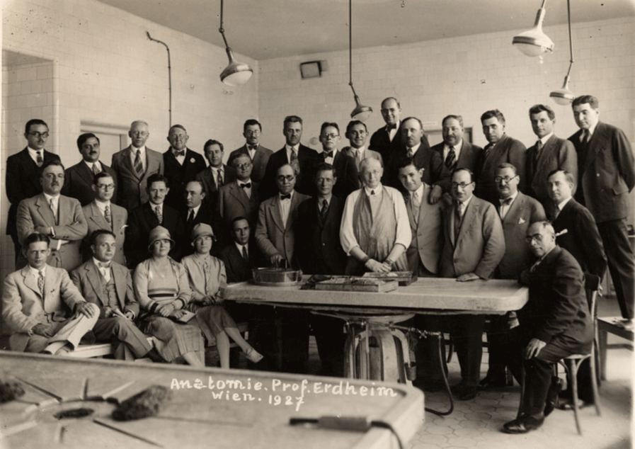
Figure 9: IMAGE erdheim 1927.
- Adam Erdheim (early photographs of Jakob Erdheim).
- Univ. Prof. Walter Ulrich MD (idiopathic medionecrosis)
- Dr. Sharon Romm (Eminent Pathologist of Vienna NCBI)
- Wikipedia Jakob Erdheim
- Erdheim Chester Disease (article in Virchow’s Archives in 1930)
- Edwin E. Osgood Collection (photograph of Professor Jakob Erdheim in Vienna, 1927)
- Harald Koeck (photographs of Jakob Erdheim Institute and Grave Vienna)
- Harald Koeck (film – The reclusive pathologist 2012)