Article Text

Abstract
Introduction Internal carotid artery termination (ICAT) and proximal A1 aneurysms can be challenging for open surgical clipping or endovascular coiling. Treatment with flow diversion covering the middle cerebral artery (MCA), an end vessel supplying a terminal circulation, has not been reported.
Methods A prospective, Institutional Review Board-approved database was analysed for patients with pipeline embolisation device (PED) placement from the anterior cerebral artery (ACA) to the ICA during cerebral aneurysm treatment.
Results Nine cases were identified, including five proximal A1, three posterior communicating artery and one ICAT aneurysm locations. Average aneurysm size was 8.3 mm (range 3–17), with 67% saccular and 78% right-sided. Primary indication for treatment was significant dome irregularity (44%), recurrence or enlargement (33%), underlying collagen vascular disorder (11%) and traumatic pseudoaneurysm (11%). Preservation of the ipsilateral ACA (with PED placed in A1) was performed when the anterior communicating artery (67%) or contralateral A1 (33%) were absent on angiography. Adjunctive coiling was done in four cases (44%). There was one major stroke leading to mortality (11%) and one minor stroke (11%). Clinical follow-up was 27 months on average. Follow-up digital subtraction angiography (average interval 15 months) showed complete aneurysm obliteration (88%) or dome occlusion with entry remnant (12%). The jailed MCA showed minimal or mild delay (primarily anterograde flow) in 75% of cases and significant delay (reliance primarily on ACA and external carotid artery collaterals) in 25%.
Conclusions Covering the MCA with a flow diverting stent should be reserved for select rare cases. Strict attention to blood pressure augmentation during the periprocedural period is necessary to minimise potential ischaemic compromise.
- coil
- flow diverter
- aneurysm
- technique
- intervention
This is an open access article distributed in accordance with the Creative Commons Attribution Non Commercial (CC BY-NC 4.0) license, which permits others to distribute, remix, adapt, build upon this work non-commercially, and license their derivative works on different terms, provided the original work is properly cited, appropriate credit is given, any changes made indicated, and the use is non-commercial. See: http://creativecommons.org/licenses/by-nc/4.0/.
Statistics from Altmetric.com
Introduction
Proximal A1, internal carotid artery (ICA) termination and distal supraclinoid ICA aneurysms with a short postcommunicating segment are challenging cases for traditional treatment with open microsurgical clipping or endovascular coiling.1–3 Initial success with proximal ICA aneurysms has led to off-label use of flow diverters for aneurysms in more distal locations, often covering branch vessels.4–9 Consensus is that directly collateralised side branches covered by a flow diverter stop filling anterograde while end vessels supplying terminal circulations continue filling anterograde;10 however, one side branch for which this theory has not been verified is the middle cerebral artery (MCA). This was a retrospective case series on the safety and efficacy in our experience with flow diversion from the anterior cerebral artery (ACA) into the distal ICA covering the M1 origin.
Methods
This was a retrospective cohort study using an Institutional Review Board-approved, prospectively collected database of aneurysm patients at a tertiary medical centre. The treatment decision was based on (a) high risk aneurysms based on history of rupture, morphology or underlying aetiology (eg, traumatic, collagen vascular disorder) (b) obligation to cross the ICA termination given the location of the aneurysm and (c) need to preserve the ipsilateral ACA because of absence of contralateral ACA or anterior communicating artery (ACoA). Patients were started on dual antiplatelet therapy with Aspirin 325 mg and Clopidogrel 75 mg daily 7 days prior to intervention. Antiplatelet therapy monitoring of P2Y12 levels was performed in cases since 2014. Single modality flow diversion was performed through a tri-axial setup as previously described11 12 and single-stage flow diversion with adjunctive coiling was performed as described separately.13 Demographics, clinical history and outcomes were collected from medical records. Anatomic and technical details were collected from intraprocedure events, angiograms and operative reports. Follow-up consisted of digital subtraction angiography (DSA) at 6, 12 and often at 24 months postembolisation. Occlusion was graded according to the O’Kelly-Marotta scale for flow diversion14 as complete, trace filling, entry remnant or aneurysm filling.
Results
In the institutional database of over 700 flow diversion cases, nine were identified where traditional treatment options with open microsurgical clipping or endovascular coiling were limited and necessitated placement of a flow diverter from the ACA into the distal ICA covering the M1 origin. These cases included five proximal A1, two ICA posterior communicating artery (PCoA) and one internal carotid artery termination (ICAT) aneurysms. One additional patient had two aneurysms with a recurrent, previously coiled ICAT and adjacent newly formed PCoA aneurysm. Average aneurysm size was 8.3 mm (±4.2, range 3–17). Morphology was 56% saccular, 33% fusiform and 11% dissecting. Most patients were female (7/9, 78%) and most aneurysms were right-sided (78%).
The primary indication for treatment was significant dome irregularity (44%), recurrence or enlargement (33%), underlying collagen vascular disorder (11%) and traumatic pseudoaneurysm (11%). Absence of the ACoA (67%) or contralateral A1 (33%) necessitated preservation of the ipsilateral A1.
The average fluoroscopy time was 62 min and radiation exposure was 3066 mGy. A single pipeline embolisation device (PED) device was used in all cases and adjunctive coiling was performed in four cases (44%). Intra-arterial verapamil was administered for spasmolysis in one case (11%), balloon angioplasty was not performed (0%) and platelet aggregation along the stent was observed in three cases (33%). Two of these were observed intraprocedurally and resolved with Abciximab administration. One patient (case 5) awoke with aphasia and hemiparesis; immediate DSA demonstrated platelet plugging at the orifice of the covered M1 which resolved with intra-arterial Abciximab and complete resolution of clinical deficits.
Two patients (22%) experienced complications resulting in permanent neurological deficit, including one major stroke that led to mortality (11%) and one minor stroke (11%) (table 1).
Case 3 was a septuagenarian with 8 mm proximal left A1 aneurysm treated with PED and coiling. Post-treatment angiography showed slight left MCA delay which resolved with blood pressure augmentation; however, hypotension during management of a retroperitoneal hematoma requiring surgical repair led to left MCA stroke ultimately resulting in death. Case 6 was a cinquagenarian smoker with 3 mm proximal right A1 aneurysm treated with PED after unsuccessful attempted stent-coiling. The patient received intra-arterial Abciximab at procedure end for delayed filling of the covered right MCA. The patient had some left upper extremity weakness on awakening, which resolved with blood pressure augmentation. On postoperative day one in the intensive care unit, the patient developed left upper extremity weakness after an episode of vasovagal hypotension that occurred during removal of the femoral sheath. MRI showed scattered MCA territory ischaemia and the patient was mRS 2 at last follow-up.
Patients were followed clinically for an average of 27 months. Follow-up DSA showed complete aneurysm occlusion in 7/8 cases (88%) at an average interval of 21 months. Follow-up DSA was performed in all living patients at 6 months, 88% at 12 months and 50% at 24 months. The covered MCA showed no delay or minimal delay (arterial phase filling) in 50% of cases (figure 1, case 4), moderate delay with primarily anterograde flow in 25% of cases (figure 2, case 5) and significant delay (reliance primarily on ACA and external carotid artery (ECA) collaterals) in 25% (figure 3, case 1).

Case 8: Cinquagenarian with (A,B) R ICA DSA showing growing residual of a previously coiled 8 mm right A1 aneurysm and (C) L CCA DSA showing absent ACoA. (D) Unsubtracted DSA during PED placement from R ACA into R ICA across M1 origin, which continued to fill anterograde, without delay, in parallel with the ACA at both (E) 6 months and (F) 12 months follow-up R ICA DSA. ACA, anterior cerebral artery; ACoA, anterior communicating artery; CCA, common carotid artery; DSA, digital subtraction angiography; ICA, internal carotid artery; PED, pipeline embolisation device.
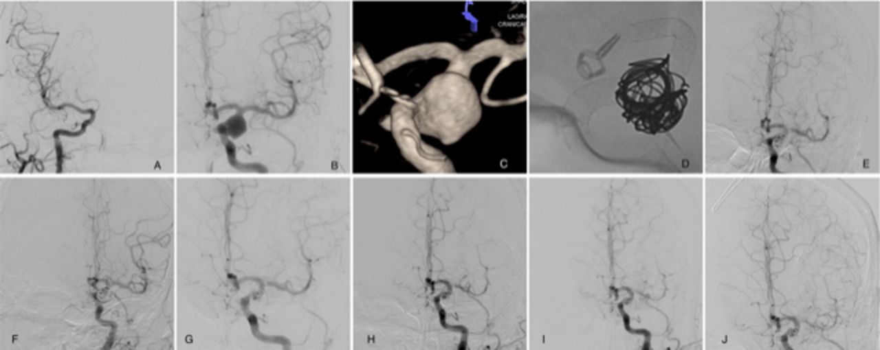
Case 5: Sexagenarian with (A) R CCA DSA showing absent ACoA and (B) L CCA DSA showing fusiform PCoA aneurysm with (C) dome irregularity and short postcommunicating landing zone in the ICA. (D) Treatment with single-stage pipeline with adjunctive coiling from the ACA into the ICA. (E) Immediate postprocedural DSA showing platelet aggregation at the M1 origin and delayed MCA filling when the patient awoke with right upper extremity (RUE) weakness (F) resolved clinically and improved angiographically after intra-arterial Abciximab administration. Follow-up DSA at (G) 2 months, (H) 6 months, (I) 12 months, after which Prasugrel was weaned and (J) 24 months showing progressive recruitment of pial collaterals from the ACA and ECA to supply the MCA territory and moderately delayed filling of the covered MCA in the parenchymal phase. ACA, anterior cerebral artery; ACoA, anterior communicating artery; CCA, common carotid artery; DSA, digital subtraction angiography; ECA, external carotid artery; ICA, internal carotid artery; MCA, middle cerebral artery; PCoA, posterior communicating artery.

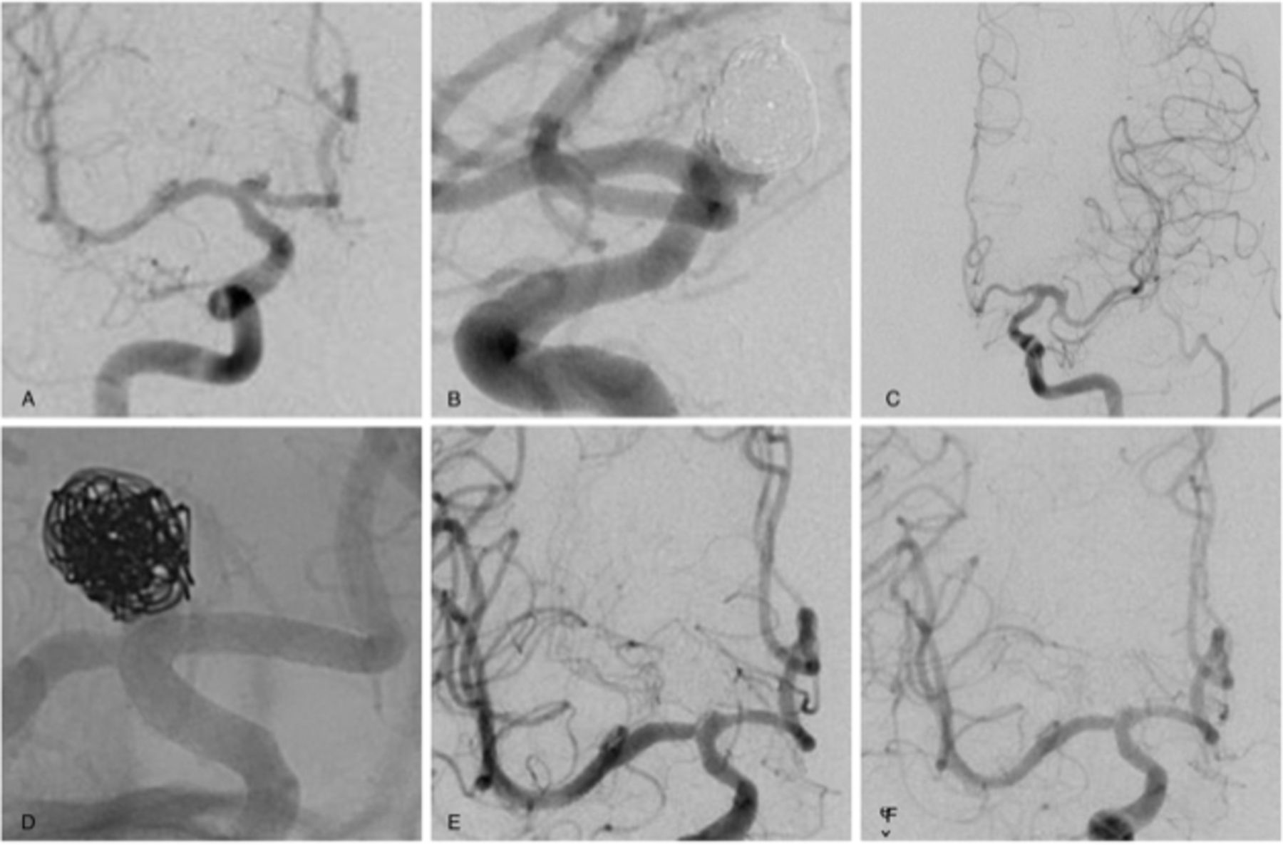
Case 1: Teenage patient with history of bicoronal craniotomy for craniopharyngioma resection followed by proton beam therapy who presented with (A) R ICA DSA showing 11 mm A1 aneurysm and (B) L CCA DSA showing hypoplastic left ACA. (C) 3-D rotational angiography shows fusiform and highly irregular morphology. (D) Unsubtracted DSA from treatment with single-stage pipeline with adjunctive coiling from R ACA into R ICA. Follow-up R CCA DSA at (E) 2 months shows dome occlusion of the aneurysm with residual neck filling and anterograde arterial phase filling of the jailed R MCA. (F) 6 months shows some ghosting across the M1, (G,H) increased ghosting at 12 months follow-up DSA after stopping Plavix with significant recruitment of pial collaterals from the ACA in the late arterial and parenchymal phase. (I) 24-month DSA arterial phase shows limited anterograde filling of the jailed MCA and (J) robust pial collaterals from ACA and ECA apparent on parenchymal phase runs. ACA, anterior cerebral artery; CCA, common carotid artery; DSA, digital subtraction angiography; ECA, external carotid artery; ICA, internal carotid artery; MCA, middle cerebral artery.
Discussion
Treatment with the flow diverting stent that covered the M1 origin was used as a last resort option with only nine cases identified in an institutional database of over 700 flow diversion cases. These nine cases were for high-risk, challenging aneurysms located near the ICA termination where traditional treatment approaches with open microsurgical clipping and endovascular coiling were limited. Morbidity was significant, with ischaemic stroke in 2/9 patients (22%) resulting in one mortality (11%). Both these cases highlight the need for improved perioperative blood pressure management to minimise the ischaemic complications of this approach. Efficacy was good, with complete aneurysm occlusion in 88% of aneurysms. No complications were observed beyond the initial 48 hours following embolisation over 27 months of clinical follow-up, even with diminution of the MCA and reliance on collateral supply from the ACA or ECA in half of cases.
Patients in this series underwent flow diversion only after extensive consideration was given to the need to treat. Proximal ACA aneurysms have a notable tendency to rupture at small sizes,2 while ICAT aneurysms commonly present with rupture in young patients,1 both factors arguing for prophylactic treatment of unruptured aneurysms. In each case, the decision to treat was also based on patient-specific factors that increased the risk of rupture, including: recalcitrant smoking, aetiological factors including trauma or connective tissue disorder, morphological factors including dome irregularity and high aspect ratio or a history of SAH from this now-recurrent aneurysm or another aneurysm.
The decision to proceed with flow diversion treatment for the patient in this series was also made after extensive discussion on the limitations of traditional open surgical or endovascular coiling options. Many have argued that open surgery is the standard of care for aneurysms in the ICA termination region. However, the intraoperative challenges of protecting the medial lenticulostriates and other perforating arteries can result in significant ischaemic complications. ICA termination and proximal ACA aneurysms are also notably thin-walled and, when projecting posteriorly or superiorly, can be embedded in parenchyma, which heightens the risk of intraoperative rupture during a surgical approach.1 2 Patient-specific factors increased the risk associated with open surgical treatment in some cases in this series, including a history of radiation to the region (Case 1), uncontrolled bleeding during prior surgeries (Case 7) and a traumatic pseudoaneurysm too friable to clip (Case 9).
Endovascular coiling of ICA termination and proximal ACA aneurysms can be challenging because regional tortuosity and the direction at which the aneurysm dome projects often prevents stable access to the aneurysm dome, risking intraprocedural rupture. Aneurysms in this region tend to be discovered and to rupture at small sizes, which increases the risk of rupture during coiling.3 Several aneurysms in this series were fusiform or wide-necked and for that reason not amenable to the traditional endovascular options of coiling, balloon-coiling or a dual-microcatheter coiling technique. There is a limited literature on stent-assisted coiling for aneurysms specifically in this location;15 however, stent placement followed by coiling increases the challenge of dome access. One aneurysm in this series (Case 6) was treated with a flow diverter only after the aneurysm dome could not be stably catheterised for attempted stent-coiling with a jailed coiling catheter.
Consideration was given to treatment with alternative flow diverter configurations. In patients with a patent ACoA, placement of a flow diverter from the MCA into the distal ICA can be used to reconstruct the ICA termination, causing gradual involution of the ipsilateral A1 and associated aneurysms.8 16 Horizontal placement of a PED at the ICA termination from the MCA into the ACA has been reported.17 Patients in this series, however, lacked a patent ACoA, preventing horizontal PED placement; several lacked a contralateral A1, heightening the necessity of preserving the ipsilateral A1.
While the PED was originally approved for proximal carotid aneurysms, the fate of covered branch vessels became a controversial subject as use expanded to off-label locations in more distal locations. Side-branches were initially grouped together and occlusion rates reported in aggregate.9 Saleme et al introduced a more sophisticated framework, distinguishing between collateralised branch vessels (eg, ACA with a patent ACoA) and those without direct collaterals (eg, anterior choroidal) in a series of 37 bifurcation aneurysms. Directly collateralised side branches showed a lack of anterograde filling on follow-up DSA in 71% of cases, always asymptomatic; side branches supplying terminal circulations continued to fill in 91% of cases and 22% of patients experienced symptoms attributed to the covered territory.10
Corroborative studies have since emerged for many of the major branch vessels that could conceivably be covered with a flow diverting stent. Fetal-type PCA always filled, while adult PCoA showed diminished or absent anterograde flow in up to 45% of cases.8 18 19 In the rare reported instances of A1 coverage, always in the presence of a patent ACoA, the A1 commonly stopped filling anterograde8 16 By contrast, two series reported 0% of covered anterior choroidal arteries stopped filling8 18 and two reported just one such instance.20 21 Mazur et al reported 11 V4 or PICA aneurysms treated with PED covering PICA, eight of which had DSA follow-up, none showing compromised flow.22 The ophthalmic artery is a unique case of a terminal vessel that showed higher rates of anterograde flow cessation of 4%–21%8 18 23 because of frequent well-developed collaterals from the ECA. The controversy regarding covered side branches has thus achieved some consensus that ‘end vessels’ supplying ‘terminal circulations’ will continue to fill while those supplying collateralised territories will often, without clinical symptoms, stop filling anterograde.
Because of the eloquent and end vessel nature of the MCA, one scenario in which this consensus has not been verified is placement of a flow diverting stent from the ACA into the ICA across the M1 origin. Our literature review identified no reports of flow diverter deployment in this configuration. Flow diversion of MCA bifurcation aneurysms, on the other hand, is increasingly common, with flow modification of covered M2 branches reported at rates higher than observed for other end vessels. In a meta-analysis of 244 MCA aneurysms, 10% of covered side branches stopped filling anterograde and 26% showed slow flow on follow-up DSA, with associated symptoms in 5% of patients.24 This may be due to diminished arterial pressures across the stent braid in more distal locations. A reluctance to cover M1 is revealed by the images showing the off-centre deployment of flow diverters to treat proximal ACA aneurysms.25 26 Van Rooij reported an A1 aneurysm treated with a flow diverter, the proximal end of which protruded into the ICA termination, after which the patient experienced a perforator infarction.26 In our experience with flow diversion of proximal A1 aneurysms, we deployed the stent entirely within the ACA whenever possible; however, in cases with insufficient landing zone, we preferred to bring the stent into the ICA rather than to end within the termination or to insufficiently cover the neck and risk stent prolapse into the aneurysm.
Our experience with flow diversion across the M1 origin leads us to a more nuanced perspective on covered side branch patency: Long-term patency depends on the extent of pial collateralisation. Some patients develop robust pial collaterals, in these cases from the ACA and the ECA and can come to rely on these primarily, leading to reduced anterograde filling of the M1 across the flow diverter and allowing endothelialisation across the M1 origin (figure 3, case 1). In these patients, ‘ghosting’ across the M1 origin may be apparent on 6-month follow-up DSA; extensive pial collaterals are then visualised at 12 months after stopping Plavix and near-complete occlusion of the M1 origin is apparent on 24 month DSA. Limited follow-up of 12 months or less is a problem shared by much of the literature on side branch occlusion after flow diversion.8 9 20 Rare instances of asymptomatic cessation of anterograde flow in side branches on long-term DSA may provoke alarm when the reality is this is a common silent occurrence beyond the window of routine imaging. We have observed this phenomenon previously other locations, such as a patient with ACoA aneurysm treated with a PED from the ipsilateral A1 to the contralateral A2, jailing the ipsilateral A2. Twelve-month follow-up DSA in that case showed no anterograde filling of the ipsilateral A2, with territorial supply by pial collaterals from the MCA and splenial collaterals from the PCA.5 Patients that are unable to recruit collaterals will continue to experience demand across the flow diverting stent at the M1 origin, limiting the degree of endothelialisation that can occur. The MCA continues to fill in anterograde fashion, with either symmetric arterial-phase filling of the ACA and the jailed MCA (figure 1, case 8) or slight delay into the parenchymal phase (figure 2, case 5).
The unpredictability in the fate of the covered MCA vascular tree after flow diversion across the M1 origin renders this approach a committed step potentially associated with higher complication rates compared with traditional flow diversion cases. In the two major complications in this series, delayed filling of the covered MCA territory was noted at end-embolisation angiography; both initially corrected with blood pressure augmentation (Case 3) or Abciximab administration (Case 6). However, both patients subsequently experienced symptomatic strokes after hypotensive episodes in the postprocedure intensive care observation; one patient experienced a haemodynamically significant groin haematoma and the other had hypotension with vasovagal episodes. These symptomatic complications were potentially avoidable with more strict blood pressure augmentation throughout the postprocedure period. Other reports of end vessel side branches covered by flow diverters have shown a clustering of complications due to acute stent thrombosis within the first 48 hours postembolisation.22 27 28 In this series, one patient awoke with right-sided hemiparesis and immediate DSA demonstrated platelet aggregation at the M1 origin (figure 2, case 5). This was treated with intra-arterial Abciximab, after which the patient recovered completely.
We continue to reserve flow diverter placement across the M1 origin as a last resort approach for highly selected complex aneurysms in the ICA termination region where traditional microsurgical or endovascular coiling options may be limited. Limitations of this treatment strategy include the committed nature in covering the M1 origin with the inability to predetermine patient-specific pial collateralisation and the inability to perform M1 thrombectomy in the event of future large vessel occlusion in an atherogenic substrate. Our protocol for managing potential future cases now includes control angiography at a full 30 min interval following device deployment and any postprocessing. We recommend prophylactic maintenance of systolic blood pressures at least 160 during the initial 24 hours postembolisation and then gradually relaxing these parameters over several days of observation in the intensive care unit. We have an abiding mistrust of P2Y12 values29 but have tested these patients since 2014. We do not necessarily switch patients with hyporesponsive P2Y12 levels to an alternative agent and none of the complications in this series were seen in such patients. However, patients in whom platelet aggregation is observed intraprocedurally along the stent should be switched to alternative antiplatelet agents such as Prasugrel or Ticagrelor. Given the absence of delayed complications after hospital discharge, we would still taper antiplatelet therapy in a standard fashion for most patients without hyper-endothelialisation on follow-up DSA.
Conclusion
This small series represents the first report of flow diverter deployment across the M1 origin. Flow diversion in this configuration can be an effective but last resort option for treating high-risk distal ICA, ICAT and proximal A1 aneurysms. Vigilance for stent thrombosis and strict blood pressure augmentation are essential for complication avoidance.
References
Footnotes
Contributors All authors contributed significantly to conception, data acquisition and analysis; all drafted and revised manuscript contents and approved the final version of the manuscript.
Funding The authors have not declared a specific grant for this research from any funding agency in the public, commercial or not-for-profit sectors.
Disclaimer No author received financial support in conjunction with the generation of this submission.
Competing interests ALC is a consultant for InNeuroCo, a consultant and proctor for Medtronic Neurovascular, MicroVention-Terumo and Stryker Neurovascular. L-ML is a proctor for Medtronic Neurovascular, consultant for Stryker Neurovascular and Cerenovus. GPC is a consultant for Stryker Neurovascular. The other authors have no conflict of interest.
Ethics approval Johns Hopkins Institutional Review Board.
Provenance and peer review Not commissioned; externally peer reviewed.
Data sharing statement There are no additional unpublished data from this study. The relevant anonymised patient level data are available on reasonable request from the authors.
Patient consent for publication Not required.