


Dear Editor,
A 29-year-old woman developed right dorsal forearm pain one month after giving birth. She stated that the pain caused difficulty while supporting the baby’s head for breastfeeding. Moreover, the patient also mentioned weakness extending into the fingers on the affected side. On physical examination, tenderness over the right dorsal proximal forearm and weakness of finger extension (muscle strength 4/5) were detected. The patient was referred for an ultrasound (US) examination for the differential diagnosis of tennis elbow. Although there was no remarkable finding on the common extensor tendon, sonopalpation over the most painful area (around 5 cm distal to the lateral epicondyle) disclosed that the deep branch of the radial nerve (DBRN) was swollen (Figure 1A;Supplementary Data) in comparison with the asymptomatic side (Figure 1B). On long-axis imaging, the nerve was diffusely enlarged along almost its whole length (Figure 1C). The maximal diameter of the affected nerve was 1.7 mm in its short axis and 1.8 mm in its long axis. In contrast, the same nerve on the asymptomatic side had a maximal diameter of 0.6 mm in the short axis and 0.7 mm in the long axis. US-guided hydrodissection with 4 mL of 5% dextrose in normal saline was administered through an in-plane approach to target the short axis of the nerve in the supinator tunnel (Supplementary Data). US diathermy and intensive forearm stretching exercises were prescribed thereafter. The patient described 80% pain relief and recovery of finger extension strength two weeks after injection, and the nerve on the affected side had a significant decrease in the size (with a maximal diameter of 1.2 mm) compared with pre-injection status.
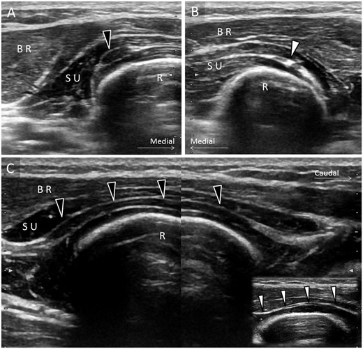
Comparative ultrasound imaging showing (A) the swollen deep branch of the radial nerve (black arrowhead) in the short axis view with a maximal diameter of 1.7 mm. B) Normal size of the nerve (white arrowhead) is seen on the asymptomatic side with a maximal diameter of 0.6 mm. C) On the split-screen view, significant enlargement (black arrowheads) with a maximal diameter of 1.8 mm is again visualized in the long axis of the nerve when compared with the nerve (white arrowheads) with a maximal diameter of 0.7 mm on the healthy side (inset). BR = brachioradialis muscle; R = radius; SU = supinator muscle.
The radial nerve separates into superficial and deep branches at the elbow [1]. Before it enters the supinator tunnel, there is an area called the “leash of Henry,” where the DBRN can be entrapped by recurrent radial vessels. After the DBRN pierces the supinator muscle, it is frequently compressed by the fibrotic arch of the superficial supinator muscle, called the “arcade of Frohse” [2]. When a peripheral nerve is compressed, it usually becomes swollen/edematous proximal to the entrapped point due to blockage of the axoplasmic flow [3]. However, here, the DBRN seemed to be diffusely enlarged without an obvious compressive point. Although there are numerous reports regarding posterior interosseous nerve syndrome [4], ours seems to be the first reported case with diffuse enlargement of the DBRN. Based on the patient’s clinical history, it was speculated that overuse of the supinator and wrist extensor muscles could have led to this compression along the whole supinator tunnel. Yet, during breastfeeding, a mother needs to eccentrically contract the wrist extensors and fully supinate her forearm in order to stabilize the baby’s head in front of her chest. This posture significantly raises the intratunnel pressure and may possibly cause nerve compression throughout a long segment. Furthermore, US imaging showed an enlarged hypoechoic portion of the DBRN without thickening of its epineurium, indicating that the compression was an acute insult. Although the case was not sent for electrophysiological examination, we speculate that a nerve conduction study would have shown conduction blockage of the DBRN. However, without US imaging, we cannot know how long the compressed segment was. As the different entrapment sites cause various neurological complaints [5], US can be used to scrutinize all possible compressive points of the radial nerve from the spiral groove to the exit of the supinator tunnel. Consistent with a recent US study [6], injection of 5% dextrose effectively relieved peripheral nerve entrapment. Because the DBRN is a pure motor nerve, it can be affected by the local anesthetics, resulting in a decrease in muscle power. Therefore, local anesthetics should not be added into the injectate as they could aggravate finger extension weakness. Lastly, physical therapy must be prescribed to stretch the tight forearm muscles and educate the patient about proper posture during breastfeeding.
Funding sources: No funding was received.
Conflicts of interest: The authors declare no conflicts of interest.
