


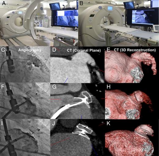
A 67-year-old woman with persistent atrial fibrillation (CHA 2 DS 2 VASc: 6 and HAS BLED: 4) who experienced recurrent stroke and intracranial bleeding on Apixaban was considered for left atrial appendage (LAA) occlusion with Amplatzer Cardiac Plug (ACP). An angiography shared multi-detector computed tomography system (MDCT) was applicable in this procedure ( Panels A and B ). Before the procedure, the dimensions of landing zone were measured at 18–21 mm in transoesophageal echocardiography (TEE), 19–21 mm in angiography, and 20–22 mm in MDCT ( Panels C , D , and E ). Then, 24 mm ACP was selected and deployed without complication. Before releasing the device, conformational change, position and anchoring of device could be assessed with angiography shared MDCT applying 640 channel double-slice technology (Aquilion ONE™; Toshiba Medical Systems, Otawara, Japan) which allows a CT scan to be evaluated during the procedure on the same table. Although angiography and TEE showed that the ACP was appropriately seated in LAA ( Supplementary material online, Videos S1 and S2 ), MDCT clearly demonstrated the configuration of device and complete closure of LAA ( Panels F , G , and H ). Then, device was safely detached from the delivery cable ( Panel I ). On 2 days after procedure, MDCT was performed to check the position of device and communication between LA and LAA ( Panels J and K ). The radiation dose and the amount of contrast via intravenous route for MDCT were 11.2 mSv and 60 mL.
The intraprocedural MDCT is able to provide more accurate information for the position of device and relationship between heart structure and device.
Supplementary material is available at European Heart Journal online.

