Main
Sir,
Wegener's granulomatosis is a systemic inflammatory disease with a broad range of clinical manifestations. The complete form is characterized by necrotizing granulomatous inflammation of the upper and lower respiratory tract, glomerulonephritis, and systemic vasculitis. However, limited disease is not uncommon, and the presenting symptoms and signs may be highly variable.
Ocular disease is the presenting manifestation in 8–16% of patients, in which necrotizing sclerokeratitis and proptosis, caused by orbital inflammation, are most common.1, 2 We describe an unusual ocular manifestation of Wegener's granulomatosis, which illustrates the diversity of the presenting signs of this disease.
Case report
A 45-year-old woman was referred with diplopia since 1 month.
Her medical history featured a superotemporal mass in the right orbit 2 years earlier. At that time, there were no abnormalities on ophthalmic examination, except a diminished tear production. Enlargement of both lacrimal glands was seen on computed tomography. Biopsy showed chronic fibrosing inflammation with areas of ‘smudgy’ necrosis and focally necrotizing vasculitis (Figure 1), but no granulomas and few eosinophils. Although the histopathologic findings were not characteristic for Wegener's granulomatosis, they were suggestive. Serum analysis for antineutrophil cytoplasmic antibodies (ANCA) showed a weakly positive p-ANCA, but c-ANCA was negative. Ear–Nose–Throat (ENT) and pulmonary examination revealed no abnormalities. After 5 months, the symptoms had disappeared without any intervention.
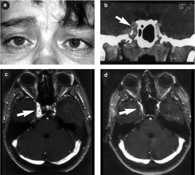
The newly developed diplopia appeared to be caused by a sixth nerve palsy of the right eye. Ophthalmic examination also showed miosis and mild ptosis of the right eye (Figure 2a). After cocaine and hydroxyamphetamine testing, the diagnosis of third-order neuron Horner syndrome was made. This time, no signs of lacrimal gland enlargement were seen, and biomicroscopy, fundoscopy, and Hertel ophthalmometry revealed no abnormalities. Computed tomography (CT) and magnetic resonance imaging (MRI) showed a retroclival mass lesion extending to the right cavernous sinus (Figure 2b and c). Again, ANCA was weakly positive but aspecific. Biopsy of the tongue and nose septum showed aspecific chronic inflammation. ENT -examination was repeated and this time a collapsed nasal bridge, crusts on the nasal mucosa, right-sided hearing loss, and a right vocal cord palsy were found. A working diagnosis of Wegener's granulomatosis was made and therapy with dexamethasone 10 mg daily was initiated. During this therapy, the patient developed a mania, upon which dexamethasone was tapered and azathioprine 75 mg twice daily and co-trimoxazol 180 mg daily were started. Follow-up MRI showed diminishing of the lesion in the cavernous sinus (Figure 2d). The diplopia disappeared, but Horner's syndrome was still present at the last visit, as well as the hearing loss and vocal cord palsy.
(A) right-sided Horner's syndrome and sixth nerve palsy (fixating with left eye); collapsed nasal bridge (printed with patient's permission). (B, C) CT and MRI showing retroclival mass lesion extending to the right cavernous sinus. (D) Follow-up MRI after treatment showing diminishing of the lesion in the cavernous sinus.
Comment
In Wegener's granulomatosis, ocular disease is the presenting manifestation in 8–16% of patients, and eventually develops in 28–87%. Necrotizing sclerokeratitis and proptosis caused by orbital inflammation are most commonly seen.1, 2 Reported neuro-ophthalmologic manifestations include palsies of the second, third, fourth, and sixth cranial nerves.3 Horner's syndrome as manifestation of Wegener's granulomatosis is extremely rare: only five cases are reported.3, 4
Diseases such as Wegener's granulomatosis that have a broad range of clinical manifestations and no known aetiology may be difficult to diagnose. According to the classification system of the American College of Rheumatology (1990, designed for categorization for clinical trials), a patient could be diagnosed with Wegener's granulomatosis if two or more of these criteria are present: (1) nasal or oral inflammation, (2) abnormal chest radiograph, (3) abnormal urine sediment, or (4) granulomatous inflammation on biopsy. The sensitivity of this classification system was 88%.1 More recently, tests for ANCA became available. The sensitivity of c-ANCA is 85–96% in patients with systemic Wegener's granulomatosis, but is less than 80% in limited disease.1 It may be negative initially and become positive later.2 Wegener's granulomatosis is defined histopathologically by three major findings: parenchymal necrosis, vasculitis, and granulomatous inflammation.1 However, this triad is not always present on biopsy of the involved tissue and therefore misdiagnosis is not uncommon.1, 2
CT and MRI may be helpful in making the diagnosis by detecting orbital masses or involvement of the sinus structures or mastoid.2
Our case illustrates the diversity of clinical manifestations of Wegener's granulomatosis, and also shows that atypical presentation, indiscriminate ANCA-test results, and aspecific histopathology may cause a diagnostic delay. Wegener's granulomatosis may be progressive and even lethal if untreated. Early diagnosis and therapy greatly improve the patient's prognosis. It is therefore important to consider the diagnosis in any patient presenting with ocular symptoms caused by inflammation or mass lesion, especially when signs of upper or lower respiratory tract, ear or kidney disease are present.
References
Harman LE, Margo CE . Wegener's granulomatosis. Surv Ophthalmol 1998; 42: 458–480.
Perry SR, Rootman J, White VA . The clinical and pathologic constellation of Wegener granulomatosis of the orbit. Ophthalmology 1997; 104: 683–694.
Nishino H, Rubino FA, DeRemee RA, Swanson JW, Parisi JE . Neurological involvement in Wegener's granulomatosis: an analysis of 324 consecutive patients at the Mayo Clinic. Ann Neurol 1993; 33: 4–9.
Nishino H, Rubino FA . Horner's syndrome in Wegener's granulomatosis: report of four cases. J Neurol Neurosurg Psychiatry 1993; 56: 897–899.
Author information
Authors and Affiliations
Corresponding author
Rights and permissions
About this article
Cite this article
Weijtens, O., Mooy, N. & Paridaens, D. Horner's syndrome as manifestation of Wegener's granulomatosis. Eye 18, 846–848 (2004). https://doi.org/10.1038/sj.eye.6701324
Published:
Issue Date:
DOI: https://doi.org/10.1038/sj.eye.6701324
This article is cited by
-
A diagnostic dilemma: a case report
Cases Journal (2009)


