
Abstract
Alginate is a polysaccharide of natural origin, which shows outstanding properties of biocompatibility, gel forming ability, non-toxicity, biodegradability and easy to process. Due to these excellent properties of alginate, sodium alginate, a hydrogel form of alginate, oxidized alginate and other alginate based materials are used in various biomedical fields, especially in drug delivery, wound healing and tissue engineering. Alginate can be easily processed as the 3D scaffolding materials which includes hydrogels, microcapsules, microspheres, foams, sponges, and fibers and these alginate based bio-polymeric materials have particularly used in tissue healing, healing of bone injuries, scars, wound, cartilage repair and treatment, new bone regeneration, scaffolds for the cell growth. Alginate can be easily modified and blended by adopting some physical and chemical processes and the new alginate derivative materials obtained have new different structures, functions, and properties having improved mechanical strength, cell affinity and property of gelation. This can be attained due to combination with other different biomaterials, chemical and physical crosslinking, and immobilization of definite ligands (sugar and peptide molecules). Hence alginate, its modified forms, derivative and composite materials are found to be more attractive towards tissue engineering. This article provides a comprehensive outline of properties, structural aspects, and application in tissue engineering.
1 Introduction
The alginate is a term which is commonly used instead of alginic acid salt, but this term is also denoted for the derivatives of alginic acid and also original alginic acid. The structural materials of alginate are the cell walls of the brown micro-algae called as Phaeophyceae, which is existing as divalent alginic acid salts and forms an intercellular gel matrix. Alginates are mainly the scaffolding polysaccharides, extracted from brown seaweeds and alginate in the commercial form are extracted from the different species such as Laminaria, Macrocystis, Sargassum,.Ascophyllum, Lessonia, Eclonia and Durvillea [1, 2]. Although the major source of alginate is brown seaweed, but it also be produced from the bacteria like Azotobacter and Pseudomonas, which is much abundantly found in the vegetative growing cells. Alginates derived from algae possesses a wide range of molecular weight, while that extracted from bacteria retain high molar masses and also high Degree of Polymerization (DP). Alginate is the most abundant biopolymer available in the whole world and marine environment after cellulose [3, 4]. Sodium salt of alginate is the major source of alginate and the other kind of alginates are alginic acid, its calcium, potassium and ammonium salts and propylene glycol alginate, which is an ester of alginic acid. It is an important biomaterial mainly applied for wound healing, wound dressing and cell culture. The grafted copolymer of alginate is a polysaccharide, which has multifunctional activity and used in diverse fields including pharmaceutical, biomedical, tissue engineering owing to its property of non-toxicity. It consist of (1–4)-bonded β-d-mannuronic acid with the blocks of α-l-guluronic acid [5, 6].The salt of Ca, Na and Ba alginate has the gelling capability because of the presence of divalent cations, highly viscous in the aqueous medium and hence extensively used in different biomedical fields mainly cell transplantation, tissue regeneration, drug delivery. Now the most evolutionary concept of alginate for the biological systems is to treat, supplement of any organ, tissue or any part of the human body [7, 8]. Alginate is an anionic biopolymer of natural origin and more suited for biomedical applications because of its many outstanding properties such as low toxicity, biodegradability, biocompatibility, comparatively low cost of extraction and processing, mild gelation and good hydrogel forming ability due to the addition of divalent cations such as Ca2+. The hydrogel form of alginate can be synthesized by different cross-linking methods and its structure are similar to extracellular matrices of living tissues. Recently more emphasis is given towards the tissue engineering, since for the last few years because of its pioneering approaches towards the healing of the injured or damaged tissues. The bone tissue of our body has a greater ability to reconstruct itself, if small damage appears, but otherwise in case of large bone defects some additional treatment is needed. Many critical diseases like osteoarthritis, cancer, osteoporosis, major bone fracture and bone infections needed extra treatment for complete recovery of tissue. Bone is considered as the second most transplanted tissue next to blood. Alginate is also commonly used as a good therapeutic material for pain relieving along with anti-inflammatory and antibacterial agent. Alginate hydrogels are specifically attractive in drug delivery, wound healing and application in tissue engineering because alginate gel retain structural similarity to the extracellular matrices in tissues [9, 10].
2 Biosynthesis of alginate
-
The commercial form of alginate mainly derived from brown algae, which include Laminaria hyperborean, Laminaria digitata, Laminaria japonica, Macrocystis pyrifera and Ascophyllum nodosum by using the aqueous solution of NaOH.
-
The extract obtained after treatment with alkali is first filtered and then either calcium or sodium chloride is mixed in the filtrate for precipitation of the alginate.
-
Then the salt of alginate is changed into the alginic acid by using dilute HCl and after proper conversion and purification the water-soluble form of sodium alginate power is obtained. The dry alginate contains 22– 30% for Ascophyllum nodosum and 25–44% for the Laminaria digitata.
-
The alginate produced from the bacterial biosynthesis has defined physical properties and chemical structures than the alginate extracted from seaweed. The alginate obtained from the bacteria is mainly from Pseudomonas and Azotobacter.
-
The different pathway of the biosynthesis of alginate follows the following steps
-
Synthesis of the precursor substrate
-
Transfer of the cytoplasmic and polymerization membrane.
-
Transfer and modification of periplasmic.
-
Transfer through the outer membrane [11].
-
3 Structure of alginate
The two important components of alginate are D-mannuronate and L-guluronate bonded by1 → 4 linkage. Dmannuronate is the major component of alginate and on fractional precipitation with Ca and Mg salts; it was regarded that alginate is a block copolymer of natural origin. The ratio of mannuronate to guluronate varies depending upon the source of the occurrence. Recently more than 200 different categories of alginates are being identified and extracted from nature. The whole family of alginate consists of linear copolymers containing blocks of (1, 4)-linked α-L-guluronate (G) and β-D-mannuronate (M). The different blocks of alginate are arranged consecutively as G residues (GGGGGG), then consecutively as M residues (MMMMMM), and then interchanging G and M residues (GMGMGM). The content of the G-block in the Laminaria hyperborean stems is of about 60%, whereas in other commercially obtainable alginates has the range of 14.0–31.0%. It was expected that only the G-blocks present in the alginate can take part in the intermolecular covalent cross-linking bonds with some divalent cations like Ca2+ and facilitates the formation of hydrogel. Hence the length of the G-block, molecular mass and M/G ratio are the important factors that affect the physical properties and capable of hydrogel formation in alginate. The alginate obtained from different source exhibits different chemical structure. The physical properties of alginate significantly regulated the rate of gel formation, drug release rate from the gel and the function of cells encapsulated in the alginate gels [12, 13].
Example
The bacterial alginate derived from the Azotobacter has higher content of G-blocks and the gel produced from it is comparatively stiffer. The structural formula of sodium alginate and alginic acid are represented in Fig. 1.

Structural formula of sodium alginate and alginic acid
4 Alginic acid
Hence all the form of alginates contains three types of blocks in their chemical structure such as G-block (poly α-Lguluronic acid), M-block (β-D-mannuronic acid) and MG block (comprising both the polyuronic acids). The different blocks of alginate are bonded with each other by 1 → 4 glycosidic linkages [14], which was shown in Fig. 2.

Different blocks of alginate bonded with 1 → 4 glycosidic linkages
5 Biocompatibility property of alginate
The property of biocompatibility of alginate has been evaluated broadly in vivo and in vitro, but still there is no consistency of the biocompatibility of the alginate and alginate based biomaterials. The degree of biocompatibility depends upon the composition of the alginate and its level of purity.
Examples
It was found that the alginates containing higher concentrations of M-blocks were immunogenic and almost ten times more powerful in persuading the formation of cytokine relatively to the alginates that contains a high % of G-blocks and other alginates have very less or almost no immunoresponse, if it is implanted in our body.
The response towards immunology at the site of implantation or injection depends upon the extend of the presence of the impurities. Since the biopolymer alginate is extracted from natural origin, therefore various kinds of impurities including heavy metals, proteins, endotoxins, and toxic polyphenolic substances are significantly present in the natural form of alginates. The purification of alginate is a multi-step extraction process and the alginates having a high degree of purity does not create any chemical reaction with the body tissue. Similarly, the negligible significant inflammatory response was realized in the commercially obtainable gel form of alginate in a high degree of purified form. The capsules of alginate poly-l-lysine (PLL) are normally used for immunoprotection of the endocrine tissues. Different kinds of alginates have different G/M ratio and the biocompatibility of the alginates depends upon the G/M ratio. The fibrotic reaction is more severe against the capsules of higher G- block alginate in comparison to the intermediate G-block alginate. Hence the capsules of high G-block alginate is much denser and adhere to the different abdominal organs, but otherwise the alginate capsules of the intermediate G- block were observed to be easily moved in the peritoneal cavity and also adhesion free of the cells. Hence the high G-block alginates are much beneficial impact for the key encapsulation and efforts has to be made to apply polycations and these cations are more efficiently interact with high G-block alginate than alginate -polylysine (PLL) [15, 16].
6 Advantages and disadvantages of alginate in tissue engineering
Although alginate possesses many unique properties that desirable for different biomedical applications and tissue engineering, but still it has many drawbacks. Therefore, the interactions in between the monovalent cations and the alginate blocks are the cause of dissolution of the gels. The non-bonded polymer blocks never degraded through the bacterial activity of the mammalian hosts. Although the gel formed is dissolved in the physiological condition of the mammals, but the molecules of alginate cannot be fully eliminated from our body, because the average molecular mass of the commercially obtainable alginates are more than the renal clearance verge of our kidney. Alginate is the only biopolymer of natural origin, which exhibits a number of unique properties and more suited for tissue engineering, but possesses some major disadvantages [17].
-
Although alginate hydrogels cannot easily undergo degradation, but rather can be easily disintegrated if it is coordinated with divalent cations and eliminates the monovalent cations existing in the body fluids. Alginate is characteristically non-degradable in mammals because of the absence of enzyme (alginase), which has the capability to break the polymer chain, but the cross-linked alginate gels in ionic form can be dissolved by the divalent cations (Ca2+) responsible for cross-linking during gel formation. The divalent cation gradually replaces the monovalent cations (Na +) in the surrounding media because of exchange reactions and stimulates the dissolution of alginate. Partially oxidized alginate might degrade in water, for which alginate is treated as a good promising material from drug delivery vehicles and cells for different applications. Alginate can be oxidized by sodium periodate, which can able to break the carbon–carbon linkage of the cis-diol group in the uronate and changes the chair conformation of alginate to an open-chain form, which supports the degradation of the backbone of the alginate. The partial oxidation of alginate cannot interfere with its gel forming ability in the presence of Ca2+ [18].
-
Another important limitation of alginate is its deficiency in the property of cell adhesion themes, which is the cause of the failure of the cell attachment resulting less cell-material interactions with both 3D and 2D surroundings. But however alginate hydrogels can promote the adsorption of minimal protein because of its property of hydrophilicity
-
.Hence the mammalian cells cannot interact with the alginate hydrogel through the serum proteins, but the interaction of cell-material or anchorage plays a crucial role in the survival of 3D and 2D cultures and orchestrates of the cellular activity such as relocation, differentiation, proliferation and apoptosis. But however high value of mechanical strain is employed on the cells entrenched in the alginate hydrogel, which may obstruct migration and elongation of the viable functioning cells.
-
Hence the alginate hydrogels unable to promote cell migration and cell adhesion and can be refined on the 2D surface or at the interior of the 3D of the alginate hydrogel form multicellular collections. Hence the use of alginate as an emerging material for the application in tissue engineering can be removed by increasing the rate of degradation through the process of chemical modification (gamma irradiation or partial oxidation) and by adding the cell-binding materials such as RGD (ArgGly-Asp)-containing proteins or peptides, which may conjugated with the biopolymer alginate to facilitate the cell adhesion. The chemical structure of RGD is shown in Fig. 3
Fig. 3 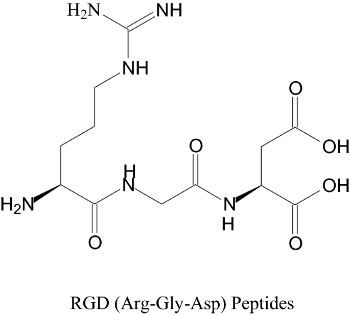
Chemical structure of RGD
-
But still we have to develop and design an alginate-based hydrogel material which can facilitate the cell and the cell anchorage of different cellular function such as elongation, proliferation, migration, and differentiation in the 3D situations should appropriate for applications in tissue engineering is now still a challenge to researchers. This problem of using alginate can be removed by inserting gelatin, which is cross-linked covalently with alginate dialdehyde (ADA).
-
ADA is actually a moderately oxidized substance of alginate, which supports the cross-linking of gelatin with alginate through the formation of Schiff’s base because of the reaction between the free amino groups present in lysine or residues of hydroxylysine amino acid present in the gelatin and the existing aldehyde groups(-CHO group) of the ADA. Due to the partial oxidation of carbon–carbon bonds present at the cis-diol group in the uronate residue of alginate, it undergoes cleavage and changes the chair conformation in the open-chain structure and support the rate of degradation of the alginate. Hence finally the property of biodegradability of the covalently bonded cross-linked hydrogel of alginate can be modified by using ADA of various grades of oxidation. It can regulate and control the hydrolysis of alginate, thereby altering the ratio of gelatin and ADA [19].

6.1 Use of alginate in tissue regeneration
Sodium alginate is categorized as a group of hydrogels, which were synthesized by the free‐radical polymerization technique using some specific water‐soluble monomers such as methacrylamide (MAm), acrylamide (Am), water‐soluble polysaccharide (SA)and N‐isopropylacrylamide (NIPAAm),which can be used in different medical purposes, regenerative medicine and is considered as the second most abundant polysaccharide available on the globe, which is mainly extracted from the seaweed. This versatile biopolymer has the composition of β-L-guluronate subunits (G- blocks) and β-D-mannuronate (M-blocks) covalently linked with 1, 4 linkage. The mechanical properties of the alginate gel formed mainly depend upon the divalent cations that are used for crosslinking with the alginate molecules. Both trivalent and divalent cations (Ca2+, Mg2+, Ba2+, Fe2+, and Al3+) are covalently linked to the G- blocks of the alginate and forms a 3D structure commonly called as “egg box”.
Example
The presence of Sr and Ba ions in the place of Ca ions in the cross-linking gel form is the cause of more rigid in structure of it. The alginate in the microcapsules form cross-linked with the divalent ions for cell immobilization. The gel strength and dimensional stability comparatively increased in case of alginate gels of high-G due to exchange of traditional Ca2+ ions with Ba2+. The use of Ba2+ decreases the size of the alginate beads and cause of decrease in permeability to immunoglobulin G. The high-M content exhibit opposite behavior in combination with Sr and Ba ions because these beads were larger surface area as compared to the beads of calcium-alginate and shows better swelling capacity, which is the cause of increased permeability. Different ions binds in different extent in different block structures in the alginate., Ca2+ was found to bind to G- and MG-blocks, Ba2+ to G- and M-blocks, and Sr2+ particularly to G-blocks [20].
Hence the substitution of different divalent ions such as, Ca2+, Ba2+ ions in place of Na+ ions of guluronic acid is the main reason that supports sodium alginate to behave as a hydrogel. The G and M blocks combined with each other in a diverse sequence or alternately settling as the divalent cation, covalently cross-linked with the polymer chains and forms a 3D structure, which is capable of binding huge quantities of drugs, water. Hence alginate serves as bioactive materials that promoting tissue regeneration. Therefore finally it was revealed that the sodium alginate can able to cross-link with the Ca2+ and improves the differentiation and proliferation of osteoblasts in vitro [21, 22].
6.2 Wound healing, wound dressing and skin repairing
The principal organ of our body is the skin, which serves as a crucial role in maintaining homeostasis and also protecting the internal part of our organs from the toxic external environment. The treatment of severe and chronic wound infection, intense burns in wound on skin requires a longer period of time. In US 6.5 million patients are severely affected by chronic wound infections along with more than 18% of the diabetic patients over the age of 65 years are suffering from foot ulcers, which are not easily healed or repaired. The process of wound healing is normally a complicated process and vastly controlled by our biological system, which includes different types of cells, i.e., immune cells, keratinocytes, endothelial cells, and fibroblasts. The wound dressings by using alginate based materials have many advantages in comparison to conventional gauze dressing because the wound exudates can be easily evaporated with preventing the entry of pathogenic bacteria and provides a moist environment in the wound areas and accelerates wound healing. During the time of propagation of wound area, the epithelialization occurs and new granulation tissue containing macrophages fibroblasts and endothelial cells has covered the surface of the wound areas by creating a completely novel extracellular matrix (ECM). That ECM formed plays a vital role for proper and rapid healing because ECM offers a suitable environment for blood vessels and sustaining cells, which provides the nutrients that required restoring the homeostasis and integrity of the tissue [23]. The extracellular matrix formed functions as a suitable pliable and porous scaffold for facilitating the diffusion of nutrients, cells and the growth factor through the wound areas. The wound-healing ability of the thin films of hydrogel produced consisting of sodium alginate (NaAlg) in 3 g/100 mL of water with 10% of the antiseptic povidone iodine (PVPI) shows a significant decrease in the wound areas to increase in re-epithelialisation even after 3 days and the complete wound healing is achieved within 12 days, which is more rapid relative to the conventional wound healing process. The povidone iodine present in the hydrogel provides antiseptic properties and prevents the infection due to bacterial activity [24, 25].
PVPI is treated as one of an exciting antiseptic agent and popularly used in wound treatment. PVPI is a complex of iodine and polyvinylpyrrolidone and soluble in water. It is a broad-spectrum antiseptic agent and highly efficient against a wide range of bacteria, fungi, viruses, yeast and protozoa. In spite of its significant antibiotic property the solution of PVPI exhibits fewer toxic effects on the human skin fibroblast and act as retarder of the cell growth at concentrations of 0.1% and 1.0% and prevents the wound healing process.. Hence, in order to overcome this toxic effect PVPI have been incorporated in the alginate matrix to form a film of NaAlg/PVPI composite. This film possesses antifungal and antimicrobial properties against Candida albicans and Escherichia coli respectively. The NaAlg/PVPI films were synthesized by taking 3 g of NaAlg dissolved in 100 mL of distilled water under continuous stirring for about 1 h at 100 °C. Then after that 0.3 g of PVPI was mixed with the prepared solution of NaAlg solution and it was allowed to dissolve at continue stirring for about 1 h at normal room temperature till to get final concentration of 10% by wt. of PVPI. Finally, the appropriate volume of glycerol was mixed with the prepared NaAlg/PVPI solution and allowed to dissolve under continuous stirring for about 1 h at normal room temperature. Then each film was cast by taking 10 ml of the solution and finally dried for 24 h under a chemical hood to get final film [26,27,28].
The ideal dressings of skin damage or injury can be done by using alginate or sometimes alginate on combination with some of the multifunctional substances such as gelatin, polyvinyl alcohol, skin fibroin, hyaluronic acid and graphene oxide and the added materials, which enhances the different essential properties such as hydrophilicity, mechanical properties, and cell adhesion to the scaffolds. For specific target applications some collagen and hormones, mainly triiodothyronine may be added. The surface-to-volume ratio of the nanofibers is much higher and is valuable for recovery of wounds. The nanofibrous scaffold of polyvinyl alginate- alcohol-gelatin on modification with collagen improvers the hydrophilicity and adhesion properties of the fibroblast cells. Therefore, it is the cause of improving the proliferation, cell viability and better bio-response for tissue regeneration to an increase in tensile strength [29].
Honey possesses excellent antimicrobial properties and the addition of honey with the nanofiber form of alginate- alcohol-gelatin is much more effective for inhabiting the growth of gram-negative bacterium such as E. coli and gram-positive bacterium such as S. aureus, but however it shows better antibacterial properties against the gram-positive bacterium. This is highly effective for the diabetic patients suffering chronic wounds. The addition of triiodothyronine, which is an active thyroid hormone with the lyophilized hydrogel containing gelatine, polyvinyl alcohol and alginate (AGPT) and plays a crucial role in the revival, repairing of different tissues. It is much more effective towards skin healing. Triiodothyronine is a active form of thyroid hormone and exhibits crucial role in the regeneration and repairing of tissues if incorporated in the scaffold. The scaffold formed has the capability of releasing hormone, which stimulates the neovasculature development, cell migration, skin cell proliferation, lamellipodia creation and cell-biomaterial interaction. Hence it enhances the healing process with the development of blood vessel and deposition of collagen. Therefore the scaffold prepared possesses huge potential to be exploited as a chronic wound therapeutic. Natural honey contains more than 200 different kinds of substances in it including glucose, fructose and frocto-oligosaccharides and popularly used for therapeutic purposes, since about 8000 years. The composition and quality of the honey depend upon the plants or trees at which the bees are fed. It contains flavonoids (including apigenin, galangin, pinocembrin, quercetin, hesperetin, kaempferol and chrysin), superoxide dismutase (SOD), ascorbic acid, reduced glutathione (GSH), catalase (CAT) and tocopherols. In addition to that products of Millard reaction and peptides are the major components of the natural honey. The majority of the compounds present in natural honey provide significant antioxidant property. Hence honey can be more efficiently used for wound healing treatment. Alginate has the unique property of gel forming ability having a hemostatic effect and therefore popularly used as a wound healing agent because of dehydration preventing and gel forming properties. The dressing based on calcium-alginate with honey, can able to absorb excess of wound exudates and produces a non-adherent gel. The gel formed facilitates wound healing, stimulates debridement, supports for preventing trauma to the wound bed and also the surrounding area of skin. Hence, due to good hydrogel forming ability, alginate on mixing with honey can be served as a promising material for quickening the wound healing with the prevention of antibiotic resistance towards wound infections and may be used as a topical ointment in the treatment of both healing and therapeutic effects. Alginate-gelatin hydrogel on combination with honey shows excellent antibacterial and biocompatible property. The gelation property of this exciting material is due to the Schiff-base reaction between the amino groups of gelatin and carbonyl (aldehyde) groups of the oxidized alginate. The C–C bonds of the cis-diol groups in the molecular chain segments of alginate undergoes cleavage due to periodate oxidation and forms reactive –CHO groups, which helps in cross-linking with the amino groups through the Schiff-base bonding [30,31,32].
Turmeric (Curcuma longa) is treated as an important and widely used spice in Indian. Recently it has been used as herbal medicines for the treatment and cure of a number of diseases, including rheumatism, anorexia, sinusitis, diabetic ulcers and cough. The major component of turmeric is Curcumin (diferuloylmethane) and the yellow colour of turmeric is due to the presence of curcumin. Hence curcumin possesses many essential properties such as antiinflammatory, anti-carcinogenic, anti-oxidant, anti-infective, anti-mutagenic and anti-coagulant effects. Therefore Curcumin exhibits significant properties of wound healing. It acts on different phases of the natural wound healing to accelerate the healing process. Curcumin has the capability of decreasing the natural response to cutaneous wounds of our body including oxidation and inflammation. The wound healing characteristics of curcumin provide its capability to improve granulation tissue formation, tissue remodeling, wound shrinkage and collagen deposition. The extracts of the turmeric plant contain curcumin, which can be loaded to form nanoparticles with sodium alginate (SA) and Carboxymethylcellulose (CMC). SA and CMC are crosslinked by using methylenebisacrylamide(MBA) as crosslinking agent and Ammonium persulfate(APS) as initiator [33, 34]. The structure of curcumin is represented in Fig. 4.

Structure of curcumin
Now high quality of advanced wound dressing materials is designed and developed by various researchers, which are easy to control and handle with the higher absorbent ability and cost effective. These materials provide a moist occlusive environment for promoting healing. These wound dressing materials are of different kinds like gel, occlusive, sponge or semi-occlusive forms. Alginate on combination with nanoparticles of Ag and Chitosan fabricates a composite polysaccharide, which possesses good antibacterial and anti-adhesive wound dressing characteristics. The wound dressing by using alginate in dry form can absorb the fluids produced from the wound to re-gel, and this gel formed provide water to the dry wound in order to maintain the physiologically moist microenvironment and reduces infection due to the bacterial activity at the site of the wound [35]. The alginate dressings can be prepared by different methods such as in ionic cross-linking with Ca, Mg, Pb, Co, Ba, Zn, Mn, Ni, Sr and Cd ions or can be used in the form of gels or in the form of freeze-dried porous sheets or fibrous dressings. Alginate dressings in the dry form absorb wound fluid and converted into a gel form and the gel form provides the dry wound a moist physiological environment with drastically reduces the bacterial infections, which causes quick granulation tissue formation and re-epithelialization. The immunogenic effect of alginate in dry form is greatly influenced by the concentration of M-blocks of alginate. This is because the alginate having higher concentration of M-block promotes the cytokine creation relative to the alginate having higher concentration of G-blocks. Before applying the alginate should be properly purified because impurities may stimulate the immunogenic response [36, 37].
The fibers of alginate on cross-linking with Zn2+ ions exhibits better result for wound dressing, because Zn2+ ion has the ability of producing antimicrobial and immunomodulatory effects with enhancing the keratinocyte migration and increases the phases of endogenous growth factors. A little fraction of Zn2+ chelated by carboxylate groups of alginate and are substituted by Na+ ions, which is the cause of the increase in electrostatic repulsion among the negatively charged COO− ions. This causes a relaxation of the polymeric network and enhances the water absorption capacity resulting the formation of hydrogel. Zinc alginate hydrogels were synthesized by internal gelation techniques and the concentration of Zn increases the mechanical property, swelling ability, antibacterial property, the rate of evaporation of water. Hence Zn alginate exhibits higher swelling behavior than Ca alginate. The blends of chitin or Chitosan with alginate and fucoidan gels provide an excellent moist healing environment and more successfully can be applied in chronic wound dressing [38].
The process of wound repairing is normally a three phase process such as inflammation, tissue formation and then remodeling. All the three processes are simultaneously operating and correlated with each other that either suppress or activate to maintain and initiate adequate repair of the wound. During this process different signaling factors, including miRNAome, transcriptome, cistrome and proteome are regulating for suitable wound repair. The Nuclear hormone receptors (NHRs) and the ligands present in its control injury and cutaneous homeostasis through migration, differentiation, modulating cellular metabolism, inflammation, apoptosis and applied effectively in dermatologies therapy [39, 40]. Because of excellent biocompatibility and less antigenicity property with most of the endogenous tissue natural collagen plays an essential role in many for surgical repair. Wound dressing by using collagen based materials have been specially applied in the treatment of ulcers and burn wounds. The powdered form of avian collagen is more effective for the treatment of chronic wound healing. This powdered form of avian collagen stimulates cellular recruitment, activates the inflammatory phase of wound healing and also promotes the growth of new tissue. Collagen on combination with alginate shows positive result in the stimulation of the inflammatory phase of wound healing and impacts required mechanical strength, which are the characteristics of collagen fibrils. The collagen dressing forms a semi-occlusive thin polymer film on the outer surface of the wound and those occlusive films protect the bacterial attack with additional mechanical trauma and provide vapor permeability and proper air to the wound area. Collagen decreases the scarring and shrinkage and enhances the rate of epithelialization. Such kind of wound dressing is extensively used in burn wounds [41].
6.3 Gelatin- alginate hydrogel for wound healing
Saarai et al. synthesized a novel hydrogel by the combination of gelatin (G) and sodium alginate (SA) in the ratio of G/SA 30/70, 40/60, 50/50, 60/40, and 70/30 for application in the wound healing applications. The hydrogel having the ratio of G/SA 30/70, 40/60, and 50/50 shows droplet morphologies, whereas hydrogels with ratio of G/SA 60/40, 70/30, and 80/20 shows fibrous morphology. The water absorption of the hydrogel was 81.86% to 60/40 SA/G, 72.52% for SA/G 70/30, 84.03% for SA/G 50/50, 90.1% for SA/G 30/70 and 87.97% for SA/G 40/60 respectively. The higher capability of the swelling behavior of the SA/G was due to the presence of hydrophilic functional groups COO– and NH2- from both the biopolymers. The hydrogel of sodium alginate and gelatin is cross-linked by addition of either CaCl2 or glutaraldehyde as cross-linking agent. The water absorption of the hydrogels retards with the rise in the concentration of cross-linking agent, which establishes high mechanical strength. The swelling behavior of the hydrogels increases with the increase in the % of gelatin in the hydrogels. The crosslinking time significantly influenced the mechanical strength of the hydrogels. The less crosslinking time brings about reduced mechanical strength with quick increase in swelling behavior, whereas the more crosslinking time brings about the reduction in the swelling capacity of the hydrogel. The SA/G hydrogel in the ratio of 50:50 was found to be best appropriate for wound dressing or wound healing having good swelling capability [42, 43]. Polymer hydrogels are treated as good materials for wound dressing for the treatment of severe wounds and tissue revival. The hydrogel containing gelatin, sodium alginate and silver nanoparticles exhibits good antimicrobial activity for tissue healing. The hydrogel form of soft tissue adhesives synthesized by using amino gelatin (AG) and aldehyde sodium alginate (ASA) exhibits excellent clinical properties for wound healing because of some of its unique properties including high capacity of water absorption, low immune response, good biodegradability, nontoxicity, good biocompatibility. The swelling behavior, gelling time and the bonding strength of the ASA/AG hydrogel varied with the variation of the concentration of amino groups in AG and aldehyde groups in ASA. The gelation time can be controlled within 18 min, if the aldehyde content in ASA is 75.24% and amino content in AG is 0.61 mmol/g. The adhesive strength of this novel hydrogel is almost similar to the adhesive strength of the commercially obtainable adhesive Ebrin glue. Hence the hydrogel ASA/AG is treated as a promising candidate material for soft tissue adhesive and a number of biomedical applications [44,45,46].
6.4 Cartilage for tissue engineering
The Articular cartilage is considered as an extremely organized and specialized important tissue in our body, which connects the end parts of long bones and responsible for functioning the joints. It delivers mechanical strength and lubrication to prevent the tensile and compressive strength, which is vital for bearing of body weight and movement. The cartilage possesses an exceptional matrix containing an interpenetrating system of collagen with negatively charged proteoglycans and chondrocytes. Since cartilage has a restricted regenerative ability, therefore it cannot be healed appropriately. Hence the repairing of articular cartilage is still a great challenge for scientist, doctors and researchers. The injury in the articular cartilage mainly due to sports injuries and the patients having severe injuries in traumatic joint has more possibility of suffering posttraumatic osteoarthritis, which is a worsening situation that causes severe pain, disability and sometimes may require a complete joint replacement. The alginate hydrogel can able to retain a large quantity of water, which facilities lubrication and retards the coefficient of friction; in addition to that, the water stored in the hydrogel plays a crucial role in the mechanical properties and chemical structure of the hydrogel [47, 48].
Due to restricted self-healing capacity of the damaged articular cartilage, millions of people throughout the world are suffering primary osteoarthritis whose severity gradually increases with increase in age. Although alginate can be applied popularly as the scaffolding substance for regeneration of tissue for cartilage, but it has some drawbacks, therefore some peptides, proteins and specific inorganic materials are added in alginate in order to reduce the deficiencies. But it was reported that a system of chitosan-alginate in porous freeze-dried scaffolds forms are developed for the treatment of articular cartilage. The composite of chitosan-alginate scaffolds supports the development of chondrocytes having a distinguishing morphology and shows excellent results in the biosynthesis of polymer collagen relative to the pure form of chitosan scaffold because of the capability of alginate for inducing redifferentiation of the 2D culture-expanded dedifferentiated chondrocytes. But in order to increase the interaction of the cell-materials, the peptide (RGD) was conjugated with the complexes chitosan-alginate-hyaluronate (CAH) and highly useful for cartilage of tissue engineering, but however characteristic spherical morphology of the chondrocytes was detected in all the scaffolds without or with RGD. It was observed that a hybrid form of hydrogelbased on alginate (ALG)-polyacrylamide on combination with nanoparticles (SiNP) of silica serves as a promising candidate material for the replacement of cartilage. It was proved that the required mechanical strength of the hybrid material is due to the structural aspects of ALG/PAAm (alginate–polyacrylamide) hydrogel. The high mechanical strength is due to the cross-linked covalent bonding in between the two intertwined polymer chain segments. The carboxyl and amine groups of both ALG and PAAm respectively interact with each other and form a cross-linked network structure, which is accountable for the load bearing capacity and mechanical strength of the hybrid scaffolds. The SiNP-ALG-PAAm hydrogel of three-dimensional structure is the cause of increasing mechanical properties and inspire the cell proliferation [49, 50].
Again, it was observed that the hydrogels produced from alginate is the cause of quick relaxation of stress and supports the promotion and repairing of the cartilage matrix by chondrocytes. The low-molecular mass of ALG/CS and high molecular mass alginate/chondroitin sulphate (CS) covalently cross-linked with the strontium of 3D microenvironment which provides relaxation moduli and elastic is the appropriate matrix for cell culture. It can able to control and regulate the metabolic activity of the living cell tissues. From earlier, the suspension of chondrocytes in combination with CaSO4 and alginate solution is injected into the molds of facial implants for the formation of preshaped cartilage [51, 52]. Another important material progressively developed is shape-memory alginate gels, which can able to construct cartilage having required dimension in vivo followed by negligibly invasive delivery. Many researchers reported that by using stem cells for repairing and regeneration of cartilage is very interesting and attractive because of the offensive and damaging procedures needed to get primary chondrocytes from different tissues. The biopolymer alginate condensed with human mesenchymal stem cells (MSCs) forms a gel bead which is cultured in a non-serum medium in combination with the transforming growth factor (TGF)-β1 dexamethasone, and ascorbate 2-phosphate even more than 7 days and established the formation of cartilage also in intense osteochondral defects [53, 54].
The combination of collagen with alginate exhibits a significant effect toward the regeneration of articular cartilage as in the form of bio-ink for 3D cell printing. The mixture of alginate hydrogel with hyaluronic acid with some biomimetic peptides forms a modified hybrid gel, which is more popularly used for increasing the interaction in between the cell–matrix and HAV peptide (histidine alanine valine) sequence and subsequently increases the cell–cell interactions. It was studied by many researchers that the combination of both hydrogels and alginate-based microspheres is found to be more effective for the controlled delivery of growth factor in tissue regeneration.
Example
There is a constructive impact of immobilizing RGD into a macro-porous form of the alginate scaffold of stimulating the TGF-β-induced human MSC differentiation. The interaction of the cell matrix supports the immobilized RGD peptide, which is the vital feature of the cell microenvironment and is the cause of good accessibility of the cell to the chondrogenic stimulating molecule (TGF-β) [55, 56].
6.5 Bone repairing
Bone is considered as a characteristic multifaceted tissue having the composition of about 70% of nanohydroxyapatite (HA) symbolized as Ca10 (PO4)6(OH)2 and approximately 30% of collagen by mass. Some noncollagenous organic matter in aqueous solution present in it in negligible amount. The ratio of volume to weight fractions of the different components such as hydroxyapatite, collagen and water is not constant and is varying with the individuals. Next to chitosan and chitin the second most widely and popularly used biomaterials in the world for bone tissue engineering is alginate because of its greater ability to form scaffolds. A good biomaterial for the treatment of bone repairing or bone healing must have to possess the following properties.
-
The biomaterials must be perfectly biocompatible having adequate surface area.
-
The biomaterials must be nontoxic in nature, having 3D structure and contains required porous and the pore size should be about 100 μm.
-
The biomaterials used must facilitate cell migration, proliferation and cell adhesion.
-
The biomaterials must be perfectly biodegradable and used as a drug carrier and growth factors
-
These biomaterials must possess the mechanical properties comparable to the cortical bone and can offer initial support at the site of the implant.
For the necessary growth of tissues, the biomaterials must have to possess increased vascularization, which is very vital for cell proliferation and migration in the required direction. The porous biocompatible and biodegradable scaffolds function as a provisional structure in the treatment of bone tissue engineering (BTE), which supports cell growth perfectly and forms a completely new bone tissue at the damaged site and simultaneously the implanted scaffold continuously degrades and finally replaces the newly formed bone tissue. Although alginate is non-toxic to host tissues or cells, but it has a deficiency of the properties of cell adhesion, therefore some specific kinds of peptides or proteins are normally added which forms conjugation with alginate and used for the design and development of tissue engineering materials. The sustained release of bone-forming peptide-1 (BFP-1),which is obtained from the bone morphogenetic protein-7 (BMP-7) produces the scaffolds and promotes the osteogenic diversity of osteoblasts. It was experimented that BFP-1 exhibits optimal result in osteogenic activity and cell viability in the hydrogel of alginate having 10 μg/mL of the peptide. Hence the peptide incorporation significantly increases the osteo-regeneration in vivo if the scaffolds were implanted into the defects of Beagle calvarial. The osteoid is the organic component of the matrix part of the bone, which consist of chondroitin sulfate and collagen-I and on injection it is deposited at the damaged part of calvarial defects, where the BFP-1 having alginate scaffolds were implanted [59, 60].
In another case it was found that gelatin and alginate both are cross-linked with each other and form a stable amide linkage in between gelatin and alginate in the presence of NHS (N-hydroxysuccinimide) and EDC(1ethyl-3-[3-dimethylaminopropyl] carbodiimide hydrochloride) in three different stages of the reaction. Now a new technique called as microwave-vacuum drying is applied for fabricating porous scaffolds having both more and less cross-linked gelatin- alginate blend hydrogels. The high and low cross-linked hydrogels in vivo and in vitro scaffolds revealed exceptional biocompatibility, cytocompatibility and bioresorbability properties, which is required for special bone implantation and repairing. In the composite of gelatin- alginate neovascularization and angiogenesis were identified, which mainly supports for distributing nutrients into the implanted cells and is the cause of enhancing regeneration of new tissues. Bone contains both organic matter and inorganic materials such as hydroxyapatite and collagen [61, 62]. Therefore, researchers are always trying to modify the materials by adding some inorganic substances in alginate in order to develop a new material having high mechanical strength, which is the major requirement of bone tissue. The addition of inorganic material is the cause of enhancing the formation of new tissue in the damaged site of the body of the patient. The different kinds of inorganic materials such as bioactive glass (BG) and a number of various categories of calcium phosphates mainly hydroxyapatite (HAp), tricalcium phosphate (TCP) are used for the formation of alginate hydrogels for designing and developing scaffolds needed for tissue engineering. In alginate-polyvinyl alcohol a bioactive phase of sol–gel-derived BG was inserted in order to develop the composite scaffold that required for bone regeneration [63, 64]. The porous scaffold of alginate gel can be fabricated by using a modern technique called as a surfactant foaming technique in which sodium lauryl sulfate was normally used as a surfactant. Because of the addition of bioactive glass as the inorganic component, the composite scaffolds obtained shows the property of osteoconductivity due to the formation of hydroxyapatites (Hap) [65].
In another study it was revealed that by incorporating tricalcium phosphate (TCP) into the oxidized form of alginate-gelatin hydrogels, the bone formation is induced due to osteoblasts, which are best applicable for BTE. The presence of BG enhances the degree and kinetics of crosslinking with the oxidized alginate-gelatin base hydrogel (ADA-Gel), hence it is the cause of improving the mechanical strength of the hydrogel scaffolds. The scaffolds containing high BG possess low release rate of protein because of the presence of more cross-linked structure, which inhabits the hydrolytic degradation and protein release rate. The Scaffolds having optimum BG (1%), with appropriate % of protein and the controlled rate of degradation facilitate better cell growth, migration, proliferation and differentiation in osteogenic, but the existence of BG supports the HAp formation onto the scaffolds and cause of osteoconductive.
Recently it was reported that the nanocomposite scaffold containing alginate, gelatin and graphene oxide (GO) possesses the better property of bone healing. The addition of GO in the scaffold of gelatine-alginate enhances the biomechanical strength [66, 67].
Example
The scaffold of hydrogel without GO have the compressive strength of 30 MPa, but whereas those containing GO have 44 MPa.
The GO increases the property of hydrophilicity of the scaffolds of the composite and exhibit slow rate of degradation (almost 30% within 28 days). Hence the nanocomposite scaffolds are the cause of improvement in the cell proliferation and attachment relative to the simple gelatine-alginate scaffold. A better cross-linked hydrogel network was developed by using Ca2+ from calcium silicate (CS) cross-linked with sodium alginate and silk fibroin (SF). The presence of calcium silicate within the hydrogel scaffolds increases the degradation rate, hydrophilicity, and bioactivity and compression resistance. Hence the hydrogel scaffolds having a higher % of calcium silicate exhibits properties of excellent biocompatibility and supports the promotion of osteogenic variation in vitro. Doxorubicin hydrochloride (Dox) is normally used in the study of drug delivery and it was examined that the composite of HA/SA/CS/Dox supports the regeneration of the bone defects. The combination of nano form of hydroxyapatite (nHA) with sodium alginate and chitosan on combination with HA shows statistically significant results of the decrease in swelling behavior and the enhancement of stability with the required mechanical properties [68]. The Synthesis of Chitosan-HA is represented in Fig. 5.

Synthesis of chitosan-HA
The calcium phosphate cement if incorporated in the alginate- chitosan complex, then it shows better results in bone healing due to the interaction between the anionic parts and cationic parts of alginate and chitosan. Again new improved bone cement may be produced by the combination of sodium alginate and citric acid (CA) and on mixing with α-tricalcium phosphate, the physicochemical properties along with the binding capacity and mechanical properties of the bone cement formed is drastically increased. Therefore the hydrogel networks of sodium alginate with citric acid impacts a very strong cohesive force due to strong chelation with the Ca2+ ions during the process of hydration after the use of the bone cement [69, 70].
6.6 Chitosan-alginate- fucoidan gel for bone tissue engineering application
In the human body bone is a hierarchical and complex tissue. The majority % of it is collagen and hydroxyapatite (HA) and both the components serve a vital role in mineral deposition, mechanical support, pH balance and finally the structural framework. The bone damage or bone defects are due to different factors such as accidents, chronic diseases and also birth defects. A number of different kinds of materials and treatment processes are available for reconstructing bone defects, which includes autograft, synthetic graft, allograft and xenograft. Although allograft and autograft are both suitable techniques for bone grafting, but there are some difficulties are associated with these such as donor site injury and the possibility of transmissible diseases.
Example
Hepatitis or acquired immune deficiency syndrome (AIDS) may be associated with allograft bone grafting [71].
Recently, synthetic bone grafts are developed and designed for fabrication of artificial bone, which should be osteoconductive, biocompatible, osteoinductive, biodegradable, outstanding mechanical strength, structurally almost similar to natural bone, easy to handle and cost effective [72, 73].
Chitin is an important natural polysaccharide and the second most valuable biopolymer after cellulose. The major sources of chitin are exoskeleton of arthropods, cell walls of yeast and fungi. Chitosan is a most important derivative of chitin and can be obtained by deacetylation of chitin in alkaline medium (NaOH) or enzymatic hydrolysis. It consists of the repeating units N-acetyl glucosamine and d-glucosamine bonded with each other by β(1, 4) linkage. The biopolymer chitosan exhibits excellent properties of biodegradation, antimicrobial activity, non-toxicity, biocompatibility and low immunogenicity. It can be synthesized in numerous forms such as gels, sponges, membranes, scaffolds and beads and possess outstanding ability of the pore forming, which is essential in the tissue engineering applications, wound healing and drug delivery. Chitosan can be combined with a number of bioceramics and biopolymers such as alginate, amylopectin, hyaluronic acid (HA), carbon nanotubes (CNTs), poly (methyl methacrylate) (PMMA), polylactic acid (PLA) and Ca3PO4. Alginate is an anionic form of linear copolymer consist of homopolymeric blocks of both (1,4)-linked α-l-guluronate and β-d-mannuronate. Like chitosan, alginate is also an outstanding biopolymer for tissue engineering applications, because of its excellent properties such as biodegradability, encapsulation capacity, biocompatibility, chelating ability and capability to synthesize various forms such as microspheres, fibers, gels, sponges and foams. The composite of chitosan-alginate is now extensively used for protein delivery, wound healing, drug delivery, and tendon and ligament tissue repairing with the creation of new bone.
Fucoidan is a sulfated form of polysaccharide, which contains l-fucose and SO42− and the major source of it is the marine brown seaweeds. Fucoidan is the cause of increase in the level of alkaline phosphatase (ALP), osteocalcin, bone morphogenetic protein-2 (BMP-2), type-1 collagen and also supports the deposition of minerals, which requires for bone mineralization. Fucoidan stimulates osteogenic differentiation in the amniotic fluid stem cells of the human body, therefore treated as a promising candidate material for regeneration of bone tissue. The mineral deposition increases about 30%, if fucoidan is present in the composite material. The composite film of chitosan- fucoidan exhibits excellent ability of wound dressing and wound healing in vivo and in vitro. Hence, taking in the consideration of the exceptional properties such as biocompatibility, antibacterial nature, biodegradation, film forming capability and the introduction of the osteogenic differentiation by fucoidan, the composite of chitosan-alginate- fucoidan is treated as an exceptional biomaterial for bone tissue engineering. The alginate-chitosan-fucoidan sponge exhibits outstanding elastic properties and retains their shape after compressive strain and bending without failure. This composite material having 10% fucoidan exhibits better hemostatic and antibacterial performances. In the last few years some significant research is conducted to develop a suitable material for artificial bone scaffolds, but presently the composite of chitosan-alginate along with fucoidan is considered as the most suitable material [74,75,76].
6.7 Tissue regeneration with cell delivery and protein
The alginate hydrogels are more popular and widely used over past several decades as a candidate material for cell populations or protein delivering, which is the cause of direct regeneration and fabrication of various organs, tissues and other different parts of our body. The wide range of applications of alginate hydrogel varies with its physical properties along with cell adhesion and improved degradation activity of the candidate materials. The alginate hydrogels are released through the process of diffusion in the porous hydrogel scaffolds having a pore size of about 5 nm. The macroporous beads with an interrelated pore structure from alginate has been synthesized by the incorporating gas pockets within alginate beads, stabilizing the gas bubbles with surfactants, and subsequently removing the gas and better serves for wound healing applications. Most of the added proteins are freely diffused from the alginate hydrogels even without the gel degradation, but the gel degradation can increase the rate of release. Large molecules have also diffused significantly and successfully delivered if the gel degradation occurs [77].
Example
The plasmid DNA in the condensed state having a pore size of about 100 nm can be easily released from the degrading alginate hydrogel and the antibodies may be released from the alginate hydrogels through the similar mechanism [78].
Cells are migrated from the alginate hydrogels and again being released, when it degrades. A number of studies identify that in nanoporous alginate hydrogels of different forms, although cell migration occurs, but it is not quantitatively analyzed. The alginate hydrogels loading with the outgrowth endothelial cells (OECs) and alginate lyase, which is a specific enzyme generated by a wide range of microorganisms such as algae. That microorganism breaks the glycosidic covalent bonds present in the alginate and stimulates the cell migration [79]. These hydrogel states of alginate to begin with encapsulate a cell in a stiff nanoporous material, which increase the porosity and decreases the strength of the material in situ after injection, because the enzyme present in it degrades the alginate hydrogel. Hence, for controlling the concentration of alginate lyase in the form of injectable alginate hydrogels is treated as cost effective and a simple, effective approach to stimulate cell release by changing the mesh size and mechanical properties of the alginate hydrogels. Hydrogels are the materials having a higher water absorption capacity because of its three-dimensional networks. Gel is a semisolid form of solid and liquid phase, where solid particles are in the colloidal state. Hygrogel is a colloidal gel, where water is a continuous phase [80].
The microbeads of alginate on incorporation with the adipose-derived stem cells (ASCs) have a capacity of delivering feasible cells that facilitates tissue regeneration. The microbeads are normally fabricated by cross-linking with Ca2+ ions in the solution of organic osmolytes in order to confirm physiological osmolality. The delivering parameters that required for implementing microencapsulated cells in tissue repairing and regeneration are still unknown, but the following parameters are till now investigated [81].
-
The effect of osmolyte on the diameter of microbead, growth factor creation and cell viability.
-
The impact of the cell concentration per microbead and the number of microbeads per unit volume on cell viability, microbead degradation and production of growth factor.
-
The capability of both non-degradable and degradable form of alginate microbeads of the neighboring cells at the site of delivery in vivo.
-
The microbeads of alginate having alginate-lyase provoke have an inflammatory response if exposed repeatedly.
The microbead of smallest diameters was attained by using glucose as the osmolyte, but the growth factor production and cell viability did not depend upon the type of osmolyte. If the number of cells per microbead or microbead number increased, then the growth factor production per cell accordingly decreased although the % of cell viability was unaltered. Hence the cell release rate changes with the number of cells per microbead and number of beads per well. The cell release rate was relatively delayed if the density of microbead is highest and the density of cells per microbead is lower [82].
6.8 Blood vessels
The blood vessels form a complex network in our body and responsible for the transportation of nutrients and oxygen to all the living tissues with the elimination of the waste metabolic products. It is the cause of trafficking of progenitor cells and stem cells, which are necessary for the growth of organs in the wound and embryo repair for adults. The production of completely new blood vessels is most vital for tissue engineering applications because the cells more than a few hundred microns forms blood vessels, which will affect hypoxia and the incomplete supply of required nutrients. Further the creation of new blood vessel called as neovascularization is an appropriate alternative for the treatment of patients, those suffering the obstruction of the rate of blood flow that caused by peripheral arterial and coronary diseases. The neovascularization may be attained due to transportation or diffusion of different types of cell in our body that distributing the angiogenic molecules, mainly genes or recombinant proteins or the combination of both.
Alginate hydrogel is a popular and widely used material for delivery of different angiogenic molecules. But the most talented use of alginate gels is to support the formation of blood vessel because of their capability for providing a localized and sustained release of the heparin, which binds with the growth factors such as vascular endothelial growth factor (VEGF). Alginate gels if injected directly into the ischemic muscle tissue, then it is the cause of a long term release of VEGF within the ischemic tissue, and creation of VEGF gradients around the neighboring tissue that can able to guiding formation of new capillary into the ischemic tissue and releasing tissue ischemia. Hence, generally VEGF plays a key role in originating angiogenesis and also starting new capillaries. In addition to that, Platelet-derived growth factor (PDGF) stimulates the maturation rate of the newly produced capillaries into superior and more functional blood vessels. There are two strategies are usually adopted for the sequential release of all these factors by using alginate gels [83, 84].
-
The PDGF is groomed and capsulated into poly (lactide-co-glycolide) PLG microspheres, which was formerly encapsulated into gels without VEGF. The structure of VEGF is shown in Fig. 6.
Fig. 6 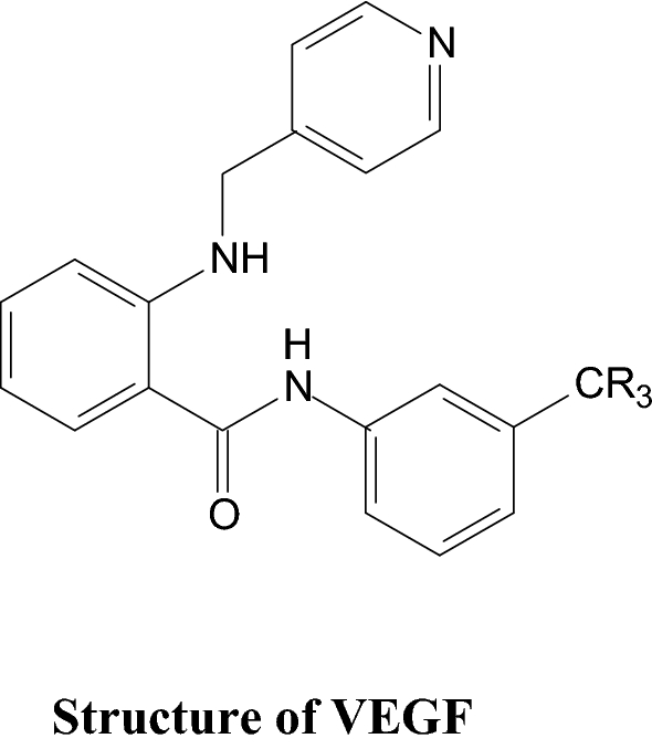
Chemical Structure of VEGF
-
In the second strategy, the more heparin bonding with PDGF, which on exploitation slower the rate of release in comparison to VEGF, basically encapsulating both these factors is free from the gels [85, 86].

7 Poly (lactide-co-glycolide)
In both the circumstances the release rate in case of VEGF was much faster relative to PDGF. The release rate of VEGF can be decelerated by summarizing this substance into PLG microspheres along with microspheres in the alginate gels. The chemical structure of PLG was shown in Fig. 7. The cell transplantation is a beautiful approach to stimulate the formation of the new novel blood vessel, predominantly when the host cells are capable of replying to the transported growth factors which are not functioning properly. The movement of endothelial progenitor cells or endothelial cells is not significant in critical clinical trials because of the huge mortality and inadequate incorporation of the transplanted cells having the vascular network of host and fewer enrollments of the host muscle cells to endorse the formation of mature blood vessel [87]. Hence the delivery of the VEGF from the vehicles of alginate-PLG in performance with the endothelial cell transplantation was observed as a considerable enhancement of the concentration of new blood vessels created by transplanting endothelial cells. Furthermore the endothelial cell transplantation in blending with the dual delivery of VEGF and the monocyte chemotactic protein-1 (MCP-1) by using the microparticles and nanoparticles of alginate enhances the formation of functional vessel and rise in the concentration of smooth muscle cell, which are capitalized and mature blood vessels in mice. The vehicles delivering alginate are able to promote actively the outward relocation of transplanted endothelial ancestor cells and their dispersal throughout the ischemic tissues has been established. The endothelial ancestor cells were properly transplanted on the vehicles that are created from RGD-alginate. This supports the cell migration and adhesion and the incorporated gels of alginate releases VEGF to facilities the distribution of the cell [88,89,90].

Chemical structure of PLG
7.1 Muscle, pancreas, nerve
The alginate hydrogels have the capacity to facilitate the regeneration and manufacturing of a wide range of different organs and tissues such as nerve, pancreas, skeletal muscle and liver. The present strategies for the regeneration of skeletal muscle include distribution of growth factor, cell transplantation or the combined effect of both these two. The alginate gels are found to be a potential material for these strategies. The collective distribution of VEGF and the insulin-like growth factor-1 (IGF-1) obtained from the alginate gels are more successfully applied for modulating both myogenesis and angiogenesis. The growth factors of both the sustainable and localized delivery are the cause of a substantial regeneration of muscle because of proliferation, satellite cell stimulation and the cellular protection from the apoptosis due to release factors. The sustainable distribution of fibroblast growth factor -2 (FGF-2) and the hepatocyte growth factor (HGF) found from the hydrogels is the cause of drastically increasing the outward migration and long-standing existence of key myoblasts into the damaged or injured muscle tissue in vivo from of the RGD-alginate hydrogels. This is the cause of massive repopulation of the host muscle tissues with the increased regeneration of the muscle fibers in the surrounding area of the wound. The better result obtained by using alginate hydrogel for repairing of the damaged and injured part of our peripheral and central nervous system. The alginate and its composites of anisotropic capillary gels if injected into severely affected cervical spinal cord in adult rats, then it can integrate the spinal cord parenchyma depriving the main inflammatory responses and focused towards axonal regrowth. It was recently reported that the new material obtained due to alginate hydrogel crosslinked with ethylenediamine in covalent linkage were much beneficial to reestablish a 50-mm gap in cat sciatic nerves and also supports the extension of regenerating astrocyte and axons reactions at the base part of transected spinal cords in the young rats [91, 92].
7.2 Liver tissue engineering
Now days liver diseases are one of the leading causes of increasing the mortality rate throughout the world and are increasing at a rapid rate in every year. For liver transplantation, the waiting period gradually increasing because of the lack of organs. The development of liver tissue engineering is the finest ways to meet the excessive demand of the liver. Liver tissue engineering means the probability of replicating partially or totally the function of the liver by treating the chronic or acute liver disorders and finally developing a perfectly functional organ, which can be utilized or transplanted as an extracorporeal device. The cells and materials from the liver tissue are always chosen for developing liver, but the problem is the inadequate availability of cells, cells from the damaged organs due to diseases and shortage of in vitro propagation. But now, a remarkable achievement was observed by using differentiation of stem cell towards hepatocytes and its use in liver tissue engineering. The spherical microcarriers of alginate were reformed with the micro and nano particles of de-cellularised liver tissue. The microcapsules of alginate–chitosan for hepatocyte culture now fully developed and better applied in liver tissue engineering. It means for developing 3D sustainable biomaterials that signifies a talented approach towards hepatic tissue can be successfully used as a substitute for the function of deteriorating liver. The alginate scaffold is usually hydrophilic in nature, interconnectivity and porous structure that enables the material for effective seeding of hepatocytes into the scaffolds (nearly about 70–90% of the initial cells) which mainly depends upon the seeding technology. During the process of seeding the applied centrifugal force increases the distribution of cell within the porous scaffolds and subsequently allowing the seeding of concentrated cell suspensions (about 1.32 cells/mL).The area of liver tissue engineering normally based upon the allocation of mature hepatocytes or stem cell derived hepatocyte within a 3D structure that able to confirm their existence and to sustain their functional phenotype. The hosting structure of the scaffold can be fabricated by embedding hepatocytes in alginate or/and gelatin or may be seeded in a previously prepared scaffold fabricated with various kinds of biomaterials. A mode of advanced technology of fabrication of the scaffold is mixing 3D bio-printing hepatocytes with the bioink and the resulting solution is printed in various forms, such as spheroids or tissuelike layers [93,94,95].
7.3 Dental tissue engineering
This is almost similar to bone tissue engineering. The repairing of both bone and dental tissue is now a challenge to the researchers throughout the globe. The use of alginate and alginate based composite materials were extensively used in the regeneration of dental tissues. The most common provocative problems in dental implantology are Periimplantitis. The RGD-coupled alginate hydrogel scaffold comprising silver lactate with stem cells was properly developed and this developed scaffold exhibit excellent antimicrobial characteristics against aggregatibacter actinomycetemcomitans in a suitable dose-dependent manner. The osteogenic mineralization of the stem cells as scaffold form was successful due to the deposition of mineral matrix.
Alginate is a polysaccharide class which is completely a biocompatible, biodegradable, slow gelling time of about 20–60 min and non-toxic biopolymer, therefore, considered as an injectable bio-polymeric material suitable for applications in dentin regeneration. But however, because of the less mechanical strength, its application in hard tissue engineering is limited. Hence, to increase the mechanical strength CaCl2 is added in the alginate, which serves as a cross-linking agent and increases the covalently bonded cross-linking resulting the increase in mechanical strength. Another limitation of this bio-polymeric material is its incapability to regulate and control its rate of degradation in vivo and its property of viscoelasticity. The alginate hydrogel delivers an appropriate substrate for releasing encapsulated GFs (TGF –β) to improve the periodontal regenerations and dentin pulp. The acid-treated alginate forms a dentinal ECM and considerably affects odontoblast-like cell differentiation. Again, the alginate scaffold on combination with nano-bioglass ceramic used as a resource material for successful artificial dental implantation [96, 97].
8 Conclusion
The research and development of alginate and its composite materials for application in tissue engineering are rapidly increasing because of its many crucial properties such as gel forming ability, biocompatibility, non-toxicity, biodegradability, slow degradation ability and abundant availability. The alginate based biomaterials are mostly used in the field of drug delivery, wound healing, bone, skin, cartilage repairing and other tissue engineering and in vitro cell culture. The major disadvantages of alginate and its derivatives using tissue engineering are its poor mechanical strength and less cell adhesion. These limitations are reduced by modifying alginate and alginate based compounds by the addition of suitable materials. The modified alginate and its composites serve as a promising candidate material for proliferation, cell adhesion and differentiation after modification by ceramics, peptides and polymers. Recently a new composite material developed by taking stem cells, alginate and ceramic materials which serve as the best promising material for artificial bone. The modern technology 3D printing of alginate bio-inks provides a desired dimension to the scaffold for controlling the size of the pores and its structure of the damaged part. A major challenge of using alginate gel is its chemical, physical and mechanical properties, which can be improved by covalently cross-linking the alginate with other biomaterials, inorganic materials, proteins, peptides and polymers. Although alginate and its composites are more fruitfully exploited in various organs and tissues such as bone, teeth, cartilage, skin, liver, but however in the present situation, it is incapable to meet all the strategy parameters simultaneously (mechanical strength, degradation or bioactivities) required for tissue engineering. Hence, the porous scaffolds and the smart hydrogels are significantly applied in the gesturing molecules such as siRNAs or DNAs. Therefore, overall it was concluded that alginate gel and composites of alginate is now going to be a promising biomaterial in the area of different tissue engineering.
References
Aprilliza HM (2017) Characterization and properties of sodium alginate from brown algae used as an ecofriendly superabsorbent, IOP Conference Series: Materials Science and Engineering, 188. International Symposium on Current Progress, 012019. http://doi.org/10.1088/1757-899X/188/1/012019
Siddhesh N, Kevin P, Edgar J (2012) Alginate derivatization: a review of chemistry properties and applications. Biomaterials 33(11):3279–3305. https://doi.org/10.1016/j.biomaterials.2012.01.007
Kanasan N, Adzil S, Mustaffa Nor A, Gurubaran P (2017) The effect of sodium alginate on the properties of hydroxyapatite. Procedia Engineering 184:442–448. https://doi.org/10.1016/j.proeng.2017.04.115
Dey P, Ramanujam R, Venkatesan G, Nagarathnam R (2019) Sodium alginate potentiates antioxidant defense and PR proteins against early blight disease caused by Alternaria solani in Solanum lycopersicum Linn. PLoS ONE 14(9):1–26. https://doi.org/10.1371/journal.pone.0223216
Venkatesana J, Bhatnagarb I, Manivasagana P, Kanga K, Se-K Kima (2015) Alginate composites for bone tissue engineering: a review. Int J Biol Macromol 72:269–281. https://doi.org/10.1016/j.ijbiomac.2014.07.00
Farokhi M, Shariatzadeh FJ, Solouk A, Mirzadeh H (2020) Alginate based scaffolds for cartilage tissue engineering: a review. Int J Polym Mater 69(4):1–18. https://doi.org/10.1080/00914037.2018.1562924
Liu M, Zeng X, Ma C, Yi H, Ali Z, Mou X, Li S, Deng Y, He N (2017) Injectable hydrogels for cartilage and bone tissue engineering. Bone Res 5:1–20. https://doi.org/10.1038/boneres.2017.14
Agarwal T, Kabiraj P, Narayana GH, Kulanthaivel S, Kasiviswanathan U, Banerjee PK (2016) Alginate bead based hexagonal close packed 3D implant for bone tissue engineering. ACS Appl Mater Interfaces 8(47):32132–32145. https://doi.org/10.1021/acsami.6b08512
Kurowiak J, Kaczmarek-Pawelska A, Mackiewicz AG, Bedzinski R (2020) Analysis of the degradation process of alginate-based hydrogels in artificial urine for use as a bioresorbable material in the treatment of urethral injuries. Processes 8:304–315. https://doi.org/10.3390/pr8030304
Urtuvia V, Maturana N, Acevedo F, Peña C, Díaz-Barrera A (2017) Bacterial alginate production: an overview of its biosynthesis and potential industrial production. World J Microbiol Biotechnol 33(11):198–208. https://doi.org/10.1007/s11274-017-2363-x
Satheeshababu BK, Mohamed I (2016) Synthesis and characterization of sodium alginate conjugate and study of effect of conjugation on drug release from matrix tablet. Indian J Pharm Sci 77(5):579–585
Hecht H, Srebnik S (2016) Structural characterization of sodium alginate and calcium alginate. Biomacromolecules 17(6):2160–2167
Najdahmadi A, Lakey JRT, Botvinick E (2018) Structural characteristics and diffusion coefficient of alginate hydrogels used for cell based drug delivery. MRS Adv 3(40):2399–2408. https://doi.org/10.1557/adv.2018.455
Orivea G, Tamb SK, Pedraza JL, Jean-P Halle (2006) Biocompatibility of alginate–poly-l-lysine microcapsules for cell therapy. Biomaterials 27(20):3691–3700. https://doi.org/10.1016/j.biomaterials.2006.02.048
Becker TA, Becker TA, Brandon T (2001) Calcium alginate gel: a biocompatible and mechanically stable polymer for endovascular embolization. J Biomed Mater Res 54(1):76–86
O’Brien FJ (2011) Biomaterials & scaffolds for tissue engineering. Mater Today 14(3):88–95. https://doi.org/10.1016/S1369-7021(11)70058-X
Spicer CD (2020) Hydrogel scaffolds for tissue engineering: the importance of polymer choice. Polym Chem 11(2):184–219. https://doi.org/10.1039/C9PY01021A
Bouhadir KH, Lee KY, Alsberg E, Damm LK, Anderson KW, David J (2008) Degradation of partially oxidized alginate and its potential application for tissue engineering. Biotechnol Prog 17(5):945–950. https://doi.org/10.1021/bp010070p
Boni R, Ali A, Shavandi A, Clarkson AN (2018) Current and novel polymeric biomaterials for neural tissue engineering. J Biomed Sci 25:90–111. https://doi.org/10.1186/s12929-018-0491-8
Mørch ÝA, Donati I, Strand BL (2006) Effect of Ca2+, Ba2+, and Sr2+ on alginate microbeads. Biomacromolecules 7(5):1471–1480. https://doi.org/10.1021/bm060010d
Kim GH, Ahn S, YunY Kim, Choc Y, Chun W (2011) Coaxial structured collagen–alginate scaffolds: fabrication, physical properties, and biomedical application for skin tissue regeneration. J Mater Chem 21(17):6165–6172
Leslie SK, Nicolini AM, Sundaresan G, Zweit J, Boyan BD, Schwartz Z (2016) Development of a cell delivery system using alginate microbeads for tissue regeneration. J Mater Chem B 4(20):3515–3525. https://doi.org/10.1039/c6tb00035e
Aderibigbe BA, Buyana B (2018) Alginate in wound dressings. PSRN Pharm 10(2):42. https://doi.org/10.3390/pharmaceutics10020042
Kanagalingam J, Feliciano R, Hah JH, Labib H, Le TA, Lin J-C (2015) Practical use of povidone-iodine antiseptic in the maintenance of oral health and in the prevention and treatment of common oropharyngeal infections. Int J Clin Pract 69(11):1247–1256
Bigliardi P, Langer S, Cruz JJ, Kim SW, Nair H, Srisawasdi G (2017) An Asian perspective on povidone iodine in wound healing. Dermatology 233(2–3):1–11. https://doi.org/10.1159/000479150
Summa M, Russo D, Penna I, Margaroli N, Bayer IS, Bandiera T, Bertorelli R (2018) A biocompatible sodium alginate/povidone iodine film enhances wound healing. Eur J Pharm Biopharm 122:17–24. https://doi.org/10.1016/j.ejpb.2017.10.004
Işıklan N, İnal M, Yiğitoğlu M (2008) Synthesis and characterization of poly(N-vinyl-2-pyrrolidone) grafted sodium alginate hydrogel beads for the controlled release of indomethacin. J Appl Polym Sci 110(1):481–493. https://doi.org/10.1002/app
JamalMohamed A, Perinbam K, Vahitha V, Devanesan S, Janakiraman KK (2020) Povidone iodine loaded film-forming topical gel and evaluation of its chemical stability. Int J Res Pharm Sci 11(1):148–153. https://doi.org/10.26452/ijrps.v11i1.1799
Varaprasad K, Jayaramudu T, Kanikireddy V, Toro C, Sadiku ER (2020) Alginate-based composite materials for wound dressing application: a mini review. Carbohydr Polym 236(1–12):116025. https://doi.org/10.1016/j.carbpol.2020.116025
Mirzaei B, Etemadian S, Goli HR, Bahonar S, Gholami SA, Karami P, Farhadi M, Tavakoli R (2018) Original Article Construction and analysis of alginate-based honey hydrogel as an ointment to heal of rat burn wound related infections. Int J Burns Trauma 8(4):88–97
Zahedi P, Rezaeian I, Ranaei-Siadat S-O, Jafari S-H, Supaphol P (2009) A review on wound dressings with an emphasis on electrospun nanofibrous polymeric bandages. Polym Adv Technol 21:77–95. https://doi.org/10.1002/pat.1625
Sun J, Tan H (2013) Alginate-based biomaterials for regenerative medicine applications. Materials (Basel). 6(4):1285–1309. https://doi.org/10.3390/ma6041285
Akbik D, Ghadiri M, Chrzanowski W, Rohanizadeh R (2014) Curcumin as a wound healing agent. Life Sci 116(1):1–7. https://doi.org/10.1016/j.lfs.2014.08.016
Tejada S, Manayi A, Daglia M, Nabavi Seyed F, Sureda A, Hajheydari Z, Gortzi O, Pazoki-Toroudi H, Nabavi Seyed M (2016) Wound healing effects of curcumin: a short review. Curr Pharm Biotechnol 17(11):1002–1007. https://doi.org/10.2174/1389201017666160721123109
Dabiri G, Damstetter E, Phillips T (2016) Choosing a wound dressing based on common wound characteristics. Adv Wound Care 5(1):32–41. https://doi.org/10.1089/wound.2014.0586
AtimAderibigbe B, Buyana B (2018) Alginate in wound dressings. Pharmaceutics 10(2):42–61. https://doi.org/10.3390/pharmaceutics10020042
Rezvani Ghomi E, Khalili S, Nouri Khorasani S, Esmaeely Neisiany R, Ramakrishna S (2019) Wound dressings: current advances and future directions. J Appl Polym Sci 136(27):1–12. https://doi.org/10.1002/app.47738
Kamoun EA, Kenawy El-R S, Chenc X (2017) A review on polymeric hydrogel membranes for wound dressing applications: PVA-based hydrogel dressings. J Adv Res 8(3):217–233. https://doi.org/10.1016/j.jare.2017.01.005
Pereira L (2018) Seaweeds as source of bioactive substances and skin care therapy—cosmeceuticals, algotheraphy, and thalassotherapy. Cosmetics 5(4):68–109. https://doi.org/10.3390/cosmetics5040068
Rieger S, Zhao H, Martin P, Abe K, Lisse TS (2014) The role of nuclear hormone receptors in cutaneous wound repair. Cell Biochem Funct 33(1):1–13. https://doi.org/10.1002/cbf.3086
Chattopadhyay S, Raines RT (2014) Review collagen-based biomaterials for wound healing. Biopolymers 101(8):821–833. https://doi.org/10.1002/bip.22486
Saarai A, Sedlacek T, Kasparkova V, Kitano T, Saha P (2012) On the characterization of sodium alginate/gelatine-based hydrogels for wound dressing. J Appl Polym Sci 126:E79–E88
Boateng J, Burgos-Amador R, Okeke O, Pawar H (2015) Composite alginate and gelatin based biopolymeric wafers containing silver sulfadiazine for wound healing. Int J Biol Macromol 79:63–71. https://doi.org/10.1016/j.ijbiomac.2015.04.048
Diniz FR, Maia RCAP, Andrade LR et al (2020) Silver nanoparticles-composing alginate/gelatine hydrogel improves wound healing in vivo. Nanomaterials 10:390–406. https://doi.org/10.3390/nano10020390
Yuan L, Wu Y, Fang J, Wei X, Gu Q, El-Hamshary H, Al-Deyab SS, Morsi Y, Mo X (2016) Modified alginate and gelatin cross-linked hydrogels for soft tissue adhesive. Artif Cells Nanomed Biotechnol 45(1):76–83. https://doi.org/10.3109/21691401.2015.1129622
Dai H, Ou S, Huang Y et al (2018) Enhanced swelling and multiple-responsive properties of gelatin/sodium alginate hydrogels by the addition of carboxymethyl cellulose isolated from pineapple peel. Cellulose 25:593–606. https://doi.org/10.1007/s10570-017-1557-6
Klein TJ, Malda J, Sah RL, Hutmacher DW (2009) Tissue engineering of articular cartilage with biomimetic zones. Tissue Eng Part B 15(2):143–157. https://doi.org/10.1089/ten.TEB.2008.0563
Singh YP, Moses JC, Bhardwaj N, Mandal BB (2018) Injectable hydrogels: a new paradigm for osteochondral tissue engineering. J Mater Chem B 6:5499–5529
Shakya AK, Kandalam U (2017) Three-dimensional macroporous materials for tissue engineering of craniofacial bone. Br J Oral Maxillofac Surg 55(9):875–891. https://doi.org/10.1016/j.bjoms.2017.09.007
Venkatesan J, Nithya R, Sudha PN, Se-K Kim (2014) Role of alginate in bone tissueengineering. Adv Food Nutr Res 73:45–57. https://doi.org/10.1016/B978-0-12-800268-1.00004-4
Fenbo M, Xiangchao P, Bin T (2019) Alginate/chondroitin sulfate based hybrid hydrogel with different molecular weight and its capacity to regulate chondrocytes activity. Carbohydr Polym 206:229–237. https://doi.org/10.1016/j.carbpol.2018.10.109
Caetano LA, Almeida AJ, Gonçalves Lídia MD (2016) Effect of experimental parameters on alginate/chitosan microparticles for BCG encapsulation. Marine Drugs 14(5):90–120. https://doi.org/10.3390/md14050090
Wang W-Z, Yao X-D, Huang X-J, Li J-Q, Xu H (2015) Effects of TGF-β1 and alginate on the differentiation of rabbit bone marrow-derived mesenchymal stem cells into a chondrocyte cell lineage. Exp Therapeutic Med 10(3):995–1002. https://doi.org/10.3892/etm.2015.2584
Moshaverinia A, Xu X, Chen C, Akiyama K, Snead ML, Shi S (2013) Dental mesenchymal stem cells encapsulated in an alginate hydrogel co-delivery microencapsulation system for cartilage regeneration. Acta Biomater 9(12):9343–9350. https://doi.org/10.1016/j.actbio.2013.07.023
Wang C, Liu Y, Fan Y, Li X (2017) The use of bioactive peptides to modify materials for bone tissue repair. Regen Biomater 4(3):191–206. https://doi.org/10.1093/rb/rbx011
Stevens MM (2008) Biomaterials for bone tissue engineering. Mater Today 11(5):18–25
Gao C, Peng S, Feng P, Shuai C (2017) Bone biomaterials and interactions with stem cells. Bone Res 5:1–33. https://doi.org/10.1038/boneres.2017.59
Tsujimoto T, Sudo H, Todoh M, Yamada K, Iwasaki K, Ohnishi T, Hirohama N, Nonoyama T, Ukeba D, Ura K, Ito YM, Iwasaki N (2018) An acellular bioresorbable ultra-purified alginate gel promotes intervertebral disc repair: a preclinical proof-of-concept study. EBioMedicine 37:521–534. https://doi.org/10.1016/j.ebiom.2018.10.055
Qu H, Fu H, Hana Z, Sun Y (2019) Biomaterials for bone tissue engineering scaffolds: a review. RSC Adv 9:26252–26262
Ghosh M, Halperin-Sternfeld M, Grinberg I, Adler-Abramovich L (2019) Injectable alginate-peptide composite hydrogel as a scaffold for bone tissue regeneration. Nanomaterials 9(4):497–512. https://doi.org/10.3390/nano9040497
Moon JJ, West JL (2008) Vascularization of engineered tissues: approaches to promote angio-genesis in biomaterials. Curr Top Med Chem 8(4):300–310
Fa-M Chena, Liu X (2016) Advancing biomaterials of human origin for tissue engineering. Prog Polym Sci 53:86–168. https://doi.org/10.1016/j.progpolymsci.2015.02.004
Rubert M, Alonso-Sande M, Monjo M, Ramis JM (2012) Evaluation of alginate and hyaluronic acid for their use in bone tissue engineering. Biointerphases 7(1):44–55. https://doi.org/10.1007/s13758-012-0044-8
Sarker B, Li W, Zheng K, Detsch R, Boccaccini AR (2016) Designing porous bone tissue engineering scaffolds with enhanced mechanical properties from composite hydrogels composed of modified alginate, gelatin, and bioactive glass. ACS Biomater Sci Eng 2(12):2240–2254. https://doi.org/10.1021/acsbiomaterials.6b00470
Degen P, Paulus M, Zwar E, Jakobi V, Dogan S, Tolan M, Rehage H (2019) Surfactantmediated formation of alginate layers at the waterair interface. Surf Interface Anal 51(11):1051–1058. https://doi.org/10.1002/sia.6691
Reakasame S, Boccaccini AR (2017) Oxidized alginate-based hydrogels for tissue engineering applications: a review. Biomacromolecules 19(1):3–21. https://doi.org/10.1021/acs.biomac.7b01331
Choe G, Oh S, Seok JM, Park SA, Lee JY (2019) Graphene oxide/alginate composites as novel bioinks for three-dimensional mesenchymal stem cell printing and bone regeneration applications. Nanoscale 11:23275–23285. https://doi.org/10.1039/C9NR07643C
Du M, Song W (2011) Fabrication and biological application of nano-hydroxyapatite (nHA)/alginate (ALG) hydrogel as scaffolds. J Mater Chem 21(7):2228–2236. https://doi.org/10.1039/C0JM02869J
Bi Y, Lin Z, Deng S (2019) Fabrication and characterization of hydroxyapatite/sodium alginate/chitosan composite microspheres for drug delivery and bone tissue engineering. Mater Sci Eng C 100:576–583. https://doi.org/10.1016/j.msec.2019.03.040
Furuya DC, da Costa SA, de Oliveira RC, Ferraz HG, Pessoa Junior A, da Costa SM (2017) Fibers obtained from alginate, chitosan and hybrid used in the development of scaffolds. Mater Res 20(2):377–386. https://doi.org/10.1590/1980-5373-mr-2016-0509
Fishman JA, Greenwald MA, Grossi PA (2012) Transmission of infection with human allografts: essential considerations in donor screening. Clin Infect Dis 55(1):720–727. https://doi.org/10.1093/cid/cis519
Zarif ME (2018) A review of chitosan-, alginate-, and gelatin-based biocomposites for bone tissue engineering. Biomater Tissue Eng Bull 5(3–4):97–109
Sezer AD, Cevher E, Hatıpoğlu F, Oğurtan Z, Baş AL, Akbuğa J (2008) Preparation of fucoidanchitosan hydrogel and its application as burn healing accelerator on rabbits. Biol Pharm Bull 31(12):2326–2333
Hao Y, Zhao W, Zhang L, Zeng X, Sun Z, Zhang D, Zhou Q (2020) Bio-multifunctional alginate/chitosan/Fucoidan sponges with enhanced angiogenesis and hair follicle regeneration for promoting full-thickness wound healing. Mater Des 193:1–27. https://doi.org/10.1016/j.matdes.2020.108863
Venkatesan J, Bhatnagar I, Kim S-K (2014) Chitosan-alginate biocomposite containing fucoidan for bone tissue engineering. Marine Drugs 12(1):300–316. https://doi.org/10.3390/md12010300
Venkatesan J, Anil S, Kim S-K, Shim M (2016) Seaweed polysaccharide-based nanoparticles: preparation and applications for drug delivery. Polymers 8(2):30–55. https://doi.org/10.3390/polym8020030
Eiselt P, Latvala RK, Yeh J, Shea LD (2000) Porous carriers for biomedical applications based on alginate hydrogel. Biomaterials 21(19):1921–1927. https://doi.org/10.1016/S0142-9612(00)00033-8
Lee KY, Mooney DJ (2012) Alginate: properties and biomedical applications. Prog Polym Sci 37(1):106–126
Miao T, Wang J, Zeng Y, Liu G, Chen X (2018) Polysaccharide-based controlled release systems for therapeutics delivery and tissue engineering: from bench to bedside. Adv Sci 5(4):1–32. https://doi.org/10.1002/advs.201700513
Campbell KT, Stilhano RS, Silva EA (2018) Enzymatically degradable alginate hydrogel systems to deliver endothelial progenitor cells for potential revasculature applications. Biomaterials 179:109–121
Lee C, Moyer HR, Gittens RA (2010) Regulating in vivo calcification of alginate microbeads. Biomaterials 31(18):4926–4934. https://doi.org/10.1016/j.biomaterials.2010.03.001
Schmitt A, Rödel P, Anamur C, Seeliger C, Imhoff AB, Herbst E, Vogt S, van Griensven M, Winter G, Engert J (2015) Calcium alginate gels as stem cell matrix—making paracrine stem cell activity available for enhanced healing after surgery. PLoS ONE 10(3):1–18. https://doi.org/10.1371/journal.pone.0118937
Kerdjoudj H, Boulmedais F, Berthelemy N, Mjahed H, Louis H, Schaaf P, Voegelde JC, Menua P (2011) Cellularized alginate sheets for blood vessel reconstruction. Soft Matter 7:3621–3626. https://doi.org/10.1039/C0SM00998A
Gao G, Hee J, Jinah L, Dong J, Lee H, Sik J, Byoung K et al (2017) Tissue engineered biobloodvessels constructed using a tissuespecific bioink and 3D coaxial cell printing technique: a novel therapy for ischemic disease. Adv Funct Mater 27(33):1700798. https://doi.org/10.1002/adfm.201700798
Campbell KT, Hadley DJ, Kukis DL, Silva EA (2017) Alginate hydrogels allow for bioactive and sustained release of VEGF-C and VEGF-D for lymphangiogenic therapeutic applications. PLoS ONE 12(7):1–15. https://doi.org/10.1371/journal.pone.0181484
Murata M, Yudoh K, Masuko K (2008) The potential role of vascular endothelial growth factor (VEGF) in cartilage. Osteoarthr Cartil 16(3):279–286. https://doi.org/10.1016/j.joca.2007.09.003
Du F, Zhou J, Gong R, Huang X, Pansuria M, Virtue A, Li X, Wang H, Yang X-F (2012) Endothelial progenitor cells in atherosclerosis. Front Biosci 17:2327–2349
Beyranvand F, Gharzi A, Abbaszadeh A, Khorramabadi RM, Gholami M, Gharravi AM (2019) Encapsulation of Satureja khuzistanica extract in alginate hydrogel accelerate wound healing in adult male rats. Inflamm Regen 39(1):1–12. https://doi.org/10.1186/s41232-019-0090-4
Bosak A, Kwan MWC, Willenberg A, La Perle KMD, Weinstein D, Hines RB, Gregory SS, Ross EA, Willenberg BJ (2019) Capillary alginate gel (Capgel™) for the treatment of full-thickness dermal wounds in a hypoxic mouse model. Int J Polym Mater Polym Biomater 68:1108–1117. https://doi.org/10.1080/00914037.2018.1534112
Lemke A, Penzenstadler C, Ferguson J, Lidinsky D, Hopf R, Brad M, Redl H, Wolbank S, Hausner T (2017) A novel experimental rat model of peripheral nerve scarring that reliably mimics post-surgical complications and recurring adhesions. Dis Models Mech 10:1015–1025. https://doi.org/10.1242/dmm.028852
Mao AS, Mooney DJ (2015) Regenerative medicine: current therapies and future directions. PNAS 112(47):14452–14459. https://doi.org/10.1073/pnas.1508520112
Andersen T, Auk-Emblem P, Dornish M (2015) 3D cell culture in alginate hydrogels. Microarrays 4(2):133–161. https://doi.org/10.3390/microarrays4020133
Byamba B, Annabi N, Yue K, T-de Santiago G, Alvarez MM, Jia W, Kazemzadeh-Narbat M, Shin SuR, Tamayol A, Khademhosseini A (2017) Bioprinted osteogenic and vasculogenic patterns for engineering 3D bone tissue. Adv Healthcare Mater 6(16):1–15. https://doi.org/10.1002/adhm.201700015
Mazza G, AlAkkad W, Rombouts K, Pinzani M (2018) Liver tissue engineering: from implantable tissue to whole organ engineering. Hepatol Commun 2(2):131–141. https://doi.org/10.1002/hep4.1136
Dvir-Ginzberg M, Bonshtein G, Agbaria R, Cohen S (2003) Liver tissue engineering within alginate scaffolds: effects of cell-seeding density on hepatocyte viability, morphology, and function. Tissue Eng 9(4):757–766
Ansari S, Seagroves JT, Chen C, Shah K, Aghaloo T, Wu B, Moshaverinia MA (2017) Dental and orofacial mesenchymal stem cells in craniofacial regeneration: the prosthodontist’s point of view. J Prosthet Dentistry 118(4):455–461. https://doi.org/10.1016/j.prosdent.2016.11.021
Sharma S, Srivastava D, Grover S, Sharma V (2014) Biomaterials in tooth tissue engineering: a review. J Clin Diagn Res 8(1):309–315
Author information
Authors and Affiliations
Corresponding author
Ethics declarations
Conflict of interest
The authors declared that there is no conflict of interest.
Additional information
Publisher's Note
Springer Nature remains neutral with regard to jurisdictional claims in published maps and institutional affiliations.
Rights and permissions
Open Access This article is licensed under a Creative Commons Attribution 4.0 International License, which permits use, sharing, adaptation, distribution and reproduction in any medium or format, as long as you give appropriate credit to the original author(s) and the source, provide a link to the Creative Commons licence, and indicate if changes were made. The images or other third party material in this article are included in the article's Creative Commons licence, unless indicated otherwise in a credit line to the material. If material is not included in the article's Creative Commons licence and your intended use is not permitted by statutory regulation or exceeds the permitted use, you will need to obtain permission directly from the copyright holder. To view a copy of this licence, visit http://creativecommons.org/licenses/by/4.0/.
About this article

Cite this article
Sahoo, D.R., Biswal, T. Alginate and its application to tissue engineering. SN Appl. Sci. 3, 30 (2021). https://doi.org/10.1007/s42452-020-04096-w
Received:
Accepted:
Published:
DOI: https://doi.org/10.1007/s42452-020-04096-w